Histological analysis of indirect somatic embryogenesis in the
Marsh clubmoss
Lycopodiella inundata
(L.) Holub (Pteridophytes)
N. Atmane
a, A.S. Blervacq
a,*, N. Michaux-Ferriere
b, J. Vasseur
aaLaboratoire de Physiologie Cellulaire et Morphogene`se Ve´ge´tales,USTL,Baˆtiment SN2,F-59655Villeneu6e d’Ascq Ce´dex, France bLaboratoire d’histologie,CIRAD-BIOTROP,BP 5035,F-34032Montpellier Ce´dex, France
Received 6 October 1999; received in revised form 1 March 2000; accepted 1 March 2000
Abstract
An efficient in vitro plant regeneration method was developed forLycopodiella inundata (L.) Holub, an endangered medicinal Lycopod (Pteridophytes). Vegetative apices were used as explant material. Nodular calluses were established after three cycles (13 weeks each) on a medium containing a few minerals and organic compounds and supplemented with 0.05mM 3-indolebutyric acid
(IBA) and 1.4mM kinetin (Kin). Propagation was achieved every 13 weeks on this callus medium (CM). When nodular calluses
were transferred on a medium supplemented with 2.5 mM IBA and 0.33mM gibberellic acid (GA3) designated as embryogenic
medium (EM), organized structures appeared and developed into plantlets. Development phases were characterized by histological studies. Some phases of zygotic embryogenesis previously described for Lycopods were observed in L. inundata. Histological analyses established that an indirect somatic embryo was derived from a single embryogenic cell by following the zygotic developmental pathway. As this phenomenon has not previously been reported in Lycopods, a comparison between somatic and zygotic embryos is discussed based upon morphology and histology. © 2000 Published by Elsevier Science Ireland Ltd. All rights reserved.
Keywords:Callogenesis; Histology; Indirect somatic embryogenesis;Lycopodiella inundata; Pteridophytes
www.elsevier.com/locate/plantsci
1. Introduction
Marsh clubmoss, Lycopodiella inundata (L.) Holub is commonly found in hygrophile and olig-otrophic environments. It is an ancestral vascular plant that belongs to the Lycopodiales order which also includes Huperzia selago and Ly
-copodium cernuum [1]. Non-regulated collecting of these plants for industrial or pharmaceutical uses endangers their existence [1,2]. The wild popula-tion is a source of spores for condoms [3,4], and is traditionally used in practical medicine [5] and homeopathy [6]. New opportunities have been opened since alkaloids from Lycopodiales were
tested for the alleviation of symptoms related to Alzheimer’s dementia [7] and as a drug against nerve agent toxicity [8].
In vitro propagation is currently initiated on ferns (Pteridophytes) from sporophyte fragments such as creeping stems. That process usually pro-duces a typical nodular callus. Meristematic nod-ule propagation is also reported as an efficient method for in vitro plant regeneration ofMatteuc
-cia struthiopteris [9]. While ferns are often propa-gated by in vitro methods, no propagation via axenic spore germination or calluses derived from sporophyte has been described for Lycopods, in-cluding L. inundata.
According to its recent protected status as an endangered plant species, and because of the phar-maceutical importance of its alkaloids, it is impor-tant to micropropagate L. inundata. Axenic spore germination was not obtained, so fragments of the sporophyte were chosen as the explant. Vegetative
Abbre6iations: CM, callus medium; EM, embryogenic medium;
GA3, gibberellic acid; IBA, 3-indolebutyric acid; Kin, kinetin; MS,
Murashige and Skoog medium; SE, somatic embryogenesis. * Corresponding author. Tel.: +33-3-20434016; fax: + 33-3-20336044.
E-mail address:[email protected] (A.S. Blervacq).
apices of L. inundata were responsive. The proto-col for propagation [10] was optimized. Organo-genesis was studied using cytological techniques and concluded to result from the direct shooting from axillary meristems [10].
In this work, we report on the establishment of nodular callogenesis for propagation. The histo-logical analysis of the initiation and development of somatic embryos is detailed and compared with zygotic embryogenesis in Lycopods. Somatic em-bryogenesis (SE) among Plantae (Pteridophytes-Spermaphytes) is discussed.
2. Materials and methods
2.1. Collection and sterilization of source material
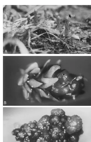
L. inundata plants were harvested from a wild population in Villepail (Mayenne, France). Vege-tative apices (3 – 5 mm) were excised from vegeta-tive stems (Fig. 1A). Apices were sterilized as previously established [10].
2.2. Callogenesis establishment
A total of five callogenesis media (CM-1 to CM-5) were used (Table 1). They contained full strength (CM-1, CM-2) or half-strength (CM-3 to CM-5) Murashige and Skoog medium (MS) [11] basal salts, 0.05 mM 3-indolebutyric acid (IBA) and 1.4 mM kinetin (Kin), and combinations of various additives (Table 1).
2.3. Indirect somatic embryogenesis establishment
For inducing SE, calluses were transferred on the medium used for direct shooting ofL.inundata
from vegetative apices [10]. This medium was com-posed of: 100 g/l NH4NO3, 100 g/l KNO3, 200 g/l KH2PO4, 50 g/l CaCl2, 80 g/l MgSO4and 1/4 MS microelements and was supplemented with 2.5mM IBA and 0.33 mM gibberellic acid (GA3). This medium is called embryogenesis medium (EM). When each embryo developed into a plantlet char-acterized by two to three leaf cycles and a few roots, it was excised and transferred to a new EM to induce growth.
All media (CM and EM) were supplemented with 20 g/l sucrose and 7.5 g/l agar (Pronatec). The pH was adjusted to 5.4 and hormones were
added to the media before autoclaving at 120°C for 20 min. All cultures were incubated at 209 1°C, in light at 50 mmol m−2 s−1 photosynthetic photon flux density and 14-h photoperiod.
2.4. Histological analysis
Calluses were propagated by subculturing onto fresh callogenesis medium once every 13 weeks. To study starch evolution during callogenesis, callus fragments were excised from the same calluses each 9 days during 13 weeks.
Calluses were transferred to EM medium to study the SE. Callus samples were collected from the same calluses once a week during 13 weeks. The samples were vacuum-infiltrated with FAE fixative (formaldehyde 37%:glacial acetic acid 100%:ethanol 50%, 6.5:3.5:100 ml) for 72 h at room temperature. Fixed tissue was dehydrated in a series of ethanol concentrations: 50, 70, 90, 100% (v/v) (30 min each bath). This material was vacuum-infiltrated with component A of JB-4 em-bedding plastic with catalyzer, and incubated at ambient temperature for 12 h. Components A and B were mixed together with catalyzer (30 min). The material was polymerized at 20°C in gelatin capsules for 50 min.
Thin sections (3 mm) were obtained using a Leica RM 2065 microtome. Sections were stained with PAS (periodic acid Schiff) or double stained with PAS and NBB (Naphtol Blue Black). PAS specifically stained polysaccharides red [12], and NBB stained soluble and reserve proteins bluish-black [12,13]. Observations were made with a light microscope.
To assess the mitotic reactivity of callus cells during culture, the surface ratio of the nucleus and/or the nucleolus was achieved after staining. Measures were established on ten cells/section, for ten sections/sample distributed all along the sample.
3. Results
3.1. Callogenesis propagation
3.1.1. Callogenesis establishment
died after 3 weeks of culture (Table 1). When the medium was relatively poor in mineral salts and in organic components (CM-5), all apices survived (Table 1). After 13 weeks of culture, the initiation of callogenesis was observed in the meristematic zone of each surviving apex (Fig. 1B). The callus was obtained only after three cycles of 13 weeks of culture on CM-5. The CM-5 medium was chosen
for callogenesis establishment and propagation, and is henceforth called the callogenesis medium (CM). Continuous propagation of callus could be obtained by subculturing on CM every 13 weeks. Initial callus (6 – 8 mm) was then subdivided into three to four small calluses (2 mm) which were separately cultured on CM for a new culture cycle.
Fig. 1. Establishment and morphological appearance of the nodular callus inL.inundata(L.) Holub. (A) Wild plant exhibiting vegetative apices (va) and sporangiferous apices (sa) in natural habitat, bar= 10 mm; (B) initiation of the callogenesis in the meristematic zone (mz) of the vegetative apex after one 13-week period on CM, bar=500mm; (C) nodular callus obtained after
Table 1
Effect of various additives and media composition on callus formation from vegetative apicesa
Micro-Macro- Additives (mg/l) % Of apices forming callus Medium tested
aEach medium was supplemented with 0.05
mM IBA and 1.4mM kinetin. CM, callogenesis media; MS 1/2, Murashige and
Skoog medium diluted twice. For each condition, 30 vegetative apices were cultured.
3.1.2. Morphological description
Since the 13th week of culture on CM (first period of the establishment), callogenesis was ob-served on the apical meristem and also on the axillary meristems situated at the basis of the four to five leaf cycles surrounding the apical meristem. During the second period, callogenesis extended along the explant only in the dorsal part. After three cycles of subculturing, calluses homoge-neously consisted of nodules that were green and compact (Fig. 1C) and further designated as nodu-lar callus. Each vegetative apex gives rise to one nodular callus (size 6 – 8 mm).
3.2. Histological analyses of callogenesis
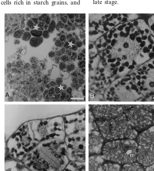
Calluses were uniformly composed of non-vac-uolated cells with prominent and central nuclei undergoing rapid cell division. Most of the cells exhibited various amounts of starch grains (Fig. 2A). Mitotic activity was intimately linked to the quantity of starch granules in the cytoplasm. Dur-ing the S phase of the cell cycle, each cell con-tained 20 starch granules, was 40 mm in diameter, and contained a nucleus of 20mm with a surface ratio of 0.25 (Fig. 2B). The increasing size of the nucleus is due to the ploidy of L. inundata
(2n=4x=156). The number of starch granules decreased to ten at the end of the telophase (Fig. 2C), then increased to 40 during the interphase (Fig. 2D). During the interphase, the diameter of the nucleus was 12 – 13 mm and the surface ratio was 0.1. When calluses were cultured more than 13 weeks on the same CM, they needed more time to reinitiate mitosis, they multiplied very slowly and did not give rise to organized structures after the transfer to embryogenic medium (data not shown).
3.3. Somatic embryogenesis
3.3.1. Morphological description
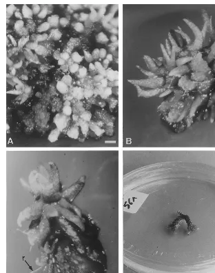
After 2 – 3 weeks of culture on EM, domes emerged non-synchronously from the callus. Within 3 – 5 weeks, they evolved to a cylindrical shape and covered the callus surface (Fig. 3A). After 6 – 8 weeks, these cylindrical structures had an enlarged base and tapered top. They acquired a ‘leaf morphology’ (Fig. 3B). Within 8 – 13 weeks, each structure developed a plantlet consisting of numerous leaves and roots (Fig. 3C). These plantlets developed dichotomies similar to the wild
L. inundata plants (Fig. 3D).
It was therefore established that organized structures could emerge from the callus surface, and undertake an autonomous development lead-ing to a normal plantlet. Characteristic stages observed such as the cylinder form, leaves and finally the plantlet were remarkably similar to those previously described in literature on zygotic embryogenesis of Lycopodiales [14 – 16], and led us to initiate an histological study for confirmation.
3.3.2. Histological analysis of somatic embryos
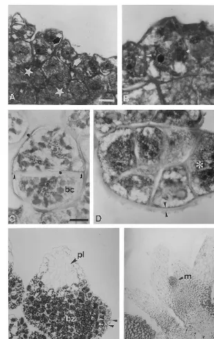
stage. The apical cell underwent one anticlinal or oblique division to produce a three-celled structure of 40 – 50 mm in size (Fig. 4C). The next divisions were periclinal to the surface resulting in the for-mation of cylindrical multi-celled structures of 70 – 80 mm (Fig. 4D). They were delimited by a thick polysaccharidic mucilage layer (2 – 3 mm in thickness) strongly stained by PAS, and presented one apical cell that was actively dividing. Within 4 – 5 weeks of culture, their growth led to a pyra-midal shape (250 mm). Since the 6th week of culture, this structure appeared lengthened, be-came bulbous at its basis, and its top was tapered (Fig. 4E). A single-celled layer, poor in starch grains and devoid in proteins as no blue stain was detected, was located at the surface and sur-rounded the structure as a protoderm. From this stage, two parts were distinct: a basal bulbous zone consisting of cells rich in starch grains, and
an apical tapered zone composed of vacuolated cells with neither starch grains nor soluble proteins located in the cytoplasm. Such a tapered zone is considered as an embryonary leaf. Between the 6th and 7th week of culture, the organized structures became more and more tapered, and showed a leaf structure (0.9 – 1 mm). They developed one lateral prolongation which helped the structure to emerge from the callus. Within the 7 – 8th week, an apical meristematic zone was noticed at the base of the embryonary leaf. This zone was consti-tuted of small cells containing proteins. At the 8 – 10th week of culture, a first leaf cycle was produced on the structure (2 – 3 mm) (Fig. 4F). Then, 3 weeks later, the second leaf cycle ap-peared. Following this stage, one to two roots were formed and the plantlet (\3 mm) began to differentiate tracheids. These events occurred at a late stage.
Fig. 2. Histological analysis of the nodular callus during the propagation phase. Sections were double stained with PAS and NBB. (A) Nodular callus contains non-vacuolated cells exhibiting various amounts of starch grains (star), bar=50 mm; (B) during S
phase of cellular cycle, a prominent nucleus is observed in a central position and the number of starch grains decreased, bar=10
mm; (C) during mitosis, and the end of the telophase, the number of starch grains is greatly reduced, bar=10 mm; (D) during
Fig. 3. Morphological appearance of the somatic embryos, derived from a nodular callus ofL.inundata(L.) Holub, during their development. (A) After 5 weeks on CM, numerous cylindrical embryos appeared on the surface of the callus, bar=500mm; (B)
after 8 weeks, a leaf morphology is observed, bar=500mm; (C) after 13 weeks, structures are isolated and exhibit numerous leaf
cycles and roots (r), bar=500mm; (D) after 23 – 26 weeks, dichotomies appear as in the wild plant, bar=20 mm.
Each callus that was initially 3 – 7.5 mm in di-ameter, gives rise to ten to 23 plantlets which are isolated and transferred onto new EM to encour-age their growth. More than 61% of plantlets were successfully acclimatized in the greenhouse. They remained green, developed new vegetative apices, roots and dichotomies. After 2 years in the green-house, all of them exhibited sporangiferous or-thotropic apices, and appeared morphologically identical to the wild plants.
4. Discussion
The first signs of callogenesis were observed after 13 weeks of culture of apices on CM medium
only in the meristematic zones. Histological analy-sis showed that the callus arose from the apical and axillary meristems. Axillary meristems also gave rise directly to shoots [10]. In this study, L.
inundata vegetative apices were transferred onto EM after 11 weeks of culture on CM. Callus only formed at the dorsal part on the vegetative ex-plant, opposite to the ventral part that forms roots.
The establishment of a homogenous nodular callus was possible only after three 13-week cycles of culture on CM. According to our histological studies and by comparison with another fern, M.
struthiopteris (L.) Todaro [9], whole callus in L.
connection was observed between the callus and organized structures.
Secondly, most of the phases described in L.
inundata are morphologically similar to those ob-served in the zygotic pathway in some Lycopods [14 – 16]. The cells designated as embryogenic cells
Fig. 4. Histological study of the somatic embryogenesis: characterization of the main developmental stages. Sections were double stained with PAS and NBB. (A) Embryonic cells (large arrows) appear at the surface of the callus. They were easily distinguishable according to their small size and colour after double staining. Inner cells show large amount of starch (star). Bar=20mm. (B) First divisions occur (ink blot) inside the structure which does not grow. A thick wall (arrow heads) surrounds
it. Bar=10 mm. (C) Three-celled stage: an apical cell undergoes mitosis (ink blot) while a basal cell (bc) is more voluminous.
Bar=10mm. (D) Cylinder stage: a thick mucilage (arrow heads) is located all around the embryo. The apical cells (asterisk) divide
actively. Bar=10mm. (E) Pre-leaf stage: a bulbous zone (bz) is characterized by starch-filled cells. The tapered zone corresponds
to the pre-leaf (pl) and contains no reserves. A protoderm (arrow heads) is differentiated and surrounds the embryo. Bar=50mm.
in our calluses have been cytologically well-defined. A three-celled structure is observed after 1 – 2 weeks of culture on EM. An asymmetrical division was reported to produce a three-celled zygotic embryo in H. selago [14], and subsequent periclinal and anticlinal divisions gave rise to a cylindrical structure. A cylindrical stage was also observed in the multi-celled embryo of L.cernuum
[15] like in L. inundata somatic embryos of 3 – 4 weeks. A cylindrical stage was also reported in SE in Asparagus officinalis L. (Liliaceae) [17].
A pyramidal stage has not been described in the literature on the zygotic pathway. We character-ized a similar stage inL.inundatasomatic embryos after 6 weeks of culture as Treub [15] described in
L. cernuum, but considered it to be a pre-leaf step in L. inundata. An inclusion, designated as the foot, was noticed inL.inundataafter 7 – 8 weeks of culture. In nature, the foot serves to hold the embryo to the prothalle [15], and such a step is also the only zygotic phase partially described in
L. inundata in the literature [16].
The differentiation of an apical meristematic zone in theL. inundataplantlet was noticed, and it is assumed that in ferns, the apical meristem is a single pyramidal cell designated as the initial cell. Mitosis occurs on all its faces. The daughter-cells surround it and may undergo mitosis. In our Lycopod, we did not observe a very distinguish-able apical cell, but instead saw a group of small cells, strongly stained with NBB (for proteins).
The size (2 – 3 mm) of the Lycopodiale gameto-phytes in natural conditions [14 – 16] does not eas-ily allow investigations, so zygotic embryogenesis is not well-defined for Lycopods. Information on zygotic embryogenesis was unavailable on Ly-copodiales since the end of the 19th century. If we hypothesize that both the zygotic and somatic pathways are similar, as is assumed for Sperma-phytes, L. inundata is a convenient material for such investigations.
As no other embryogenesis system exists in Pte-ridophytes, we focus on the other models available
in Spermaphytes (Gymnosperms and
An-giosperms) to highlight common or specific fea-tures of somatic embryogenesis among Plantae. Such a SE in a Lycopodiale member exhibits characteristics slightly different from those de-scribed in Spermaphytes. In Encephalartos cycadi
-folius, Encephalartos dyerianus, Encephalartos natalensis [18,19] and Ceratozomia mexicana [20],
some primitive Gymnosperms, the first division of the embryogenic cell is asymmetrical, and pro-duces an embryonary mass with a pseudo-suspen-sor. Polyembryonary is often observed and differs from our observations on Lycopodiella. However, they all need an extremely long time to cover all the steps for in vitro propagation.
In Angiosperms, a protoderm differentiates dur-ing the globular stage. In L.inundata, the globular stage is compared with the cylindrical structure where no histo-differentiation occurs. The proto-derm appears later, simultaneously with the estab-lishment of bipolarity (tubercle-leaf). Such polarity is quite different from the Angiosperm heart-em-bryo stage as L.inundataexhibits polarity with the starch-filled cells. Vascular tissues were present in earlier stages in Angiosperms than in L. inundata, where tracheids appear later in the plantlet (with the second leaf cycle).
Usually, it is stated that somatic embryos are derived either from a unique embryogenic cell or from a cluster of embryogenic cells well-character-ized in cytology [12,21,22]. Cytological studies al-low embryogenic cell characterization. When Spermaphyte embryos are of unicellular origin, the embryogenic cells are isolated from one another by an important modification of their cell walls, in particular gelification of the middle lamella, and undergo polarized division, thus forming globular proembryos [21,22]. In this work, we provide evi-dence for the unicellular origin of embryos. More-over, embryogenic cells exhibit cytological features common to Lycopodiales (Pteridophytes) and some Spermaphytes.
In conclusion, this first report on SE in L.
inundata (Lycopodiales, Pteridophytes), gives new insights to compare developmental biology espe-cially in embryogenesis. From our knowledge, SE has not previously been described among Pteri-dophytes. Moreover, considering the callus propa-gation phase, and the Lycopod alkaloid functions for pharmaceutical purposes, sufficient masses of nodules could be produced for alkaloid extraction and characterization. Preliminary results on L.
inundata alkaloids were encouraging (Atmane and Fliniaux, unpublished data).
Acknowledgements
Conserva-toire Botanique National de Bailleul (Bailleul, France). This research was partly supported by a ‘Contrat Plan-Etat-Re´gion’ to the Laboratoire de Physiologie Cellulaire et Morphogene`se Ve´ge´tales (Villeneuve d’Ascq, France). We thank Dr A. De-ram and Professor H. Van Dijk for critically read-ing this manuscript.
References
[1] R. Prelli, Guide des Fouge`res et Plantes Allie´es, Lecheva-lier, Paris, 1990, pp. 65 – 77.
[2] G.H. Parent, Disparition et survie des Lycopods, Bull. Nat. Belg. 45 (1965) 506 – 556.
[3] K. Berkefeld, Eine Nachweismo¨glichkeit fu¨r Kondombe-nutzung bei Sexualdelikten, Archiv. Kriminol. 192 (1993) 37 – 41.
[4] U. Schippmann, Plant uses and species risk — from horticultural to medicinal plant trade, in: J. Newton (Ed.), Planta Europa Proceedings of the First European Conference on the Conservation of Wild Plants, Ip-swich, London, 1995, pp. 161 – 165.
[5] J. Bruneton, Pharmacognosie, Phytochimie et Plantes Me´dicinales, Lavoisier, Paris, 1993.
[6] H. Du Prat, Traite´ de Matie`re Me´dicale Home´opathique, vol. 2, Baille`re, Paris, 1981, pp. 900 – 901.
[7] X.C. Tang, Huperzine A (Shuangyiping) : a promising drug for Alzheimer’s disease, Acta Pharmacol. Sin. 17 (1996) 481 – 484.
[8] J. Grunwald, L. Raveh, B.P. Doctor, Y. Ashani, Hu-perzine A as pretreatment candidate drug against nerve agent toxicity, Life Sci. 54 (1994) 991 – 997.
[9] R.C. Thakur, Y. Hosoi, K. Ishii, Rapid in vitro propaga-tion of Matteuccia struthiopteris (L.) Todaro — an edible fern, Plant Cell Rep. 18 (1998) 203 – 208. [10] N. Atmane, Multiplication d’une Lycopodiale me´dicinale
menace´e de disparition [Lycopodiella inundata (L.) Holub] par les techniques de culture in vitro et inte´reˆts
pour ses alcaloı¨des endoge`nes, Ph.D. Thesis, University of Sciences and Technology of Lille, France, 1999, pp. 1 – 124.
[11] T. Murashige, F. Skoog, A revised medium for rapid growth and bioassays with tobacco tissue culture, Phys-iol. Plant. 15 (1962) 473 – 497.
[12] N. Michaux-Ferrie`re, H. Grout, M.P. Carron, Origin and ontogenesis of somatic embryos inHe6ea brasiliensis
(Euphorbiaceae), Am. J. Bot. 79 (1992) 174 – 180. [13] D.B. Fisher, Protein staining of ribbonned epon sections
for light microscopy, Histochemistry 16 (1968) 92 – 96. [14] H. Bruchmann, Die Keimung der Sporen und die
En-twicklung der Prothallen von Lycopodium cla6atum L., L. annotinum L. und L. selago L., Flora 101 (1910) 220 – 267.
[15] M. Treub, Etudes sur les Lycopodiace´es. A. Le prothalle de Lycopodium cernuum L., Ann. Jard. Bot. Buitenz 4 (1884) 107 – 138.
[16] K. Goebel, Uber Prothallien und Keimpflanzen vonLy
-copodium inundatum, Bot. Zeit. 45 (1887) 161 – 192. [17] S. Rivie`re, Les activite´s me´riste´matiques durant
l’ontoge-ne`se d’une plantule de Monocotyle´done a` germination hypoge´e : l’Asparagus officinalis L. (Liliaceae), C. R. Acad. Sci. Paris 277 (1973) 293 – 296.
[18] A.K. Ja¨ger, J. Van Staden, Somatic embryogenesis in
Encephalartos cycadifolius, Plant Cell Rep. 15 (1996) 437 – 440.
[19] A.K. Ja¨ger, J. Van Staden, Somatic embryogenesis and organogenesis inEncephalartos dyericanusandE.natal
-ensis, Plant Cell Tissue Org. Cult. 45 (1996) 99 – 102. [20] V.M. Chavez, R.E. Litz, J. Marquez, Histology of
so-matic embryogenesis of the cycadCeratozomia mexicana
var.robusta(Miq.) Dyer, Plant Sci. 108 (1995) 191 – 200. [21] J. Loiseau, N. Michaux-Ferrie`re, Y. Le Deunff, Histol-ogy of somatic embryogenesis in pea, Plant Physiol. Biochem. 36 (1992) 683 – 687.
[22] N. Michaux-Ferrie`re, J. Schwendiman, Histology of so-matic embryogenesis, in: Y. Dattee, C. Dumas, A. Gal-lais (Eds.), Reproductive Biology and Plant Breeding, Springer, Berlin, 1992, pp. 247 – 259.