Vol 6, No 2, April - June 1997
The
Importance
of Fresh
Purposes
Rino Pattiata,l'2,3 Ikuya Nonaka 3
Fresh Frozen Section 73
Frozen Section
in
Muscle Biopsy
for
Diagnostic
Abstrak
Penggunaanpotong bekupadabioPsi otot telah dilakukanpada berbagaiinstitut sejaktigapuluhtahunyang lalu. BanyaklæIainan neuromuskuler baru ditemukan sejak penggunaan potong beku, larena beberapa l<elainan patilogis otot hanyi dapat ditimukan pada potong beku termasuk miopati mitokondial dan berbagai miopati kongenital nonproiresif. -D"ngo, ditemuk)nnya a*tropi
aan dystrophin-associated glycoprotein (DAG) maka kzlainan distrofi otot lebih dikenal. Deigoi p"rgg:unoon distrofin dan
DAi
secara imunohistokimia, diagnosis distrofi otot dapat ditegakkan lebih alaffat. Melihatft^"iuo"'y"iJ
ada ini, maka sudah waktunya penggunaan teloik potong beku pada biopsi otot diterapkan di Indonesn.Abstract
to introduce muscre histochemistry of freshfrozen muscre
specim""";:i:::;;ffi'^o^"8
atrhis progress' it is timefor Indonesia
Keywords: histochemistry, immunohistochemistry, muscular dystrophy, congenital myopathy.
Muscle biopsy is a diagnostic tool which provides us the knowledge to detect abnormalities in muscles. The
pathological
findings reflect
the
pathogeneticmechanism whether
it is
myopathic, neuropathic or both. In the last two decdes, we have seen a renewed and growing interest in muscles because of theapplica-tion of new techniques, including histochemsitry,
im-munohistochemistry, electron microscopy,
and molecular biology. Thus we have detected anumberof
newmusc
and understoodt
pathophys
scle pathologies. sy is arel
e procedure. Tol.
Department of Anatomic pathology, Faculty of Medicine, University of Indonesia/Cipto Mangunkusumo National Central General Hospital, Jak)rta, Indonesia2.
International Center of Medical Research, Genetic3
';::3?:;iT,
biopsy specimen, histochemical methods are needed.
In Indonesia muscle biopsies were embedded in
paraf-fin without histochemical treatment.
In this paper we
will
describe the technique of fixation e biopsy histochemically as well ically. and underline rhe pitfallsMUSCLE BIOPSY AND
THE STAINING
Muscle biopsy
is a
relative simple procedure. How_ ever, one must make:îJT:Jïlocalanesrh":tî;:
we may use
se
neraliopsy any
mu
ld
tobest site for biopsy, because 1)
it
is easy to reach themuscle tissue; 2)
it
contains abundant peripheral ner-ves and muscle spindles; 3) typeI,2A,
and 28 fibers are distributed in a mosaic pattern; and 4) there is littlefibrous interstitial tissue.
l2
The biopsy is done as follows: after cleaning with an
antiseptic, the skin is infiltrated by local anesthesia.
It
is important not to infiltrate the muscle, because the local anesthetic agent is highly toxic to the muscles, thus inducing muscle fiber necrosis. A skin incision of about 3-4 cm is performed along the directionof
themuscle fibers. After retraction of the skin, the fascia is
incised to expose the muscle fascicles. Then a segment of the muscle is cut off by a pair of scissors. The fascia is sutured and the skin is closed.
The muscle specimen should be frozen as quickly as possible. According to our experience, we can delay
it till
30 minutes. We do not use direct immersion inliquid
nitrogen, because nitrogen gas bubbles sur-rounding the specimenwill
appear and retard thecool-ing process, thus causcool-ing artefacts. The specimen is placed on a piece
of
cork with tragacanth gum, fixed in isopentane, and cooled with liquid nitrogen.If
necessary, before fixing the specimen, a part of themuscle is fixed for electron microscopy.
The
specimenis
then readyto
be
stained.In
our laboratoryin
Japan,we
perform a batteryof
his-tochemical stainings including hematoxylin and eosin (H&E), modified Gomori trichrome (MGT), reduced nicotinamide adenine dinucleotide reductase(NADH-TR),
succinic dehydrogenase (SDH), periodicacid-Schiff
(PAS),oil
red
O,
adenosine triphosphatase (ATPase),non
spesific esterase (NSE), acid phos-phatase, alkaiine phosphatase, acetylcholine esterase,cytochrome C-oxydase (CCO), adenosine monophos-phate (AMP) deaminase, phosphorylase, phosphofruc-tokinase
(PFK),
menadione-linked alpha-glycerol-phosphate dehy drogenase. 2H&E is the most important staining, because most of the pathological information can be traced by this stain-ing. We can differentiate the fibers with H&E staining; type 1 fiber is stained darker than type 2. However, we need additional staining
for
further differentiation. With simpleH&E
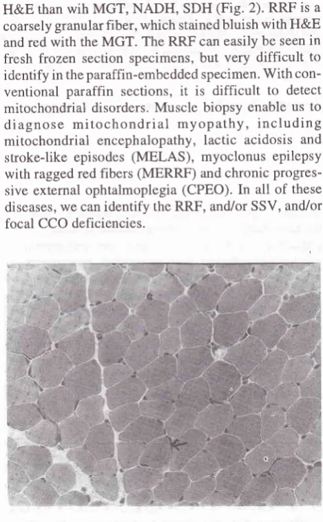
staining, we may overlook several abnormalities such as nemaline bodies, cytoplamic bodies, central core, which are commonly seen incon-genital non-progressive myopathies. (Fig. 1)
In mitochondrial myopathy, some important findings
[image:2.595.305.540.92.278.2] [image:2.595.308.543.328.707.2]are red ragged fibers (RRF), strongly SDH blood ves-sels (SSV) and
focal
deficiencyof
cytochrome C-oxydase activities. RRFis
staineddifferently
withFigure l. Myotubular myopathies, H&E staining, 40x, H&E = hematoryclin & eosin
H&E than wih MGT, NADH, SDH (Fig. 2). RRF is a coarsely granularfiber, which stainedbluish with H&E
and red with the MGT. The RRF can easily be seen in fresh frozen section specimens, but very
difficult
to identify in the paraffin-embedded specimen. With con-ventionalparaffin
sections,it
is
difficult
to
detect mitochondrial disorders. Muscle biopsy enable us todiagnose
mitochondrial
myopathy, including
mitochondrial encephalopathy,lactic
acidosis and stroke-like episodes (MELAS), myoclonus epilepsy with ragged red fibers (MERRF) and chronic progres-sive external ophtalmoplegia (CPEO).In
all of these diseases, we can identify the RRF, and/or SSV, and/or focal CCO deficiencies.Figure 2a. RRF, H&E staining, 40x, RRF = red raggedfibers,
Vol 6, No 2, April - June 1997
[image:3.595.39.273.92.313.2]Fresh Frozen
Section
75Figure 2b. RRF, MGT staining, 40x, RRF = red raggedfibers, MGT= modirt.ed Gomori trichrome
Modified Gomori trichrome is an important staining to identify some specific structure. With this staining we
can
seethe
myelinated fibers,
nemaline bodies,bodies,
tubular
aggregates
and . Nemaline body myopathy can easily be fresh frozen sections,but difficult
in paraffin sections after formalin fixation. Various con-genital nonprogressive myopathies have been foundsince 1963, when Shy et al first described the nemaline myopathy.' Before that time a number
of
muscular disorders in paraffin sections might be overlooked. NADH-TR staining also helps us to detect myofibrillar organization and to classify muscle fibers. Almost all patientswith
pathological abnormalities may show structural disorganization with this staining. NADH is ingi:
be-cause
the
core
region lacks
mitochondria.
Thesease all family members of the patient must be care_
fully
assessed.blood vessel in rnitochondrial rnyopathies.
Also important is rhe lipid staining such as
oil
red O.The excess accumulation
of lipid
dropletsin
musclefibers is demonstrated in mitochondrial myopathy and
lipid storage myopathy.In paraffin embedded sections,
we cannot detect the
lipid
in
the specimen, because lipid is soluble in the alcohol, used either forembed-ding or for deparaffinization.
If
the lipid infiltrates theinterstitium, we can still see the shape of the lipid cell.
Because the accumulation of
lipid
droplets in musclefibers is so tiny, we cannot identify it even in well fixed frozen sections.
Non specific esterase (NSE) may help to identify small angular
fibers
after
denervation.The
enzyme is elevatedat
the
neuromuscularjunctions
andin
lysosomes. Acid phosphatase is increasedin
degene-rating and regenerating fibers especially in the former
with active phagocytosis, becuase this enzyme is
high-ly
concentr4tedin
lysosomes andin
macrophagis. Alkaline phosphatase is sometimes increased in early stage regenerating fibers.Cytochrome C-oxydase, is an enzyme in mitochondrial electron transport system and
its
activityis
demons_y.
I
"i",
inc
MELAS, these enzyme deficient fibers are scattered
y. The staining confirm the
as
well
as the presenceof
y specimens. In a few cases the histopathological
findings
showa
CCO focal deficiency without any RRF changes (Fig.3). [image:3.595.298.529.511.705.2]ATPase provides us many information. To complete the staining, it takes at least 2 hours. We can differen-tiate the fiber type into type
l,2A,2b
and 2C fibers(fig.a). In normal human muscles, type 1, 2A, and2B
fibers are almost equally distributed
in
a mosaic pat-tern, so a disturbed mosaic pattern is a pathognonomic finding of a neurogenic process. There is a reciprocal relationship between phosphorylase and the oxydative enzymes. In general, typeI
and 2 fibers are recognizedby
the
different
concentrationof
ATPase, phos-phorylase, and the other oxydative enzymes. However,in
some fibers this quantitative differentiation is un-clear. lOther stainings which may provide us some diagnostic clues are phosphofructokinase and menadione linked
alpha- glycerophosphate dehydrogenase.
IMMUNOHISTOCHEMISTRY
Fresh frozen tissue is also applicable for
immunohis-tochemistry
to
detect specific protein defects which play important rolesin
the pathogenesisof
diseases,while it is not traceble from paraffin embedded tissue.
It
is not always easy to make a diagnosis of musculardystrophy, from clinical and morphological findings;
for
instance, diagnosing
limb girdle
musculardystrophy and mani-festing carriers of Duchenne mus-cular dystroptry
(lltp)
by clinical andhistopathologi-cal findings alone.''o To differentiate the disease, an immunohistochemical staining by using antidystrophin
antibody gives a
lot of
help. Dystrophinis
a protein productof
the gene.v and is almost completely absent in the sarcolemma in DMD muscles, minimal to patchyin Becker muscular dystrophy and in the mosaic pattern of manifesting DMD carrier. In other progressive
mus-cular
dystro-phies
the dystrophin
is
positively
.
/.to.tt
'
present.McDonaldl2reported
a
case whoseEMG
showedneurological changes, but histopathological findings suggested a myopathic process. Dystrophin staining
helped
to
make
the
final
diagnosis. Even
in a
myopathic process, the biopsy may show a neurogenic
pattern for several reason. First, segmental
degenera-tion of indivi-dual muscle fibers might be followed by segme-ntal
re-
innervation (myopathic
reinnerva-tion).t3 Secondly, in chronic myopathies, splitting
of
muscle fibers may give the false appearance of a group
atrophy.l4
Conu"rr.ly,
myopathic histological pat-terns may be-seen in chronically denervated muscles."Conflicting EMG
andbiopsy results
have been describedin
some genetic neuromuscular disordersincluding Emery-Dreyfuss muscular dystrophy, limb
girdle
muscular dystrophy, Charcot-Marie-Toothsyndrome and spinal muscular atrophy."
Another importance
of
dystrophin staining is,it
can determine certain typesof
muscular dystrophies asproposed by the workshop sponsored by the European
Muscular Centre. There main muscular dystrophies can be diagnosed by the dystrophin staining, namely
Duchenne muscular dystrophy,
Becker
musculardystrophy
.'
and the ruling outof
limb girdle muscular to-ttoy stropny.
The advantages
in
muscular dystrophy diagnosis is based on the discovery of structure and function of the [image:4.595.56.546.516.722.2]dystrophin associated
glycoprotein(DAc).
Dystro-phin is associated with a large oligomeric complex ofVol 6, No 2, April - June 1997
sarcolemmal proteins, comprised
of
a i56-kdglyco-protein (156DAG), a 43-kd prorein (43DAP), a 50-kd
glycoprotein (50DAG). a 35-kd
glycoprotein
(35DAG) and a 25-kd prorein (25 DAP). tÔ The
abnor-malities of 50DAG, for example, is pathognonornic for
the severe childhood autosomal recessive muscular
dystrophy (SCARMD).i7
This
diseaseis
highly
prevalent
in
North Africa, but also reportedin
othercountries" The overall clinical symptoms mimic DMD.
V/ithout
immunohistochemical
staining on the
sporadic patient it may be difficult to obtain a definite diagnosis.
The discovery of utrophin has stimulated us to
recon-sider the treatment of DMD. If the secretion of utrophin
could be increased
by
a genetic or pharmacologicalmanipulation, it could be Lreneficial for the prevention
of muscle degeneration. l6
THE ADVANTAGES OF FRESH FROZEN SEC.
TION
ON MUSCLE BIOPSYAs
mentioned above, there are many advantages,beginning from an accurate diagnosis to the
pathophy-siology
of
muscle diseases by performing a muscle biopsy in fresh frozen sections.It
is notdifficult
and easy to handle. The cooperation between the clinician and the pathologist is necessary to promote theproce-dure. Without clinical and Iaboratory data, it is hard to
make a pathological diagnosis. Some special
skill
isnecessary to perform the biopsy
in
orderto obtain
enough samples
for
pathological examinations. Themost important thing
is
howto
process the musclebiopsy specimens for histochemical examination.
One of the advantages of fresh fiozen sections is that
the clinician
will
get a tentative answer within 2 hours after the muscle biopsy.After fixation we cut
thefrozen section with a cryostat and then perform the 3
most important stainings,
H&E,
MGT
and NADH, SDH and CCO. The staining can be finishedin
60 minutes. The final pathological diagnosis will be givenafter routine histochemical and immunohistochemical
stainings, usualy 2-7 days after the muscle biopsy. If
it
is an urgent case, all the histochemical stainings can be done
on
the same day.If
there isstill
enough tissue,one can make formalin fixed and paraffin embedded
sections which may help to identify infiltrated cells in
more details. For routine examinations, formalin
fixa-tion can be ommited.
The important stainings in Indonesia are H&E, MGT, NADH, SDH, CCO and ATPase. From these six
stain-ings, we can obtain approximately 997o
of
definiteFresh Frozen
Section
77pathological diagnoses. Other stainings
to
becon-sidered in Indonesia are PAS, oil-red-O, NSE, and the
phosphatases. Stainings
like
PFK,
phosphorylase,AMP
deaminase areonly
exceptionally necessary,rnaybe once or twice a year.
As
described above,it's
tirneto
introduce the freshfrozen section technique
for
muscle biopsyin
In-donesia. Many abnormal findings can be highlightedby histochemical stainings using the fresh frozen
sec-tions. We
will
get more appropriate diagnoses and thus better treatrnent and follow up of the patient. Research work would be stimuiated and cooperationwith
the Eyckman Institute invoiving mitochondrial diseasescould be started. However,
if
westill
depend oncon-ventional paraffin-embedded sections,
we
will
staybehind and miss many important findings"
A comparative study
on muscle biopsy betweenfor-malin and paraffin embedded sections and fresh frozen
specimens has proven the superiority of the latter.
ACKNOWLEDGEMENT
Dr. Rino Pattiata holds the Monbusho scholarship from
the
Governmentof
Japanto
work
in
the ICMR
laboratory, Kobe University School of Medicine.
REFERENCES
1. Dubowitz V. Muscle Biopsy. 2nd Ed. London: Baillere Tindall, 1985
2. Nonaka I. Muscle biopsy for clini cians (in Japanese). Tokyo: Nihoniji-shinpo, 1994
3. Shy GM, Engel WK, Somers JE, Wanko T. Nemaline myopathy: a new congenital myopathy. Brain
1963;86:799-810
4. Bodenstein JM. Congenital myopathies. Muscle Nerve 1994:17:l3l-44.
5. Arikawa E, Hoffman EP, Kaido M, Nonaka I, Sugita H,
Arahata K. The frequency of patients with dystrophin abnor-malities
in
alimb-girdle
population. Neurologyl99l;41:1491-6.
6. Middleton L, Moser H. Rare neuromuscular disease. In: Emery AEH, editor. Diagnostic criteria for neuromuscular
disorders. B aarn: European Neuromuscular Centre, 1994:9-l3
7. Hoffman EP, Kunkel LM, Angelini C, Clarcke A, Johnson
M, Harris JE. Improved diagnosis
of
Becker musculardystrophy : dystrophin testing. Neurology I 989;39 : I 0 I I -7
8. Norman P, Coackley J, Thomas N, Harper. Distinction of
Becker from limb-girdle muscular dystrophy by means of
dystrophin cDNA probes. Lancet 1989;l:466-8.
9.Hoffman EP, Brown RH, Kunkel LM. Dystrophin: the
protein product of the Duchenne muscular dystrophy locus.
10. Jennekens FGI, ten Kate LP, de Visser M, Wintzen AR.
Duchenne and Becker muscular dystrophy. In: Emery AEN,
editor. Diagnostic criteria for Neuromuscular disorders. Baam: European Neuromuscular Centre, 1994: 9-13.
ll.
Bushby KM. Limb girdle muscular dystrophy. In: EmeryAEN. editor. Diagnostic criteria for Neuromuscular
disor-ders. Baarn: European Neuromuscular Centre, 1994:25-31.
12. McDonald TD, Medori R, Younger DS, Chang HW, Mineti
C, Uncini et al. Becker dystrophy of spinal muscular atrophy
-dystrophin studies resolve conflicting results of
electromyography and muscle biopsy. Neuromusc Dis
1991; l:195-200.
13. Hernriksason KG, Stalberg E. The terminal innervation
pat-tern in polymyositis: a histochemical and SFEMG study.
Muscle Nerve 1978;l :3-12.
14. Bradley WG, Jones MZ, Mussini JM, Fawcett PR.
Becker-type muscular dystrophy. Muscle Nerve 1978; I :1 I 1-32.
15. Achan AN, Anderson MS. Myopathic changes in
amyotrophi c lateral sclerosi s. Neurology 197 4;24:47 7 -88.
16. Matsumura
K,
Campbell KA. Dystrophin-glycoproteincomplex: its role in the moiecular pathogenesis of muscular
dystrophies. Muscle Nerve 1994; I 7:2- 1 5.
17. Matsumura K, Tome FMS, Collin H, Azibi K, Chaunch M,
Kaplan JC. 50kd dystrophin-associated glycoprotein in