Microbiological
physiology,
structure,
diagnosis
and
future
perspectives
of
Chlamy dis
p n
eumo
nine
infection
Ariati
Safiriani
Abstrak
Chlamydia pneumoniae (C. pneumoniae) adalah smtu patogen intraseluLar obligat yang baru diidentifikasi pada tahun 1989. Bakteri
ini
digolongknn sebagai spesies baru dari Chlamydia, berdasarknndefnisi
uLtastruktur dan analisis homologi asam deoksi ribonukleat(DNA).
C. pneumoniae memiliki siklus hidup bifasik yang unik, dengan dua bentuk yang berbeda, yaitu badan elementer dan badan retikuLat. Patogenini
dapat mensintesis DNA, RNA dan proteinnya sendiri; namun tidak memilikijalur
metabolik untuk mensintesisATP.
PenggolonganC.
pneumoniae diantara patogenyang "baru
muncul", mungkin disebabkanoleh
karena diagnosisnya yang cukupsulit.
Diagnosis laboratorium berdasarkan pada kuhur, identffikasi spesimen biologis menggunakanantibodi monoklonal, "polymerase chain reaction" (PCR), dan metoda-metoda seroLogis lainnya; tidak cukup banyak tersedia. Beberapa spesies Chlamydia yang berbeda
juga
memiliki persama.an antigenik, sehingga dapat menyebabkan spesifisitas yang rendah. Berbagai tes diagnostik mutakhir, dengan beberapa kelebihan dan kekurangannya akan dibahas lebih terinci pada makalahini.
Perspektif masa yanq akan datang pada metoda diagnostik; seperti standarisasi PCR, kloning dan sekuensing gen omp-4 dan omp - 5, serta p enemuan vaksin ; sedang dalam proses pengembangan lebih lanjut.Abstract
Chlamydia pneumoniae (C. pneumoniae) is a unique obligate intracellular pathogen which has been recently identif.ed
in
1989. The patlrcgen was classified asa
new speciesof
ChLanrydia, basedon
its ultastructural definition and deoryribonucleic acid (DNA) homology analysis.It
has a distinctive biphasic life-c1'cle, with nvodffirent
forms, the Elementary Body and the Reticulate Body. The pathogen could synthesize its own DNA, ribomtcleic acid (RNA) and protein, butit
lacks the metabolic pathway of producing adenosine triplnsplmte(ATP).
The classificatiort of C. pneumoniae among "new and emerging" pathogens is probably due 1o therather
dfficult laboratory
diagnosis. lnboratory
diagnosis basedon
the culture, identificationof biological
specimens using monoclonal antibodies, polymerase chain reaction (PCR) and serological metlnds, are not widely available.Dffirent
Chlamydial species, whichare
very similar genetically, could also causelow
specificityin the diagnostic
tests.
Several advantages and disadvantages of the recently developed diagnostics test are expLicated. Sonte future perspectives in the diagnosis methods, such as the standardization of the PCR assays, the cLoning and sequencing of the onrp-4 and omp-5 genes, and vaccine development, are currently in progress.Keywords: C. pneumoniae, ultrastructure, biphasic lift-cycLe, Laboratory diagnosis
Chlamydia pneumoniae (C. pneumoniae),
which
wasfirst
described
in
1986,
is an
obligate
intracellular
pathogenand a common
causeof
respiratory infection.Many
reportshave indicated
the importance and the
relevance
of
C.
pneumoniae
not only
in respiratory
tract
infection,
but
also
in
extrapulmonary
diseases.l'2'3'a'5Antibody
prevalence
ratesin Western
countries
reaches
50Voin the
âdult population
andremain
high
in
old
age,
suggesting
a
high rate of
reinfection.
Diagnosisis hampered by
the
requirementfor
specializedculture
techniques
and reliance upon
expensive serological
tests
or
Polymerase
ChainRespiratory Department Hoechst Maion Roussel Indonesia, Jaknrta, Indonesia
Reaction (PCR).
The wealth
of
knowledge conceming
this
pathogen has increased
dramatically
in
the last
decade, setting up theway to
further exciting research
lines.History of
anew
pneumonia
agent
There are three species
of
Chlamydia:
C. pneumoniae,C. trachomalis and
C. psittaci. Therecognition
of
C.Safiriani
history
of
bird contact.T
In
1965,
during a trachoma
vaccine
trial
in
Taiwan,
an
atypical
strain
of
Chlamydia was
obtained
from
the conjunctiva
of
aschool
child,
and identified
on
the
yolk
sac chick
embryo egg number
183.The
strain
was namedTW-183.
A
further step
was
madein
1983,when
anotherisolate
antigenically similar to TW-183
was
obtainedfrom
the pharyngeal swab
of
a university
student
in
Seattle.
suffering
from
pharyngitis. The strain
was namedAR-39
(Acute
Respiratory).bBetween 1985 and 1986,
two studies
were
publishedon
the clinical
relevance
of
respiratoly
infections
sustained
by
unusual
strain
of
C.
psittaci.s''
Suikkuin
1985,
reported an epidemic of
mild
pneumoniadiscovered
during
a
chest radiographic survey of
young adult
in
Finland. The
followingyear,
Graystonrlescribed an
isolation
of
a C. psittaci
strain
within
apopulation
of
university
students withrespiratory tract
infèctions.
Following
ultrastructural
definition
andDNA
homology
analysis.in
1989, anew
classificationof
athird
,p".i"i
of
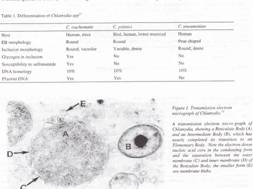
Chtamyttia, was recognized.roTable I . Diil-erentiatio n ot Chlamydia spptl
Med J Indones
Within the three Chlamydia species,
there are
somedifferent
characteristics, such as the host rangeof
each species, themorphology of
theElementary
Body
(EB)
and
the
inclusion
body,
their
susceptibility
tosulfonamide,
the
DNA homology and
the
plasmid
DNA
(table
l).
Microbiological physiology
and
structure
The modality
of
replication
is
a
characteristic
of
the genusof
Chlamydia, and
the
three
speciesshow
the samecomplex
cycle.
Chlamydia
has abiphasic
life-cycle.
TheElementary
Body (EB)
is the smaller
(300-400 nm), extracellular,
infectious
form
and
theReticulate
Body (RB)
is the intracellular,
non-infectious, metabolically active
form.
The
RB is
abigger
form
(800-1000 nm)
andit
is
capableof
binary
division.
The
Intermediate Body
(IB)
is
anintermediate
fbrm between
the
RB and the
EB.
It
hasa
condensed
DNA, hence
it is
also called
the CondensingForm (Figure
l).rr
C. tachomatis C. 1-tsittaci C. pneumoniae
Host
EB morphology Inclusion morphology Glycogen in inclusion
Susceptibility to
sulfonamide
YesDNA homology lOVo
Plasmid
DNA
YesBird, human, lower
mamtral
HumanRound
Pear-shaPedVariable,
dense
Round, denseHuman, mice
Round
Round, vacuolar
Yes No
No
lÙVo
Yes
No No to% No
Figure
L
Transmissiort electron micrograph of Chlaml'dia'| tA
Iransmission electron nticro-graphof
Chlamydia, showing a Reticulate Body (A) and an Intermediate Body (B), u'hiclt has
nearly
completedits
transitiort
to
anElementary Body. Note the electron-dertse
[image:2.595.81.577.353.724.2]Chlamydia
is
a
nonmotile and nonpiliated
micro-organism.
It
has surface projections
which
allow
nutrient
uptake
from
the host
cytoplasm.
,?
vivo,
the
infectious
EB
attachesto
microvilli of
susceptible hostcells
and
actively
penetratesthe
host
cells.
EB
thenreorganizes
into
metabolically active, dividing
RBswhich
areable
to
synthesizeDNA, RNA
andprotein,
but
could
not
produce
ATP. The
reason
why
Chlamydia
is
called
as an energy parasitesis,
it
lacksthe
metabolic
pathway
of
producing
ATP,
andtherefbre
it
is
dependent on the hostcell
fbr
its ATP.r2
Ninety two
hoursafter infection, the
hostcells
releasethe
EB which could
further
int'ect other host cells.r3C hlamy dia p n
eumoniae
infection
C. pneumortiae
infection
is
usually
asymptomatic or
minimally
symptomatic.
C.
pneumoniae
commonly
causes
upper (pharyngitis, sinusitis, and
otitis) and
lower respiratory
tract
intèction
(pneumonia,
acutebronchitis,
exacerbations
of
chronic
obstructive
pulmonary
disease/COPD).
C. pneumoniae
is also
linked
to
some
other chronic
diseases,
such
asatherosclerosis,
coronary
heart
disease,
arthritis,
G u i I I ain -B arre sl,ndrome, and erythema nodosum.s' I a' I 5' I 6
C.
pneumoniae
conld
be found worldwide,
and
it
ismore
common
in
tropical
countries.
The
sero-prevalence
peak
is
in
adr-rltsover
70
years.
Male
ismore commonly intècted than
fèmale.
There
is
no seasonalperiodicity for
C.pneumoniae
infection.
Itsinfection
seems
to
be
both endemic and
epidemic.Epidemic infection
seemsto
occur
in
cycles
with a
periodicity of
3 to4
years, and theygenerally
last 6 to8
months.li
Laboratory
Diagnosis
of
C.pneunrcniae
The
first
isolate
of
C.pneumoniae
wasobtained
from
egg
yolk
sac
culture
of
the conjunctival swab
of
aTaiwanese child.3
However,
theegg yolk
sacculture
method showed
a
low
sensitivity
for
C.
pneumoniaeisolation.
Several
cell lines
havebeen
used,such
asthe
HeLa 229
and
McCoy cells, and
showed
bettersensitivity
than the eggyolk soc.''
Othe.
cell
lines, thehuman
respiratory
cell
lines,
HL,
Hep-2
andH-292
cells
present
a
high
sensitivity
fbr
C.
pneunortiue
isolation
and propagation. The
culture
could be
confirmed
by
fluorescent-antibody
st4ining
with
species-specific monoclonal anti-body.re
Specimenfor
C.pneumoniae
culture can be obtainedfrom
manysources,
including
pharyngeal
swab,
sputum,bronchial aspirate
or
bronchoalveolar
lavage
andpleural
fluid.
The handling and
storageof
specimen,require particular
care dueto the
thermolability of
theorganism. For
transportation, specimen should
beplaced
in
appropriate transport media,
stored at4
0 Cwithin 24
hours
or
frozen
at -70 o C.Direct
antigen
detection using monoclonal
antibody
immunofluorescence
test has
also been developed to detect C.pneumoniae
from
the specimen.
Pharyngealswab. as
well
as
gargle
specimen, has been used
asrespiratory
specimen.20In
acuterespiratory infection,
sensitivity
of
the test
is
around
2OVowith
relatively
high
specificity.
Non-specific background staining
of
the
mucous
and other
secretion
may
cause
falsepositive results.re
Recent reportsindicate
the possibleuse
of
PCR
in the
diagnosis
of C.
pneumoniaeinfection.
PCR method
is now
more cofirmonly
usedin
research.le'21Many
serologic
tests measuringspecific antibody
titer
have been
developed.
The
first
serological
testdeveloped
for
diagnosis
of
chlamydial infection
wascomplement
fixation
(CF)
test, basedon the
detectionof
the
lipopolysaccharide
antigen.
Nevertheless, CF
test
are
not really
species-specific.
The test can
not
distinguish
different
antibodies
of the
threeC hLamy di
al
species. 22The microimmuno-fl
uorescence test(MIF)
has become theserological
"gold
standard"for
C. pneumoniae
infections. This
test
is
highly
specific
and sensitivewhen
comparedwith culture.
It
allows
the determination
of
specific
immunoglobulin
G,
M
and
A
(IgG,
IgM,
and
IgA)
serum
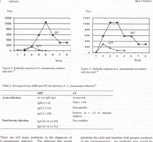
fracrions.reThe
antibody pattern
in
responseto
the primary
and ,secondary
infection is
shownin
Figure
2 and 3.te
Grayston et
al.
have proposed acriteria
for
serological
diagnosis
of
C.pneumoniae
infection
that have
beenused
by
many clinicians (Table
2).23 For
acuteinfection,
thepatient should
have a four-fold increasein
the
IgG
titer,
asingle
IgM
titer
>
1:16, a singleIgG titer
I
1:512or
a singleIgA
titer
2
l:256.
Pastor
pre-existing
infection
is defined
as anIgG
>
l:16
and!
1:512;
or
an
IgA
) l:16
and
<
1:256. Routine
Safiriani
Titer
Med J Indones
H
/
\
\
IgGr
/ /\tevt
\-"/
\--t
67
Week1200
1 000
800
600 400
200
0
Titer
1200
1000
800
600
400
200
0
7
[image:4.595.81.572.54.509.2]Week
Figure 2. Antibody response to C. pneumoniae primary
infection.te Figure infection IgG.te3. Antibody response to C. pneumoniae secondary
Table 2. Serological tests (MIF and CF) for detection
of
C. pneumoniae infection23MIF' CF
Acute infection
PasUchronic infection
4x rise IgG/ IgA
IgM
>
1:16IgG
>
l:512IgA>
1:256IgA
>l:16
<l:256IgG >1: 16
(l
:5124x titer rise Titer >
l:64
Non-specific
Positive
in < l/3 of
infectedsubjects
Not available
There
are
still many problems in
the
diagnosis of
C.pneumoniae
infection.
The infection that
would
produce adequate antibody
response,
is
only the
severe, deep
and localized
one.
Diagnostic
tools
arenot
widely
available;
and
different
Chlamydial
species, which are
very simrlar genetically,
causelow
specificity
in the
diagnostic
tests.
Furthermore,
thepresence
of
lipopolysaccharida
and the
detection
of
DNA
by
PCR,
could not
differentiate between
acute,persistence
or
chronic active infection. The
development
of a
rapid and
specific method for
identification
of
C.
pneumoniâe
will
provide
themeans
for
an
accurate diagnosis
and an appropriate
therapy.C.
pneumoniae tr eatment
C. pneumoniae
is
an obligate intracellular
parasite.To
inhibit
growth,
antimicrobial agents
need
topenetrate the
cells
andinterfere
with protein synthesis
of
the microorganism.
An
antibiotic
that would
beeffective
Torchlamydial infection
must
have a
goodintracellular,
tissue
or
secretion penetration, and
low
minimal inhibitory
concen-tration
(MIC). The active
antibiotic classesfor
Chlamydia infection are macrolides(erythromycin,
roxithromycin, clarithromycin
andazithromycin), tetracyclines,
quinolones (levofloxacin,
sparfloxacin), and chloramphenicol (used
mainly for
Salmonella typhi
treatment).Treatment
with penicillin
(BJactam),
amynoglycosides
and cephalosporin
arenot
adequate
for
the
infection;
because those
antibiotics do not enter the host
cells
adequately,
andtheir
target
is the
bacterial cell wall (a structure
lacking
in
C hlam ydi ae).2sRoxithromycin is
a newermacrolide generation which
has been developed
by its
prototype,
erythromycin.
The
addition
of
the
ether oxime
ring
gives
somebioavailability, and spectrum
of
activity
increases.Roxithromyein has
a
broader antibacterial
spectrumwhich
coversboth
typical
andatypical
pathogenslike
C.
pneumoniae, Mycoplasma pneumoniae,
andLegionella
spp.
The
antibiotic has
a
high
tissue penetration and also ahigh intracellular
concentration,which is
eradicate
the
intracellular
pathogens
studies
done
by
manyresearcher
efficacy.27'28'teFuture
perspectives
Future
development
in
diagnosis, especially
thefinding
of
better
diagnostic markers
for
chronic
infection, is essential.
Even though the
developmentof
the PCR test has
made
a better
detection
of
the organismin
the
specimen,it
doesnot
always correlatewith
the
culture
and serological
diagnosis. Although
PCR
assays have been usedfor
different
target genes,but neither the
DNA
extraction method
nor
the
assayare
standardized.
A
commercially
standardizedavailable
DNA
amplification
test
is being
developedand
urgently needed. The omp-4
and
omp-5 genes,
which
code
for
surface exposed immunogenic
96-98kDa proteins, are specific
for
C.
pneumoniae.
^[\e
cloning and
sequencing
of
those
genes,may
lead
tonew insight
tn"for
the developmentof
thenew
diagnostic tests.""A
possibility
of
a
vaccine development
could be
theideal
answerto the
C.pneumoniae
infection. A
major
outer
membrane
protein
(MOMP)
of
Chlamydia
has beenstudied
for
developing
vaccines.The
MOMP
isfound
on the outer membraneof
bothEB
andRB,
andinduces
neutralizing
antibody.
The MOMP
seems to bepartly
immunogenic
andno
specific differences in
the
amino
acid
sequences
between
different
C.pneumoniae isolates
have been found.
However,
despite years
of
effort,
an
effective
vaccine for
Chlamydia
hasnot
existed.3lCONCLUSION
Currently, there
are
no highly specific and
sensitivediagnostic
tools
for
C.pneumoniae
infection
that
arewidely
available.
In
addition, there
is
no
standard consensuson
serological
criteria
for
defining
the
C. pneumoniae infection. The developmentof
diagnosis andtreatment
will
lead
to
more
accurate
use
of
antimicrobial
agents.Further
studies
in
the
develop-ment
of
diagnosis,
treatment and prevention
of
C.pneumoniae infection,
are therefore,urgently
needed.Acknowledgements
The
author
would
like
to
thank Galatia Chandra
andGeorge
Kiongdo,
MD
(Hoechst
Marion
RousselIndonesia) and Septelia
Inawati Wanandi,
MD,
PhD(Biochemistry Department,
Faculty
of
Medicine,
University
of
Indonesia)
for
their
support
during
the preparationof
the manuscript.REFERENCES
1.
HahnDL,
Saikku P, LeinonenM.
Chlamydia. smoking and heart disease. Ann Int Med 1992; 117:.171.2.
AllegraL, Blasi
F, CentanniS.
Acute exacerbationsof
asthma in adults: role of Chlamydia pneumoniae infection. Eur Respir J 1994;7:2165-8.
3.
Beaty
CD,
Grayston
JT,
Wang
SP.
Chlamydia pneumoniae, strain TTVAR, infectionin
patients with chronic obstn:ctive pulmonary disease.Am
Rev Respir Dis 1991 ; l,l4: 1408-10.4.
Blanchard
T,
Bailey
R,
Holland
M.
Chlamydia pneumoniaeand
atherosclerosis (letter).Lancet
1993; 341:825.5.
Braun J, Laitko S, Treharne J.A
new causative agentof
reactive arthritis and undifferentiated oligoarthritis. Ann Rheumat Dis 1994; 53:100-5.6.
AllegraL.
Historyof Chlamydia pneumoniae (TV/AR).
In: Allegra
L,
Blasi F, editors. Chlamydia pneumoniae, Milan: Springer-Verlag; 1999. p. l-8.7.
Smadel JE. Atypical pneumonia and psittacosis.J
Clin Invest 1943;22:57-65.8.
Saikku P, Wang SP, KleemolaM.
An epidemicof
mild pneumonia due to unusual strain of Chlamydia psittaci. JInfect Dis 1985; l5l:832-9.
9.
GraystonJT,
Kuo
CC,
Wang SP.A
new
Chlamydia psittaci, strain TWAR, isolated in acute respiratory tract infections. N Eng J Med 1986; 315:l6l -8.10. Grayston JT, Kuo CC, Wang
SP. Chlamydia pneumoniae'sp. nov.
for Chlamydia
s2
strainTV/AR.
Int
J
Syst Bacteriol I 989; 39:88-90.11.
Murray PR. Chlamydiae.In:
Murray PRet
al., editors. Medical Microbiology Mosby; 1990.12.
Moulder
JW.
Comparativebiology
of
intracellular parasitism. Microbiol Rev 1985; 49:298-337.13. Orfila
J.
Chlamydia pneumoniaemicrobiology.
In:Allegral, Blasi
F,
editors.
Chlamydia pneumoniae infection. Milan: Springer-Verlag; 1995. p. 3-9.14. Danesh
J,
CollinsR,
PetoR.
Chronic infections andcoronary heart disease:
is there a link ?
Lancet 1997; 350:430-6.15.
Sundelof
B,
Gnarpe
H,
Gnarpe
J. An
unusual manifestationof
Chlamydia
pneumoniae infection:25
26
27
28
29.
30
31
l6
l't
18.
19
20
21
22
23
24
Safi.riani
Juvonen J,
JuvonenT,
LaurillaA.
Can degenerativeaortic valve stenosis be related to persistent infection ?
Ann Intern Med 1998; 128:741-4.
Thom
DH,
GraystonJT.
Infectionswith
Chlamydiapneumoniae strain TWAR. Clin Chest Med
l99l;
12'.'245-56.Kuo CC, Chen HH, Wang
SP.
Identihcationof
a new groupof
Chlamydia psittaci strain called TWAR. J ClinMicrobiol 1986; 24:1034-7.
Blasi
F,
CosentiniR.
Diagnosisof
asthma: laboratoryprocedures
for
identifying respiratory pathogens. Eur Respir Rev 1996; 6:38'.235-9.Oheme
A,
Rosler
J.
Demonstrationof
Chlamydiapneumoniae
in
water usedfor gargling.
Infection 1992;20 (l):56.
Gaydos
CA,
Quinn
TC,
EidenJJ.
Identificationof
Chlamydia pneumoniae
by DNA
amplificationof
the l6SrRNA gene. J Clin Microbiol 1992; 30:796-800. Manie TJ, Grayston JT, Wang SP. Pneumonia associatedwith
theTWAR
strainof
Chlamydia. Ann Intern Med1987;106:507-ll.
Grayston JT, Campbell
LA,
KuoCC. A
new respiratory tract pathogen:
Chlamydia pneumoniae strain TWAR. Jlnf Dis 1990; l6l:618-25.
Verkooyen RP, Hazenberg
MA,
Van HararenGH.
Age-related interference
with
Chlamydia
pneumoniaemicroimmunofluorescence serology
due
to
circulating rheumatoid tàctor. J Clin Microbiol 1992;30:.1281-90.Med J Indones
Grassi GG. Pharmacological and pharmacokinetic basis of Chlamydia pneumoniae treatment. In: Allegra
L,
Blasi F, editors. Chlamydia pneumoniae. Milan: Springer-Verlag;1999. p.62-69.
Young RA, Gonzales JP, Sorkin
EM.
Roxithromycin : areview
of its
antibacterialactivity,
pharmacokineticproperties and clinical efficacy. Drugs 1989; 37:8-41. Karalus NC, Garrett JE, Lang SD, Leng RA, Kostalas GN, Cooper
BS, et al.
Roxithromycin 150mg bid
versusamoxycillin 500 mg/clavulanic acid 125 mg
tid for
thetreatment of lower respiratory tract infections
in
general practice. Infection 1995;23 Suppl 1: l5-20Chatzimanolis E, Marsan
M,
Lefatzis D, Pavlopoulos A. Comparisonof
roxythromycinwith
co-amoxiclav in patientswith
sinusitis. Antimicrob Chemother 1998:'4l
Suppl B:81-4.Dautzenberg B, Scheimberg
A,
BramillaC.
Comparisonof two oral
antibiotics, roxithromycin and amoxycillitt/clavulinic acid
in
lower respiratory tract infections. Diag Microbiol Inf Dis 1992; 15 Suppl 4:85- 89.Gaydos
C.
Chlamydia pneumoniae and coronary heart disease: association and biological plausibility. Proceedings of the Australasian Chlamydia Consortium; 1999 June1l-12; Sydney, Australia.
Leinonen
M, Laurila A,
LaitinenK.
Immunology of ChLamydia pneumoniae.In:
Allegral,Blasi F,
editors.C hlamydia pneumoniae. Milan: Springer-Verlag; 1999. p.