Incidence and associated factors of posterior capsule opacification
in pseudophakic patients at Cipto Mangunkusumo Hospital
Keywords: incidence, intraocular lens, posterior capsule opacification
pISSN: 0853-1773 • eISSN: 2252-8083 • http://dx.doi.org/10.13181/mji.v24i3.1199 • Med J Indones. 2015;24:176-82 • Received 03 Feb 2015 • Accepted 27 Apr 2015
Correspondence author: Sita P. Ayuningtyas, [email protected]
Copyright @ 2015 Authors. This is an open access article distributed under the terms of the Creative Commons Attribution-NonCommercial-ShareAlike 4.0 International License (http://creativecommons.org/licenses/by-nc-sa/4.0/), which permits unrestricted non-commercial use, distribution, and reproduction in any medium, provided the original author and source are properly cited.
Sita P. Ayuningtyas, Tjahjono D. Gondhowiardjo
Department of Ophthalmology, Faculty of Medicine Universitas Indonesia, Cipto Mangunkusumo Hospital, Jakarta, Indonesia C l i n i c a l Re s e a rc h
ABSTRAK
Latar belakang: Posterior capsule opacification (PCO) merupakan konsekuensi yang paling sering dijumpai pasca- operasi katarak, yang dapat menyebabkan penurunan tajam penglihatan. Insidens PCO di Indonesia belum pernah dilaporkan sebelumnya. Penelitian ini bertujuan untuk menilai insidens kumulatif PCO dan faktor-faktor yang berhubungan dengan insidens PCO di Rumah Sakit Cipto Mangunkusumo, Jakarta.
Metode: Penelitian ini merupakan penelitian deskriptif retrospektif pada pasien yang menjalani operasi katarak senilis selama tahun 2010. Data dikumpulkan dari rekam
medis pada tahun 2013, meliputi karakteristik demografis,
jenis operasi, karakteristik lensa intra-okular (LIO) yaitu materi, optic edge design, dan diameter. Dilakukan pencatatan waktu diagnosis PCO pertama kali (dalam bulan), dan tajam penglihatan terbaik dengan koreksi (TPDK) sebelum operasi, saat diagnosis PCO ditegakkan dan dua minggu setelah tindakan laser Neodymium-doped yttrium aluminium garnet (Nd:YAG) (dalam desimal).
Hasil: Penelitian ini melakukan observasi rekam medis pada 578 mata (485 pasien) katarak senilis. Insidens kumulatif PCO dalam tiga tahun sebesar 8,82% (51 mata). Tindakan
fakoemulsifikasi dilakukan pada 496 (85,8%) mata. Median
waktu saat diagnosis PCO ditegakkan adalah 21 bulan (rentang
1-34 bulan), dengan rerata TPDK 0,50 ± 0,26. Umur (<65 dan >65 tahun) tidak berhubungan dengan insidens PCO. Proporsi PCO yang lebih tinggi ditemukan pada jenis LIO akrilik hidrofilik (10,7%) dibandingkan LIO akrilik hidrofobik (6,2%). Setelah
tindakan pasca-laser Nd:YAG TPDK meningkat menjadi 1,00.
Kesimpulan: Insidens kumulatif PCO selama tiga tahun adalah 8,82% (51 mata); tidak terdapat faktor yang berhubungan dengan terbentuknya PCO, namun persentase PCO yang lebih tinggi ditemukan pada LIO akrilik hidrofilik dibandingkan LIO akrilik hidrofobik.
ABSTRACT
Background: Posterior capsule opacification (PCO) is the
most common postoperative consequence of cataract surgery which may cause visual acuity reduction, yet the incidence in Indonesia has not been reported. The objectives of this study were to evaluate three years cumulative incidence
of PCO and factors associated with PCO formation at Cipto
Mangunkusumo Hospital, Jakarta.
Methods: This was a retrospective descriptive study on patients with uneventful senile cataract surgery during year 2010. All related data were retrieved from medical records in year 2013, which included patient demographics, type
of surgery, intraocular lens (IOL) characteristics (material, optic edge design and diameter). Moreover, time to first PCO
diagnosis (month), and best corrected visual acuity (BCVA)
pre-operatively, at time PCO was diagnosed and two weeks after
Neodymium-doped yttrium aluminum garnet (Nd:YAG) laser were noted (decimal).
Results: A total of 578 eyes (485 patients) were involved
in this study. Three years cumulative incidence of the PCO
was 8.82% (51 eyes). Phacoemulsification surgery was
performed in 496 (85.8%) eyes. The median time to PCO
diagnosis was 21 months (range 1 to 34 months), mean of BCVA was 0.50 ± 0.26. Age (<65 and >65 years old) was
not associated to PCO. Higher incidence of PCO was found in patients using hydrophilic acrylic IOL (10.7%) than
in hydrophobic acrylic (6.2%). After Nd:YAG laser was performed, BCVA was improved to 1.00.
Posterior capsule opacification (PCO) is the most
common postoperative consequence of cataract surgery.1 A cohort study by Fong, et al2 reported that three years cumulative incidence of PCO was 38.5%
(95% CI = 36.1-40.9) among 1,495 patients.
Posterior capsule opacification causes a reduction in
visual acuity and contrast sensitivity, as well as glare and monocular diplopia1,3,4 which might reduce the
patient’s quality of life and become an economic burden to the healthcare system.5 Factors related to PCO formation mostly were the type of cataract surgery, intraocular lens (IOL) design and patients with systemic diseases. Opening a blocked visual axis
with Neodymium-doped yttrium aluminum garnet (Nd:YAG) laser posterior capsulotomy will improve the visual acuity. However, in Indonesia, such procedure requires a referral to a tertiary hospital, which further increases the cost. Laser procedure itself has an inherited risk of complications such
as IOL damage, increase of intraocular pressure,
cystic macular edema, anterior vitreous surface disruption, and retinal detachment.1,6
Eventually, Department of Ophthalmology, Cipto
Mangunkusumo Hospital (CMH) Jakarta had done a retrospective study by Anggraini and Hutauruk7 and found that the incidence of PCO
was 9.2% (47 of 513 eyes) in 2003 (unpublished
data). Using modern cataract phacoemulsification machine, phacoemulsification has been performed
increasingly with a better result regarding to visual acuity, small corneal incision, rapid wound healing
and less complications. Also various better IOL
material and design had been used in our hospital.
However, there was no report of PCO incidence
since then. The objective of this study was to
evaluate three years cumulative incidence of PCO and factors associated with PCO formation.
METHODS
This was a retrospective descriptive study conducted in 2013. All data were retrieved from medical records period of January to December 2010. The inclusion criteria were all patients who had uneventful senile cataract surgery in
2010 at Department of Ophthalmology, Cipto
Mangunkusumo Hospital (CMH), Jakarta,
Indonesia. The exclusion criteria were patients with abnormalities other than PCO, which influence visual acuity, incomplete medical
record data and post-operative follow-up of less than one month. A total sampling method was used in selection of the subjects. This study has
been reported to CMH and confidentiality of
subjects was guaranteed.
Collected data were focused on patient
demographics, type of surgery (extra capsular cataract extraction [ECCE], phacoemulsification and small incision cataract surgery [SICS]), IOL
characteristics (material, optic edge design
and diameter), time to PCO diagnosis (duration between cataract surgery and diagnosis of PCO,
in months). Best corrected visual acuity (BCVA) in decimal was noted at one month post-operatively,
at time when PCO was diagnosed, and two
weeks after Nd:YAG laser posterior capsulotomy.
BCVA was classified based on the International Classification of Diseases-10 Clinical Modification
(ICD-10 CM) revision.
Analysis was performed using SPSS 11.0 and
Microsoft Excel 2011. We analyzed the proportions of PCO and compared it within phacoemulsification
groups using Chi-square test.
RESULTS
There were 1,665 cataract surgeries performed during January–December 2010 in CMH. Ninety percent (1,498 cases) had a complete medical record, but only 1,104 cases (73.7%) could be accessed. Finally, 852 eyes met the inclusion criteria. Two hundred and seventy four cases (274)
or 32.2% were excluded due to abnormalities other than PCO (138 cases) and 136 cases had
incomplete required data. A total of 485 patients
or 578 eyes (67.8%) were analyzed in this study.
Demographic data of the patients can be seen in Table 1.
Pre-operative BCVA, one month post-operative BCVA and type of surgery performed can be seen in Table 2.
Cumulative incidence of PCO and clinical characteristics of PCO patients
PCO is the most common consequence after
cataract surgery. Among 578 eyes, only 51 eyes
had PCO. Cumulative incidence of PCO was 14 (2.42%) eyes at first year, 34 (5.88%) eyes in
Variable n (%)
Age (median;range) 65 (42 - 87) years
Table 1. Patient demographics and clinical characteristics of pseudophakic patients {n=485 patients (578 eyes)}
Table 3. Clinical characteristics of PCO eyes (n=51)
Table 2. Clinical characteristics of pseudophakic eyes (n=578)
Variable Variables
Pre-op BCVA (in decimal) 0.07 (0.00-0.90) No visual impairment (> 0.5) 127 (22.0%) Mild visual impairment
Blind category 3 (< 0.05 – ≤0.02) 129 (22.3%) Blind, category 4
(< 0.02 – light perception)
133 (23.0%) Post-op BCVA at 1 month (in decimal) 1.00 (0.30-1.20)
No visual impairment (> 0.5) 577 (99.8%) Mild visual impairment
*BCVA = best corrected visual acuity
*ECCE = extracapsular cataract extraction
*SICS = small incision cataract surgery
*Nd:YAG = Neodymium-doped yttrium aluminium garnet *BCVA = best corrected visual acuity
*PCO = posterior capsule opacification The mean age of PCO patients was 63.57 ± 8.20
year old, younger than other pseudophakic
patients. Formation of PCO was most common in SICS eyes, but the number of SICS was only five
compared to the most common surgical technique,
which was phacoemulsification (in 496 eyes). The most common symptom of PCO was blurred vision, and median time to PCO diagnosis was 21
months or 1.75 years with a range of one to 34 months (Table 4).
Nd:YAG laser posterior capsulotomy is needed to improve the visual acuity by opening the visual
axis. Table 5 shows that Nd:YAG laser posterior
capsulotomy was only performed in 72.5% of eyes. After Nd:YAG laser posterior capsulotomy was performed, the BCVA showed better result with a range of 0.70-1.00(Table 5). No complications were found on two-weeks follow-up.
Variable
Age 63.57 ± 8.20 years old
Surgical technique
Phacoemulsification 49 /496 (9.9%)
ECCE 1/ 77 (1.3%)
SICS 1/ 5 (20.0%)
BCVA at PCO diagnosis ( in decimal) 0.50 ± 0.26 No visual impairment (> 0.5) 34 (66.7%) Mild visual impairment
Blurred vision 39 (76.5%) Blurred vision and diplopia 2 (3.9%)
Foggy view 2 (2.0%)
Blurred vision and glare 1 (3.9%)
No symptoms 7 (13.7%)
Time of PCO diagnosis 21 (1 - 34) months
*PCO = posterior capsule opacification *ECCE = extracapsular cataract extraction
*SICS = small incision cataract surgery *BCVA = best corrected visual acuity
Variable
Nd:YAG performed 37 (72.5%) Nd:YAG not performed 14 (27.5%)
BCVA at PCO diagnosis
Intraocular lens characteristics
Many efforts had been done to minimize PCO
formation, so that studies on the possible cause
are very important. Several types of IOL material and design may be related to PCO formation1. This study found hydrophilic acrylic IOL was the
most used. The foldable design of this material is appropriate with the width of incision in
phacoemulsification. In 2010, hydrophobic acrylic
was still minimally being used due to the more
expensive price. This material type of IOL was
mainly used in patients who had certain health insurance in our hospital. Square optic edge was the majority of the optic edge design and all eyes
used one piece IOL (Table 5).
Phacoemulsification was the most common type of surgery performed in this series. To minimize
bias from different types of surgery, cross
tabulation of IOL characteristics was made only in phacoemulsification group. However, p-value
was not tested due to unequal sample between
groups. No significant difference was found in the percentage of PCO between age groups. Higher percentage of PCO was found in hydrophilic
acrylic than in hydrophobic acrylic. Percentage
of PCO among type of IOL on optic diameter and
optic edge were almost the same (Table 6).
DISCUSSION
Our study revealed that modern cataract surgeries with a better IOL design and material may decrease the incidence of PCO formation
from 9.2% in our previous unpublished data in year 2003, to 8.82% in year 2010. The use of cutting-edge technology in cataract surgery has resulted in a less complication rate in long-term follow-up for pseudophakic eyes. Following
the excellent results, phacoemulsification has
become the standard of care for cataract surgery in our hospital. This study also showed that most of our cataract patients’ condition was in blind category four and they were relatively young. Shah, et al8 in collaboration with World Health Organization (WHO) analyzed 11,408 patients
who have had cataract surgery from 50 countries in 2008 also found that the median age was 65 years, majority had severe visual impairment, which was mostly related to senile degenerative cataract and strong correlation into ultraviolet
exposure.
Variable Frequency(%)
IOL material
Polymethylmethacrylate (PMMA) 204 (35.3%) Acrylic
Hydrophilic acrylic 309 (53.5%) Hydrophobic acrylic 65 (11.2%)
IOL design
1-piece 578 (100.0%)
3-pieces 0 (0.0%)
Optic edge design
Square 378 (65.4%)
Round 200 (34.6%)
Table 5. IOL characteristics in pseudophakic eyes (n=578)
*IOL = intraocular lens
*Chi Square test. NT: not tested for p value
*IOL = intraocular lens
*PCO = posterior capsule opacification
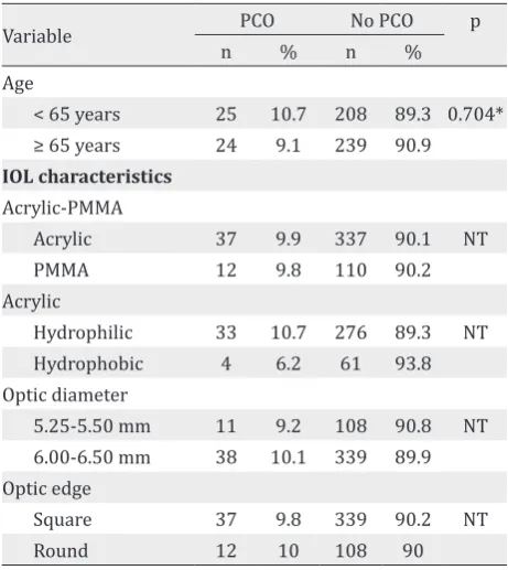
Variable PCO No PCO p
n % n %
Age
< 65 years 25 10.7 208 89.3 0.704*
≥ 65 years 24 9.1 239 90.9
IOL characteristics
Acrylic-PMMA
Acrylic 37 9.9 337 90.1 NT
PMMA 12 9.8 110 90.2
Acrylic
Hydrophilic 33 10.7 276 89.3 NT Hydrophobic 4 6.2 61 93.8
Optic diameter
5.25-5.50 mm 11 9.2 108 90.8 NT 6.00-6.50 mm 38 10.1 339 89.9
Optic edge
Square 37 9.8 339 90.2 NT
Round 12 10 108 90
Table 6. Cross-tabulation of age and IOL characteristics with PCO incidence in phacoemulsification group (n=496)
Phacoemulsification was the most common
surgical technique performed in this study (85.8%) and our result resembles the result of Gollogly, et al9 study, which reported that
99.5% out of 8,012 cataract eyes underwent
phacoemulsification from 2005 to 2011 in
advantages such as faster achievement of good visual acuity and wound healing, closed system surgery and a safe surgical technique.10 It was
showed in this study that post-operative BCVA achieved optimal target visual rehabilitation. A regular follow-up post-operatively is important to evaluate surgical success and to observe the
possibility of further complication and PCO is the
most common.
Three-year cumulative incidence of PCO reported
by Fong, et al2 was 38.5% (95% CI = 36.1-40.9)
among 1,495 patients, and was higher than the result of our study. This difference may be caused
by larger sample size, different study design, and
a long and equal follow-up time in their study. Variability in the follow-up period may result in
a bias in determining the PCO incidence in our study. We assumed that patients with only one
month duration of follow-up will not have any
visual problem. This condition might also reflect
a lower incidence in our study.
We found that PCO formation was found mostly
in younger patients among all our pseudophakic
patients. Age is an important factor to PCO formation according to Wormstone, et al11 study.
They had concluded that younger age had a potency of faster epithelial lens growth.11 Epithelial
lens migration and proliferation from equator to
central may block visual axis and causes reduction
in visual acuity.1 Most of our PCO patients experienced blurred vision. Their visual acuity
was reduced to 0.5±0.26, but still in the category of mild visual impairment. It was comparable with Van Bree, et al3 study (0.20 ± 0.23 logMAR or 0.64 ±
0.60) and Anggraini and Hutauruk7 reported (0.47 ± 0.46). Decreasing BCVA to 0.5 will significantly
cause visual impairment to the patient. In this
study, duration to PCO diagnosis was 21 months or
1.75 years, which was comparable to Khan, et al12 and Khanzada, et al13 which reported a duration of
23 months (two until 24 months) and 2.06 years, respectively.
Treatment with Nd:YAG laser posterior
capsulotomy in PCO is needed to clear the visual axis. In this study, not all patients underwent
Nd:YAG laser, not only because of weakness in our management protocol, but also due to patients’ refusal of having a laser treatment due
to accepted BCVA or financial problem. During a posterior capsulotomy, by amplification and
focusing of 1,064 nanometers (nm) infrared light, which electrons are ripped away from nuclei, forming energy plasma and corresponding shock wave. The plasma formation is known as optical
breakdown and cut the target. When pressure
wave created on the anterior vitreous side of the capsule, the laser breaks the posterior capsule.14 In this study, after the opening of visual axis, the
BCVA achieved 1.00, which was the same as BCVA on one month post-operative. This result was similar to Van Bree, et al3 (0.001 ± 0.12 logMAR or 1.0) and Khanzada, et al13 studies (74.4% cases
achieved 6/9 to 6/6).
Several studies related to risk factors of the
formation of PCO had been done and some studies had concluded that age and IOL
material and design might be associated with
PCO formation.1,13,15 In 2003, Anggraini and
Hutauruk7 reported that majority of patients used polymethylmethacrylate (PMMA) IOL (97%) and only 2.7% used acrylic IOL, at CMH Jakarta. In 2010, a transition into acrylic IOL had occurred. Acrylic IOL was used in the majority of cases i.e. 374 (64.7%), whereas PMMA IOL in 204 of cases
(35.3%).
Other studies reported that surgical technique,
including cortical cleaving hydrodissection and
rotation, cortical clean up, in-the-bag fixation of IOL, and smaller capsulorhexis size than optic diameter of IOL seem to correlate with PCO
formation. Those procedures’ factors could be
minimized by doing phacoemulsification.5,16 In this study, the cross tabulation evaluated IOL factors in phacoemulsification group only. It was considered to minimize bias due to surgical technique difference. P-value was not analyzed because of the unequal sample size between groups of IOL characteristics. Acrylic material particularly hydrophobic has been identified as a preventing factor in PCO formation. Linnola,
et al17 found that acrylic had stronger adhesion with fibronectin and laminin compared to PMMA and silicone. It caused stronger adhesion of IOL
and posterior capsule, therefore it would inhibit epithelial lens migration and proliferation. Hydrophobic acrylic has better capsular biocompatibility than hydrophilic acrylic. In
this study, a higher proportion of PCO was
found in hydrophilic acrylic than hydrophobic acrylic. Several studies also reported higher
(42.0%-64.4%) compared to hydrophobic acrylic (8.9%-34.4%).2,18,19
Optic diameter is still a mater of debate in the prevention of PCO formation. In this study, percentage of PCO in a small and a large diameter
seems to be the same. Meacock, et al20 reported
that optic diameter of 6 mm (1.5%) had lower
proportion of PCO than 5 mm (6.9%). In contrast,
Nishi and Nishi21 reported that larger optic diameter inhibit IOL adhesion to posterior
capsule and capsular bend formation.
Capsular bend formation is identified as an important factor in prevention of PCO. Square optic edge IOL may form a good capsular bend,
which therefore inhibit epithelial lens migration to posterior capsule. In this study, a higher
percentage of PCO was found in round optic edge IOL, but the difference was very small. It was similar with the percentage of PCO in PMMA and
acrylic. Most of PMMA had round optic edge while most of acrylic had square optic edge. Several
studies also reported higher percentage of PCO
in round optic edge (13.0%-38.7%) than square optic edge (1.4%-3.4%).22-24
In conclusion, three years cumulative incidence
of PCO in Cipto Mangunkusumo Hospital, Jakarta was 8.82%, with median time to PCO diagnosis of 21 months (1.75 years). There was no defined related factor to PCO reformation, but higher incidence of PCO was found in hydrophilic acrylic IOL than in hydrophobic acrylic IOL. Nd:YAG
laser posterior capsulotomy was performed in 72.5% of eyes and showed good results.
Acknowledgment
The authors gratefully acknowledge the full
support of Dr. dr. Widya Artini, SpM(K), the Head of Ophthalmology Department, Faculty of
Medicine, Universitas Indonesia, Jakarta.
Conflicts of Interest
The authors affirm no conflict of interest in this
study.
REFERENCES
1. Raj SM, Vasavada AR, Johar SRK, Vasadava VA, Vasavada
VA. Post-operative capsular opacification: a review. Int J
Biomed Sci. 2007;3(4):237-50.
2. Fong CS, Mitchell P, Rochtchina E, Cugati S, Hong T, Wang JJ.
Three-year incidence and factors associated with posterior
capsule opacification after cataract surgery: The Australian
prospective cataract surgery and age-related macular
degeneration study. Am J Ophthalmol. 2014;157(1):171-9.
3. Van Bree MC, van den Berg TJ, Zijlmans BL. Posterior
capsule opacification severity, assessed with straylight
measurement, as main indicator of early visual function
deterioration. Ophthalmology. 2013;120(1):20-33. 4. Wakamatsu TH, Yamaguchi T, Negishi K, Kaido M,
Matsumoto Y, Ishida R, et al. Functional visual acuity after neodymium:YAG laser capsulotomy in patients
with posterior capsule opacification and good
visual acuity preoperatively. J Cataract Refract Surg. 2011;37(2):258-64.
5. Awasthi N, Guo S, Wagner BJ. Posterior capsular opacification: a problem reduced but not yet eradicated. Arch Ophthalmol. 2009;127(4):555-62.
6. Khanzada MA, Jatoi SM, Narsani AK, Dabir SA, Gul
S. Is the Nd:YAG laser a safe procedure for posterior
capsulotomy? Pak J Ophthalmol. 2008;24(2):73-8.
7. Anggraini N, Hutauruk J. Insiden dan karakteristik
posterior capsule opacification pasca ekstraksi katarak
dengan implantasi posterior chamber-intraocular lens: Studi deskriptif. Departemen Ilmu Kesehatan Mata Fakultas Kedokteran Universitas Indonesia. 2005. Indonesian.
8. Shah SP, Gilbert CE, Razavi H, Turner EL, Lindfield RJ.
Preoperative visual acuity among cataract surgery patients and countries’ state of development: a global
study. Bull World Health Organ. 2011;89(10):749-56. 9. Gollogly HE, Hodge DO, St Sauver JL, Erie JC. Increasing
incidence of cataract surgery: population-based study. J Cataract Refract Surg. 2013;39(9):1383-9.
10. Bobrow JC, Blecher MH, Glasser DB, Mitchell KB, Rosenberg LF, Reich J, et al. Surgery for cataract. In:
Skuta GL, Cantor LB, Weiss JS, editors. Basic and clinical
science course: lens and cataract. San Francisco:
American Academy of Ophthalmology; 2011. p. 91-160. 11. Wormstone IM, Wang L, Liu C. Posterior capsule
opacification. Experimental Eye Research.
2009;88:257-69.
12. Khan MY, Jan S, Khan MN, Khan S, Kundi N. Visual outcome after Nd:YAG capsulotomy in posterior capsule
opacification. Pak J Ophthalmol 2006;22(2).
13. Khanzada MA, Gul S, Dabir SA, Jatoi SM, Narsani
AK. Comparative incidence of posterior capsular
opacification in AcrySof and PMMA intraocular lenses. Int J Ophthalmol. 2009;2(2):150-3.
14. Aslam TM, Devlin H, Dhillon B. Use of Nd:YAG laser
capsulotomy. Surv Ophthalmol. 2003;48(6):594-612.
15. Lundqvist B, Mönestam E. Ten-year longitudinal visual function and Nd:YAG laser capsulotomy rates in patients
less than 65 years at cataract surgery. Am J Ophthalmol.
2010;149(2):238-44.
16. Moulick CP, Rodrigues CFEA, Shyamsundar LCK.
Evaluation of posterior capsular opacification following phacoemulsification, extracapsular and small incision
cataract surgery. MJAFI. 2009;65:225-8.
17. Linnola RJ, Sund M, Ylönen R, Pihlajaniemi T. Adhesion
of soluble fibronectin, laminin, and collagen type IV
18. Kugelberg M, Wejde G, Jayaram H, Zetterström
C. Two-year follow-up of posterior capsule opacification after implantation of a hydrophilic or hydrophobic acrylic intraocular lens. Acta
Ophthalmol. 2008;86(5):533-6.
19. Saeed MU, Jafree AJ, Saeed MS, Zia R, Sheikh IM, Heravi
M. Intraocular lens and capsule opacification with
hydrophilic and hydrophobic acrylic materials. Semin
Ophthalmol. 2012;27(1-2):15-8.
20. Meacock WR, Spalton DJ, Boyce JF, Jose RM. Effect of optic size on posterior capsule opacification: 5.5 mm
versus 6.0 mm AcrySof intraocular lenses. J Cataract Refract Surg. 2001;27(8):1194-8.
21. Nishi O, Nishi K. Effect of the optic size of a single-piece acrylic intraocular lens on posterior capsule opacification.
J Cataract Refract Surg. 2003;29(2):348-53.
22. Buehl W, Findl O, Menapace R, Sacu S, Kriechbaum K,
Koeppl C, et al. Long-term effect of optic edge design in an acrylic intraocular lens on posterior capsule
opacification. J Cataract Refract Surg. 2005;31(5):954-61. 23. Kohnen T, Fabian E, Gerl R, Hunold W, Hütz W, Strobel J, et al. Optic edge as long-term factor for posterior capsule opacification rates. Ophthalmology. 2008;115(8):1308-14. 24. Hayashi K, Hayashi H. Posterior capsule opacification in
the presence of an intraocular lens with a sharp versus