Jurnal Biofisika 7 (1): 1-6
Department of Physics, Faculty of Mathematics and Natural Sciences, University of Indonesia
2)
Department of Physics, Faculty of Mathematics and Natural Sciences, Bogor Agricultural University
ABSTRACT
The apatite crystal has been growth on pores polyglycolide through precipitation of supersaturated calcium phosphate carbonate solution. The influence of magnesium on the precipitation was observed. Polyglycolide was prepared by solid state reaction of sodium chloroacetate at 192°C. To obtain pores materials, polyglycolide was pressed into pellet and washed with water to eliminate NaCl, a byproduct of sodium chloroacetate polymerization. Calcium phosphate carbonate solution (calcium: phosphate: carbonate = 0.25 M : 0.25 M: 1 M was precipitated on this pores polyglycolide. Solution with various magnesium concentrations (0, 0.5, 1, 1.5, and 2 M) was added during the precipitation. Samples were characterized by x-ray diffractometer (XRD) and scanning electron microscope (SEM). Most of XRD profiles showed that the samples contained of amorphous and crystalline phase of calcium phosphates as well as polyglycolide .The increase of magnesium ions in solution influenced the crystallization process in which it caused the decrease of amorphouse phase content and eliminated the presence of octa caluim phosphate phase.
Keyword: polyglycolide, apatite crystal
ABSTRAK
larutan mempengaruhi proses kristalisasi yang menyebabkan terjadinya pengurangan fase amorf dan menghilangkan kehadiran fase okta kalsium fosfat.
Kata kunci: polyglycolide, kristal apatit
INTRODUCTION
Bone is a natural composite material, composed of about 60 wt% mineral, 30 wt% organic matrix, and 10 wt% water1. Furthermore as a living tissue, bone consists of about 15 wt% living cells. The bone organic matrix is primarily as type I collagen2., and bone mineral is a mixture of may incorporate and/or substitute Ca2+ and PO43- ions in lattice producing apatite crystal with various composition and crystallinity.
In this work, to mimic the natural bone as a mixture of polimer, amorphous phase and crystalline phase, synthetic polymer polyglycolide (PGA) was mineralized by presipating Ca2+, and PO4 various high concentration of Mg2+ ion. It was expected that the CO3
concentration was 0.5 M, 1.0 M, 1.5 M, and 2.0 M and resulting sample A into sample A1, A2, A3, and A4.
X ray diffraction analysis to each samples was carried out by
means of Phillips difractometer with Cu Kα radiation (λ = 1.54056 Ǻ)
generated at 40 kV and 10 mA. The measurements were performed with
diffraction angle (2θ) in the range of 5° to 80°. Further evaluation of
surface morphology to each sample was performed by using JEOL SEM (scanning electron microscope).
RESULTS AND DISCUSSION
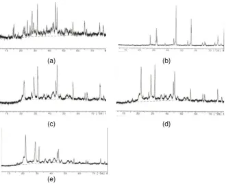
In general, X ray diffraction pattern of all samples (Figure 1) consist of continuous curve and discrete lines that represent of amorphous and crystalline phases in the sample. This continuous curve with maximum
peak at about 2θ = 30° indicates the existence of amorphous calcium
phosphate4,5 and might be also calcite hydrate. Discrete lines showed the present of a mixture of calcium phosphate and sodium chloride crystals.
Most samples consist of hydroxyapatite (2θ at about 27°, 32°, 44°), apatite carbonate type B (2θ at about 32°, 33°, 48°), calcite (2θ at about 22°, 29°, 43°), sodium chloride (2θ at about 31°, 45°, 56°, 75°), and
polyglycolide (2θ at about 21°, 29°, 36°, 39°, 47°). Most of glycolide
patterns have wide FWHM that indicated this phase in the form of microcryatalline. It is interesting that only sample A (Ca2+: PO43-: CO32+ = 0.25 M : 0.25 M : 1M) indicates the existence of octa calcium phosphate
(2θ at about 16°, 23°, 26°). The addition of magnesium changes the XRD
pattern, particularly for apatite crystal. If we consider the peak at 2θ =
27°, it appears that the increasing magnesium concentration will reduce the apatite content in the samples. However in the mean time, it will not influence the calcite content.
Polyglyclide is obtained through solid state reaction of halogenoacetate at specific temperature. In this work, it is determined by differential scanning calorimeter which the result is 192°C. It should be noticed that every batch of halogenoacetate, each will have specific polymerisation temperature.
(a) (b)
(c) (d)
(e)
Figure 1 X ray diffraction pattern of samples produced by concentration solution proportion Ca2+, PO4
3-, and CO3 2+
ions A) as 0.25 M : 0.25 M : 1.0 M (a) with additinal concentration Mg2+ ions for A1) 0.5 M (b), : A2) 1.0 M (b), A3) 1.5 M (c), and A4) 2.0 M (d).
Electron micrograph of the porous polyglycolide resulting from solid reaction was shown in Figure 2P. It can be seen that the pore sizes were not homogenous, the pore width was in the range of 0.5 – 1.5 µm. Figure 2A is a micrograph of a sample before the addition of magnesium. The crystalline pattern is clearly seen as rods. There is no significant change on the morphology after the addition of 0.5 M magnesium. The morphological change can be recognized as the magnesium increases to 1.0 M, 1.5 M, and 2.0 M (Figure 2d, 2e, and 2f).
P A
A1 A2
A3 A4
Figure 2 Scanning electron micrograph of polyglycolide (a) and samples produced by concentrationsolution proportion Ca2+, PO4
3-, and CO3 2+
ions as 0.25 M : 0.25 M : 1.0 M (b), with additional concentration solution Mg2+ ions for A1) 0.5 M (c), A2) 1.0 M (d), A3) 1.5 M (e), and A4) 2.0 M (f).
CONCLUSION
From this work it was found that calcification occured on pores polyglicolide in the form of amorphous and crystalline phase if the solution consisted of Ca2+, PO4
3-, and CO3
ions. As an inhihibitor in the formation of apatite crystal, high concentration of CO3
also affecting the production of calcite crystal. At the mean time the addition of Mg2+ ion solution prevent the growth of apatite and calcite and stabilized the amorphous phase. The influence of Mg2+ ion in preventing the growth of apatite was more evident relatively.
(a) (b)
(c) (d)
REFERENCES
1. Mickiewicz RA. Master Degree Thesis. Massachusetts Institute of
Technology. 2001
2. Hench LL, Jones RJ (Editors). Biomaterials artificials organs and
tissue engineering,Woodhead Publishing Limited. 2005
3. Baig AA, Fox JL, Wang Z, Higuchi WI, Miller SC, Barry AM, Otsuka
M. Calcif. Tissue Int. 1999; 6.
4. Termine, Lundy.Calc. Tiss. Res. 1974; 15: 55-70.
5. Harper RA, Posner AS. Mat. Res. Bull. 1970; 5: 129-136. 6. Neuman WF, Mulryan BJ. Calc. Tiss. Res. 1967; 7: 133-138. 7. Gron P, Spinelli M, Trautz O Brudevold F. Arch. Oral Biol. 1963; 8:
215-223.