Jurnal Biofisika 7 (1): 7-12
Department of Physics, Faculty of Mathematics and Natural Sciences, Bogor Agricultural University
2
Department of Physics, Faculty of Mathematics and Natural Sciences, University of Indonesia
ABSTRACT
In this study the thermal effect on apatite crystal was observed. Apatite crystal was synthesized by using eggshell as the source of calcium. To obtained calcium powder, chicken eggshell was calcined at 1000C for 5 hours. Calcium solution was precipitated into diammonium hydrogen phosphate ((NH4)2HPO4) solution in
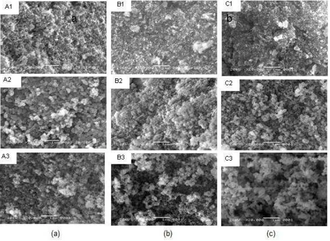
nitrogen atmosphere at 37C to yield apatite. Apatite was further calcined at 800 and 900C. The apatite was characterized by X-ray Diffractometer (XRD) and Scanning Electron Microscope (SEM). The XRD profiles of uncalcined apatite showed that the samples contained only crystalline phase of hydroxyapatite and a small amount of type B carbonated apatite. Through out the calcination, the carbonate contain is released so that the samples contained of hydroxyapatite only. The degree of crystallinity increases with the raising temperature. The SEM micrograph also shown that the grain size of apatite is bigger as the temperature is increased.
Keyword; thermal effect, apatite crystal, calcination
ABSTRAK
Efek termal pada kristal apatit diamati pada penelitian ini. Kristal apatit disintesis dari cangkang telur sebagai sumber kalsium. Serbuk kalsium diperoleh dari kalsinasi cangkang telur ayam pada suhu 1000 oC selama 5 jam. Larutan kalsium dipresipitasi ke dalam larutan diammonium hydrogen phosphate ((NH4)2HPO4)
INTRODUCTION
Calcium phosphate is known as the major compound forming bone mineral. The high demand of calcium phosphate or the bone implant initiates several researchers to conduct observations in order to meet the need1. Naturally, calcium phosphate exists in the form of biological apatite. This has a similarity with calcium deficient hydroxyapatite. The deficiency of calcium in natural apatite occurs as the existence of several foreign ions in body plasma. Synthetic hydroxyapatite is known has an ability to form chemical bond with the host hard tissue. Several factors that have an effect on the bioactivity of hydroxyapatite are the starting materials, impurity contents, crystal size, morphology2. It is believed that the use of natural raw material will be accepted better by the host hard tissue due to the similarity in the physical and chemical characteristic. Chicken eggshell consist of calcium carbonate up to 94-97% wt. The calcination of chicken eggshell results calcium oxide3. In this study, controlled precipitation is performed in order to yield hydroxyapatite. Chicken eggshell is used as the calcium source. To ascertain the termal effect, the samples then calcined at 800 and 900C.
EXPERIMENTAL
Calcium in this study was obtained from chicken eggshell. It was calcined at 1000C for 5 hours. Atomic Absorption Spectroscopy (Hitachi Z8230) was performed in order to determine calcium content in calcined eggshell. Apatite was synthesized through stirred precipitation of calcium into diammonium hydrogen phosphtate ((NH4)2HPO4) (MERCK). There
Tabel 2 Sample code
Ca/P concentration (M/M) Temperature (0C) Sample code
0.01/0.006
Figure 1 5 XRD spectra of (a) sample A1-A3, (b) sample B1-B3 dan (c) sample C1-C3.
The present study confirms the use of chicken eggshell as a potential source of calcium for synthetic calcium phosphate. The XRD spectra indicate that the samples do not contain calcium oxide that is formed during the calcination of chicken eggshell.The synthesizing results a calcium phosphate in the form of both hydroxyapatite and type B carbonated apatite. However, at higher concentration (Sample C), type A carbonated apatite also appears. Calcination of both sample A and B releases carbonate contain in them so that there are no carbonated apatite in both samples after calcination on 800 and 900C. Contrary, carbonate still exists in sample C although it has been calcined up to 900C . This finding therefore gives additional information of previous study which observes that loss of carbonate during heating is time
dissapearance of tricalcium phosphate will reduce the mechanical strength of the apatite6.
Thermal effect of apatite can be roughly estimated through the total inverse broadth of a diffraction peak as a size/strain parameter (crystallinity)7,8. Heating treatment at 110C already yields a high cystalline apatite. The broadening peak does not indicate the presence of amorphouse phase but rather the microcystalline. This is supported by the cystallite sizes which are 21.23, 16.73, and 16.69 nm for sample A, B and C, respectively. The crystallite size is obtained from simple Scherrer’s formula . The cystallinity tends to increase following the annealing treatment. The sharpening peaks at 2 around 20-35 indicate this occurance. Although there is an increase if both crystallinity and crystallite size, there is no structural change in lattice as there is no significant peak shiftness.
Figure 2 Morphology of (a) sample A1-A3, (b) sample B1-B3 dan (c) sample C1-C3.
CONCLUSION
It is to be hoped that the use of bio-based calcium source will increase the biocomptibility of calcium phosphate. Precipitation of calcium into diamonium hydrogen yields a mixture of hydroxyapatite and carbonated apatite. Carbonate will release during the heating treatment. However, the high concentration of starting materials enables the remaining of carbonate in the sample although it has been annealed up to 1000C. Thermal stability of samples is indicated by disappearance of the decomposition of hydroxyapatite in to a mixture of hydroxyapatite and
tricalcium phosphate in the annealed samples. The annealing also yields the increasing of both crystallinity and crystallite size.
REFERENCES
1. Kokubo T, Kim HM, Kawashita M. Biomaterials. 2003; 24 : 2161-2175. 2. Santos MH, et al. Mater. Res. 2004; 7: 625-630.
3. Prabakaran K, Balamurugan A, Rajeswari. Bull. Mater. Sci. 2005; 28: 115-119.
4. Barralet JE, Best S, Bonfield W. J. Mater. Sci. 2000; 11: 719.
5. Barralet JE, Knowles JC, Best S, Bonfield W. J. Mater. Sci. 2002; 13: 529-533.
6. Pattanayak, et al. Trends Biomater. Artif. Organs. 2005; 18: 87-92. 7. Baig AA, et al. Calcif. Tissue Int. 1999; 64.
8. Doi Y, Moriwaki Y, Aoba T, Takahashi J, Joshin K. Calcif. Tissue Int.