144
Suyatna etal
Med J lrulonesIdentification
of
C5*
extraband of butyrylcholinesterase and two
protein
bands
cathodic to
it
F.D. Suyatna.,
R. Setiabudy-,
O.Tjandrat,
E. HerwanalAbstrak
Pemeriksaan dengan elektroforesis padn plasma manusia menunjukkan 4 pita protein butirilkoLinesterase (BC\E), yaitu C 1, C2, Ct, C4 dan pada sejumlah individu juga saatu pita tambahan, C5* . Selain itu selaLu ditemukan juga adanya pita protein lain yang disebut
pita
"5".
Untuk mengetahui apaknhpita
C5* dan"5"
berhubungan dengan protein BChE, kami melakuknn analisis imunologi dan pemetaan peptida terhadap protein-protein ini. Cara imunologi dilakukan dengan membangkitknn antibodi poliklonal terhadap pita-pita protein tersebut (Sr, Sz Ct danCi
) dan direaksikan dengan protein plasma yang ditransfer pada kertas nitroselulosa. Antibodi individuaL tersebut bereaksi dengan semuapita protein yang
diteliti
termasukCt,
),ang merupakan isozimdari
BC\E,
yang menunjukkan bahwa pita-pita protein tersebut mempunyai epitop yang sama. Beberapa pita protein yang terdapat pada posisi katodik terhadap S 1 juga bereaksi dengan antibodi, yang menunjukkan bahwa mereka mungkin merupakan fraksi protein BC\E juga. Bila pita protein individual tersebut dicemakan dengan toksin S. aureus V6 dan a-kimotripsin, mereka menunjukkan derajat kesamaan 1,angtinggi
daLampola
peptidanya. Penelitianini
menunjukkan bahwapita
protein 51, 52 dan C5* merupakanpita
protein
BCLE.(Med J Indones 2001; 10: 144-9)
Abstract
Electrophoresis of human plasma yields 4 butyrylcholinesterase (BCLE) protein bands, i.e. C 1, C2, C-j, Ca and in some individuals also an
extraband C5'. In addition to thnt other protein bands called "S" bands are also invariably detected. In order to know whether the C5* and the
"5" bands
are related to the BChE protein, we have carried out immunological and peptide mapping studies on these proteins.The immunology approach was done by raising polyclonal antibodies against each protein bands (S 1, 52 Ca and C5* ) and reacted to the plnsma protein bands transferred on nitrocellulose papers. Indivîdual raised antibodies recognized all protein bands studied including the Ca, an isoqme of BCLE, indicating that the protein banls contain similar epitopes. Several protein bands cathodic to S 1 also reacted with the antibodies, suggesting that they are probably fractions of the BC6E protein, as welL. When individual protein bands were digested with S. aureus V6 toxin and a-chymotrypsin, they revealed a striking simiLarity in peptide pattern among each other. These studies indicate that the S 1.52 and C5' protein bands belong to the BC6E protein. (Med J Indones 2001;
t0:
144-9)Keywords: Butyrylcholinesterase, C5* variant, immunodetection, peptide mapping.
Butyrylcholinesterase
(EC 3.1.1.8,
BChE)
in
the serum catalysesthe hydrolysis
of
the muscle relaxant
agent succinylcholine which
is
administered during
general anesthesia. Patientswith
atypical type
of this
enzymesshow abnormal response
to
succinylcholine,
i.e.
: prolonged
apnoea
which may last to,
I
hour, rather than the usual 3-6min.r
Electrophoresis
of
blood
plasma, yields
4
protein
bands,i.e.
C1, Cz,Ct
and Ca andin some individuals
also an extra-band
C5.
Based
on the
absenceor
the+
Departuænt of Pharmacology, Faculty of Medicine, (Jniversity oJ Indonesia, Jakarta, Indonesi
t Department of Pharmacy, Faculty of Medicine, Tarumanagara U niv e rs ity, J aka rta, Indone sia
f
Department of Pharmacology, Faculty of Medicine, Trisakti Unive rs i N, Jakarta, I ndone s iapresence
of
the
extra band,
an
individual
can
be groupedinto
a C-s- or â C5+ variant.2During
her experiment
using
amide
electro-phoresis,
Simpson2
discovered
protein
bandswhich
were
called
"storage" bands in addrtion
to
the Cr-C-sbands.
The "storage"
bands, designated as"S"
bands were
seen
in
the electrophoresis
of
plasma samples that had been storedat -20oci
they were not
apparent
in fresh
samples.
In
our
study
recently
published.-'we
also carried
out
apopulation
study to
determine the frequency of the Cs+variant present
in a
Vol 10, No 3, July
-
September 2001METHODS
Human
blood
samples
of
Javanese
ethnic
werecollected from donors
in
the
Indonesian
Red
Cross Service.3The
plasmaobtained
wasdirectly
assayed orstored
at -20oC
until used. The distribution
of
C5+variant
was determinedby
meansof
apolyacrylamide
gel
electrophoresis
(PAGE)
essentially
as described
by Laemmli,a using
a7.5
Vo(w/v)
acrylamide
slab geland a 3 Vo
(wlv)
acrylamide stacking gel
in a
vertical
electrophoresis
equipment, except
that SDS
andmercaptoethanol
were omitted.
The proteins were
visualized
by
fast
red.
esults showedthat
among398
individuals,
there
82
(2lTo) of C5*
variant.3Fig.
1 depicts the
C5-
and
C5*variants
asshown by
acrylamide
electrophoresis.
The
protein
bands investigated were the Ca and C5 bands and the upper 2"S" bands
(Sr
and 52) cathodicto
C5.Raising
of
Polyclonal Antibody
In
order
to
investigate whether the Sr,
52 and C5 extrabands were immunologically related
to
the
BChE
protein
bands,
polyclonal antibodies were
raised. againstthe
Ss, Sz, Co and Cs*protein
bands.In
brief,
the
procedure
was
carried
out as
follows:
afterpreparative electrophoresis
and visualization
of
theprotein
with fast red, the 4
protein
bands were scrapedout
of
the gel
by
means
of
a scissor. The
protein
Lrandswere
destainedby
incubation
in
SDS
3
Vo andisopropanol
50Voat 37oC for
24
hours.-sThe
solution
was discarded and theprotein
bands were incubatedin
0.1
N
NaOH, overnight at 37oC.
The
content
of the
protein
in
the
eluates
were found to
be
negligible;
protein content
of
the
eluate
was determined
asdescribed
by
Peterson.6
Each protein band
washomogenized
with ultraturrax, mixed
and vigorously
vortexed
with
complete
Freund adjuvant
beforesubcutaneously
injected
between
the
scapulas
of
arabbit.
Booster injections using incomplete
adjuvantwere performed
in
weeks
2,
4
and
6 aftu
the first
injection.
Five days
after the
lastinjection, blood
waswithdrawn via
earveinof
the rabbits.TSince there were 4
protein
bands studied 4 rabbits were needed toraise
aspesific antibody
against each band.The
blood
were centrifuged at
low speed
for
15min
and the
plasma obtained
were
stored
at
-20oC
until
used.ldentification ofprotein
bands
145Western Blot Analysis
The
antibodies obtained
were
used
to
examinewhether
the
51,
52,Ca
and Cs
bands
were
immuno-logically identical. After
electrophoresis,
the
protein
was transferredonto
aZeta-probe@blotting membrane
by
meansof
a
Mini
Trans-blot (BioRad)
equipment set at 100 V,250
mA for 6 hours.
Immunodetection
of the
protein
bands wasperformed using the
method as describedby
Towbin et
al.8Peptide
Mapping
Peptide patterns of the
protein
bandswere
investigatedusing
toxin
of
Staphylococcus
aureus
V3
and
cx-chymotrypsin. Intragel protein
digestion
was carried
out
in a
7.5
Voof acrylamidee
in the presence
of
2
ugof toxin
of
S.
aureusVs or
û,-chlmotrypsin. After
electrophoresisthe
peptideswere visualized
by
silver
staining.loAcrylamide
andbis
acrylamide were
purchasedfrom
Nicalai
Tesque (Japan),TEMED, ammonium
persulfate,sodium dodecyl sulfate,
glycine,
Tween-2O
and Zeta-probe@filter
paper
were from BioRad (USA).
Fast red,silver nitrate, albumin, a-chymotrypsin
andbuffer
salts were from Sigma Co.
(USA).
Staphylococcusaureus
VB
protease
was from Pierce Chemical
Co.USA.
RESULTS
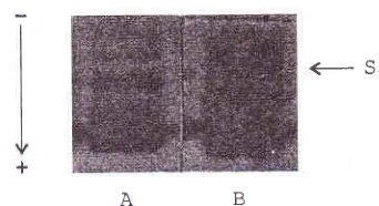
Fig. I
shows
the protein pattern
of
BChE after
gel electrophoresis; gel Aof
Cs- and gelB of
C5* variants.The most
two
prominent
bandsin
the lowest position
146
Suyatna et alBChË, bands.
The similarity
in
peptide pattem
wasexamined
in
all4
bands (C+, C5. S1 and S2).(--
S' [image:3.612.74.245.112.205.2]AB
Figure
l.
PoLl,acryl.amide patternof
hutnan plasma.Electro-plnresis
vvithout SDS and mercaptoethanol was performed ttsinga
7.5Vo (wh)auylamide
sl.ab geL anda
3Va (w/v) acrylantide stackin7 gel in a vertical electrophoresis equiptnenl. Tlrc antourrt o.f samples applied onto the gel was 2 1tl and theproteins vvere visualized by fast red azo dye
Gel. A :
Ci
varianl;
Gel B : C5* varianl, An arrow indicatesthe positiorr
for
S 1 The following proteins bands in downward di.rection are respectively the 52, C5* and C4Western
blot
Frg.
2
showsprotein
bands
of
Cs*variant
blotted on
nitrocellulose
papers
after
immunodetection.
As
weMetl J Indones
can see, individual
antibody
that was raised against S1,52 and C5* cross reacted
wlth
the bandCa
and severalbands cathodic
to
S1(nitrocellulose
paperno.1,2,3,4).
The
antibodies also recognized
severalbands
anodicto
Ca. Theseprotein
bands,
which were
ill
bordered were presumably the Cr, Cz, andCr
bandsof BChE.
The
serumof
nonimmunized rabbit did
not
reactwith
any
of
the BChE protein bands (nitrocellulose Bl).
This result
suggests thatthe
S;, 52 and Cs* bands (and someprotein
bands cathodic
to
it)
contained similar
epitopes
belong
to theBChE protein
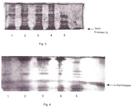
(Ca).Peptide
mapping
Figs. 3
and4
show peptide pattern
of
Sr
Sz,Cs*
and C+bands
after
digestion
with
the toxin
of
S.
aureusand
a-chymotrypsin, respectively.
As
can be seen, thepeptide pattem
of all
protein bands shows
striking
similarities
among each other.
The
immunodetection
and peptide mapping
analyses
strongly support
the suggestionthat
the
C5
extra band,
S1and
52 proteinbands are isozymes or fragments
of BChE
protein. +Ca
--1'
Figure2. Immunodetection
of
51.Sr,
C.r* and Caprotein bands. Irùividual antibodt,was raised against each protein band atttl reacterl to the serumproteizi ofCi
variantblottedonanitrocellulosepaper. S1; laneJ;52:
Lane2; C5' : Iane 3; Ca: lane 4' Lane BI was protein blotted-nitroceliulose paper reacted with serum of a non imtnunized rabbit'VoL 10, No 3, July
-
September 2001 Identification of protein bandst47
<
toxin S.aureus VeFig.
3g-cr-chymotrypsin
Fig.
4Figure 3-4. One dimensiotml peptide maps of S 1 (lane I ), Sz(lane 2), C r* (Iane 3 ) and Ct (lane 4).
Proleitt bands of interest were cut oL.l of the gel by
a
scissor after electro-phoresis, applied onto the second gel and digested intrageltt itlt 2 Stg of toxin of S. aureus Vs(Fig. 3) or a-chymotrypsin (Fig. 4),
Larte 5 v:as the peptide pattern front a non-BChE protein band.
Arrovvs indicate the position
for
toxin of S.aureus Vsand a-chymotrypsm,DISCUSSION
The
Cs*
variant indrviduals
were
associatedwith
the increasedactlvity of
BChE3'rr
and alower
meanadult
weight.r2
The
presence
of
an
extra
band
in
theelectrophoresis
of
plasma
BChE
of
individuals
with
Cs*
variant
has been a subjectof investigation.
Two
genetic
locr,
CHE1
and
CHE2
have
beenproposed
in
the production
of
plasma
BChE.
CHE1 encodesfor the "normal"
enzyme(Cr.
Cz.Cr
and
C+isozymes)
and
CHE2
which
is
not
allelic
to
the CHEr I 3is associated
with
the expressionof
C5 band.l I ,14Soreq
et
al'-
using
in
situ
hybridization
of
human chromosomes suggestedCHE1 locus
on
chromosome3
and
CHE2
locus on
chromosome
16 and that
theCHE2locus
might
be
a pseudogene expressedonly
in
Cs* variants.
However,
according
to
genetic linkage
studies,rs
the CHE2 locus
is linked to
the y-crystallin
gene cluster (CRYG)
on
chromosome
2.
Other
linkage
studiesr6
reported
a
loose linkage
between [image:4.612.67.517.100.471.2]148
Suyatna et alHowever, recent determination
of
the structure
of
theBChE
gene demonstratedthat
BChE is
encodedby
asingle gene corresponding to the E1 locus.rT
It
hasbeen
suggestedthat the
C5+variant could be
ahybrid
molecule
formed by the
association
of
BChE
uct
of
E1)
with
a different protein (product of
or
amodification of
BChE
by
a neuraminidase-l9nzyrne.
Plasma
BChE
on
electrophoresis showed
severalprotein bands, namely, Cr,
Cz,
C:,
C+and C5
.
In
addition
to
that, there
are severalsmall protein
bandst
ra band.
These protein
as
"S"
bands,2might
partial
proteolysis2r and/or
electrophoresiscondition.
In
the present study, wewant
to find
out whether or not the
"S"
bands are theBChE protein. According
to
Simpson,athere were
4"S"
bands observedafter gel
electrophoresisof
frozensamples.
In
the
presentstudy, we could
only
detect 2"S"
bands which
appearedconsistently
in
fresh
or
frozen
samples. Some bands
of
higher
molecular
weight were
faint or
could
not be visualized at
all.Considering
this, we
decided
to
carry out
experimentto
thetwo "S"
bandsjust
cathodic to
the C5* band anddenoted
it
as 51 and
52 bands, respectively.
Our
results demonstrated
that an antibody
raised
againstindividual
band
(S1, Sz, C+or
Cs)
reactedwith
otherprotein
bands.
The antibody
also recognized protein
bands
other than the
4
bands studied
including
theprotein
bands
in
location where the Cr,
C2,and Cj
bands
assumedto be.
Theseresults indicate
that
theprotein
bands 51,52
and C5+contain similar
epitopesto the
BChE protein
band, Ca.In
other studies, the 4protein
bandsof
interest (Sr, Sz,C5 and Ca)
were
scraped
out,
and were
digestedintragel
in
a secondgel
with
S. aureus Vstoxin
ands-chymotrypsin.
The
S.aureusprotease cleaves
at
thecarboxyl terminal
side
of
aspartic and
glutamic
acidresidues,zz
whereas u-chymotrypsin cleaves
at
thecarboxyl
terminal
side
of
tryptop.han,
tyrosine,phenylalanine
andleucine
residues.23As
we can seein Figs.
3&
4
theresulting mixtures
asrepresented
by
peptide bands
in
gel
electrophoreto-gram,
is strikingly similar
after proteolysis
by
specificproteases.
On the
other hand,
a
non-BChE
protein
band
scraped
out
of
electrophoresed plasma
stainedwith
Coomassie
blue, gave different pattem.
Theseresults
show strong
evidence
that the protein
bandsMed J Indones
investigated
are
identical,
i.e.
they
belong
to
theBChE
proteins.From this
study
we
can conclude
that the
C5* bandsand
the
"S"
bands (S1
and
Sz)
cathodic
to
the
Cs+bands are
identical
to theBChE protein
band (C+)Acknowledgement
We would
like
to thank Professor BertN.
LaDu
Jr.MD,
PhD
for
the scholarship granted toDr. F.D.
Suyatna andthe late
Dr.
S.
Primo-Parmofor
the valuable
technicalguidance.
This
study
was
partially funded
by
thezuSBIN IPTEKDOK
I
1996-1998,Ministry
of Researchand Technology, Republic
of
lndonesia.REFERENCES
2.
Lockridge
O.
1992. Genetic variantsof
human serumbutyrylcholinesterase influence
the
metabolismof
themuscle relaxant succinylcholine.
In:
SartorelliAC
(ed).Pharmacogenetics
of
drug metabolism. Pergamon Press,New York, pp. 15-50.
Simpson NE. Polyacrylamide electrophoresis used for the
detection of C5t* cholinesterase in Canadian Caucasians,
Indians, and Eskimos. Am J Hum Genet 1972; 24:
317-21.
Suyatna
FD,
SetiabudyR,
HerwanaE,
'ljandra
O.Butyrylcholinesterase and C5t variant in a Javanese ethnic
group
in
Indonesia. Int J Clin Pharmacol Ther 2000; 38:339-44.
Laemmli
UK.
Cleavageof
structural proteins during theassembly of the head bacteriophage Ta. Nature 1970; 227:
680-5.
Ball EH. Quantitation of proteins by elution of Coomassie
Brilliant BIue R from stained bands atter sodium dodecyl
sulfate-polyacrylamide gel electrophoresis. Anal Biochem
1986;155:23-7.
Peterson GL. A simplification of the protein assay method
of Lowry et al. whicb is more generally applicable. Anal
Biochem 1977 ; 83: 346-56.
Suyatna
FD,
Van
VeldhovenPP,
Mannaerts GP.Polypeptide pattern
of
isolated sarcolemma from normaland ischemic (autolytic) rat myocardium.
Act
BiochemIndon 19891 l:24-34.
Towbin H, Staehelin T, Gordon J. Electrophoretic transfer
of
proteinsfrom
polyacrylamide gelsto
nitrocellulosesheets : Procedure and some applications. Proc Natl Acad
Sci USA 1979; 76: 4350-4.
Cleveland
DW,
Fischer SG, KirschnerMW,
LaemmliUK.
Peptide mappingby
limited proteolysisin
sodiumdodecyl sulfate and analysis by gel electrophoresis. J Biol
Chem 1977; 252: 1102-6.
Heukeshoven
J,
Dernick
R.
Increased sensitivity forCoomassie
staining
of
sodium dodecyl
sulfatepolyacrylamide gels using Phast System Development
Unit. Electrophoresis 1988; 9: 60-1.
11.
Vol 10, No 3, July
-
September 2001Harris
H,
HopkinsonDA,
RobsonEB, Whittaker
M. Genetical studies on a new variant of serum cholinesterase detected by electrophoresis. Ann Hum Genet Lond 1963;26:359-82.
Chautard-Freire-Maia
EA,
Primo-Parmo SL, Picheth G, LourencoMAC,
Vieira
MM.
The
isozymeof
serumcholinesterase and adult weight. Hum Hered 1991;41: 330-9.
Whittaker
M.
Cholinesterase(Vol.
11).In:
BeckmanL
(ed). Monographs in human genetics. Karger, Basel, 1986.Soreq
H,
ZamirR,
Zevin-SonkinD,
ZakutH.
Human cholinesterasegenes localized
by
hybridization
tochromosomes 3 and 16. Hum Genet 1987;77:325-8. Eiberg
H,
Nielsen LS, Klausen J, DahlenM,
KristensenM,
BisgaardMI, Moller N,
et al. Linkage between serumcholinesterase
2 (CHE2) and y-crystalline gene cluster
(CRYG):
Assignmentto
chromosome2. Clin
Genet1989;35: 313-21.
Lovrien EW, Magenis RE, Rivas ML, Lamvik N, Rowe S, Wood J et al. Serum cholinesterase E2 linkage analysis : Possible evidence
for
localizationto
chromosome 16. Cytogenet Cell Genet 1978; 22: 324-6.Arpagaus
M, Kott M,
Vatsis KP, Bartels CF, La Du BN, LockridgeO.
Structureof
the genefor
humanbutyryl-Identification of protein
bands
149cholinesterase. Evidence for a single copy. Biochemistry
1989: 29: 124-31.
Scott EM, Powers RF. Properties of the C5 variant form of human serum cholinesterase. Am J Hum Genet 1974;26:
189-94.
Ogita
Zl.
(1975)In
: Isozymes,
Vol.
II.
physiological Function (Markert CL, ed) pp 289-314, Academic Press, New York.Harris H, Hopkinson DA, Robson EB. Two-dimensional electropharesis
of pseudocholinesterase components in
normal human serum. Nature 1962 196: 1296-8. Lockridge O, La DuBN.
Loss of the interchain disulfide peptideand
dessociationof
the
Tetramer following limited proteolysis of native human serum cholinesterase. J Biol Chem 1982; 257 : 12012-8.Drapeau
GR,
Boily
Y,
HoumardJ.
Purification andproperties
of
an extracellular proteaseof
staphylococcusaureus. J Biol Chem 1972;247:6'720-6.
Andrews AT. Peptide mapping. In: Gel electrophoresis
of
proteins (BD Hames,D
Rickwood eds), 2nd ed. Oxford University Press, Oxford, 1990, pp. 301-19.l8
19
20
21 t2.
13
t4
15
22
23 16