An Enhanced Iris Segmentation Algorithm Using Circle Hough Transform
Ifeanyi Ugbaga Nkole
1, Ghazali Bin Sulong
2, Saparudin
3Faculty of Computer Science and Information System
1Department of Computer Science and Communication:
ugbagaifeanyi@yahoo.com
and
nuifeanyi2@live.utm.my,
2
Department of Computer Graphic and Multimedia. Email:
ghazali@spaceutm.edu.my
Universiti Teknologi Malaysia,
Iris segmentation is the most contesting issue in the iris recognition system, since the result of this stage will mar or break the iris recognition system effectiveness. Therefore, a very careful attention has to be paid in the segmentation process if only an accurate result is expected; this depends on the accuracy of the detected pupil center. In this paper we proposed a new method to localize the center of the pupil which is concentric with the iris image by employing 8-neighbourhood operators. This parameter is then fed to a Circle Hough Transform to enhanced iris segmentation processing speed and accuracy. The occlusions due to eyelids and eyelashes noise are detected by applying canny edge operator. The experiment is conducted using 320 iris images from CASIA standard dataset, and the result shows that the proposed method had a high accuracy rate.
Keywords: Iris segmentation, Iris recognition, 8-neighbourhood operator, Circle Hough transform, and Canny edge detection.
1.0 INTRODUCTION
In the recent years, biometrics has become the major pivot in security authentication model because of the nature of the pattern of the organs been used, and iris being part of the human eye is one of the physical organs used for this purpose due to its unique nature since no two same iris exist even from the same individual. The iris recognition system stores the patterns of the iris in the human eye after a scan for the collection of the individual iris pattern comparing it with the one in the database for security surveillance. This system involves four main stages; iris segmentation, normalization and feature extraction and matching. All these stages are so important but the segmentation is the most crucial of the system because any failure in the proper segmentation will lead to erroneous data going in as input to the next stage which will eventually result to a failed system. Infact for an effective and accurate iris recognition system, the segmentation stage have to be successful.
1.1
BackgroundThe iris segmentation begins with finding an iris in an image, demarcating its inner and outer boundary at the pupil and sclera, detecting the upper and lower eyelid boundaries if they occlude. Inaccuracy in detection, modelling and representation of these boundaries: eyelashes, pupil and eyelid can cause different mappings of this iris pattern in its extraction description; such differences could cause failure to match [1]. While [4] in their work, applied sobel operator on the eye image before applying the Hough transform for the pupil detection. Then used median filter to remove spurious noises and then used histogram equalization to enhance the contrast of the image before drawing different circle using the pupil center assuming the circle with the maximum change in intensity of the circular boundary of the iris. But [5] based on the assumption that the pupil is the darkest portion of the eye and it is the center of the iris, then used average square shrinking approach to localise the center of the pupil. Furthermore, [6] assumed that the bright spot in the pupil as the center of the pupil which is so how deceiving for segmentation of iris from other eye components. Thus because of the importance of this stage [7] came out with a method details of how exclude these occlusion either eyelids, eyelashes, and reflection that was not removed in the segmentation stage in the matching result stage of the iris recognition process.
The rest of this paper is organised as follows: section 2 - localization of the pupil image, which is followed by section 3 - the detection of the iris boundary itself, then section 4 - the removal of the occlusion due to eyelids and eyelashes. The section 5 - shows the experimental result and finally section 6 - concludes the research paper.
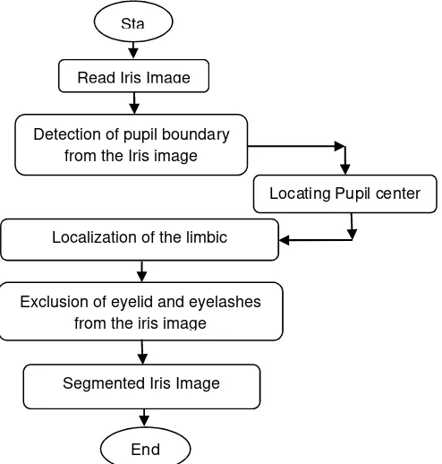
Fig. 1: The research design of iris segmentation.
2.0 PUPIL DETECTION ALGORITHM
The inner boundary of the iris can be detected by finding the pupil, which is assumed to be the darkest portion of the eye and ALGORITHM 1 used to detect the pupil because of the smaller pixels intensity values which are not uniformly distributed across the pupil.
ALGORITHM 1: ENHANCED PUPIL BOUNDARY DETECTION ALGORITHM
Step 1: Transform the original grey scale eye image into a binary image by applying global threshold Sta
Read Iris Image
Detection of pupil boundary from the Iris image
Locating Pupil center
Localization of the limbic boundary
Exclusion of eyelid and eyelashes from the iris image
Segmented Iris Image
to segment the pupil whose greyscale values are much smaller than other eye components, this threshold computes the constant of the threshold using the image space; transforming an image f to a binary image g as thus
g x, y
10
if f x, y
otherwise
T
T
is the threshold value.
Step 2: Inversion of the dark colour pupil to white to make it visible since it is the area of interest.
Step 3: Smoothen the blurred circular boundary of the pupil image by applying averaging filter for easy detection of the pupil boundary.
Step 4: Find the center of the Pupil by using ALGORITHM 2
(a) (b)
Fig. 2: Pupil localization. (a) Original Iris image. (b) Binarised image.
The pupil center is located using the following procedure:
ALGORITHM 2: FINDING PUPIL CENTER ALGORITHM
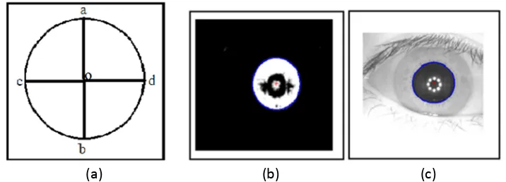
Step 1: Scan through the binary image starting from the top left horizontally, using 8-neighbour moving left to right, mark the first and last 8-neigbour encountered whose value is 1, that should form a vertical bisector of the pupil circle at 0∘ and 180∘ respectively and this correspond to the points a and b in the fig. 3a.
Step 2: Scan vertically through the image this time, using the same connected component in the same direction, also mark the first and last 8-neighbour encountered whose value is 1 that should form a vertical bisector of the pupil circle at 270∘ and 90∘ respectively and this correspond to the points c and d in the fig. 3a.
Step 3: Connect these two opposite point; whose midpoint O in fig 3a becomes the assumed center of the pupil and half of the length of the opposite connected pixel is an assumed radius of the pupil.
Step 4: Use the assumed center and radius detected to localize the pupil circular shape.
(a)
(b) (c)
Fig. 3: Pupil localization. (a) Four points of the assumed detected pupil circle image. (b) Detected binarised image boundary. (c) Detected pupil image boundary.
The Hough transform can be used to determine the parameters of a circle when a number of points that fall on the perimeter are known. And when these parameters are known it makes the computational speed faster. A circle with radius r and center (a, b) is defined by the parametric equations
x a rcos θ y b rsin θ
When the angle θ sweeps through the full 360 degree range the points (x, y) traces the perimeter of a circle. It is a bit more difficult to detect the outer boundary of the iris because of the similar intensity pixels values between the iris and sclera, but we can localise the iris boundary as in ALGORITHM 3.
ALGORITHM 3: DETECTING IRIS BOUNDARY ALGORITHM
Step 1: Remove the noise on the eye image by applying minimal blurring to avoid making the boundary difficult to visualize
.
Step 2: Apply 5 by 5 average filter for smoothing the image on the original intensity image.
Step 3
:
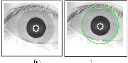
Feed the pupil center parameter as detected in section 3 to circle Hough transform to detect the iris boundary.(a) (b)
Fig. 4: Iris Localization: (a) Original Iris image. (b) Detected Iris boundary
4.0 EXCLUSION OF THE EYELIDS AND EYELASHES
Having located the assumed center of the pupil as in section 3, the eyelid is excluded with the following procedure as below.
ALGORITHM 4: DETECTTION AND REMOVAL OF THE EYELIDS ALGORITHM
Step 1: Apply Canny edge operator on the eye image
.
Step 2: Locate four different edge point pixels on the top (left and right) and bottom (left and right) respectively lying on the edge map of the eyelids detected by Canny edge operator.
Step 3: Use the center of the pupil which is also the center of the iris, to locate the curvilinear eyelids shape and excluded it from the iris.
The eyelashes detected are removed from the iris by the steps in Algorithm 5
ALGORITHM 5: DETECTION AND REMOVAL OF THE EYELASHES
Step 1: Search for pixel values that are lower than the iris pixel values at the top and bottom of the iris image.
Step 2: Set the pixel values to the same value of the iris pixels in the neighbourhood
.
Step 3: Repeat the two steps above until all the eyelashes on this area are detected and removed, thus getting rid of the eyelashes
.
5.0 EXPERIMENTAL RESULT
(c)
(d)
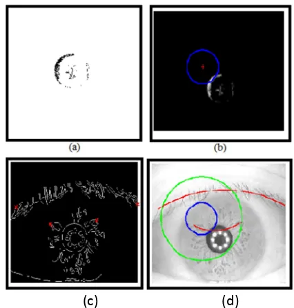
Fig. 5: Good Iris segmentation process: (1) Binarised eye image. (b) Detected Pupil. (c) Detected eyelids and eyelashes. (d) Iris Segmentation.
(c) (d)
Fig. 6: Poor Iris segmentation process: (a) Binarised eye image. (b) Failed detected Pupil. (c) Bad detected eyelids and eyelashes. (d) Failed Iris Segmentation
Table 1: Percentage results of the Iris Segmentation stages.
Stages of Segmentation Percentage Accuracy
Percentage Error
Pupil Localisation
Iris detection
Eyelids and Eyelashes Occlusion
Complete iris segmentation
99.10%
98.10%
99.40%
98.90%
0.90%
2%
0.6%
We observed that the reason for small percentage error in all the stage of the iris segmentation in Table 1 above is because of poor image quality and low pixel count recorded from some eye images. This caused a high intensity values close to the boundary of the pupil and making the detection of such case difficult as shown in Fig. 6. Most of the eye images that have such problem but with more pronounced low intensity boundaries were detected perfectly. There were also some constricted pupil which failed and causing the detection of the iris impossible too. Meanwhile, 2% error of the bad iris detected in the table was accounted for some eye images which has little variation of intensity value between the iris and sclera boundary making it very hard to detect the exert iris of the eye image
Furthermore, the 0.60% error on the eyelids was due to illumination on the eye image causing the intensity values of the lower cornea and iris to be evenly distributed. Notwithstanding the error rate shown Table 1, the overall result reveals a higher accuracy rate as shown in Table 2 and the speed of the segmentation process is also improved, since center of the pupil was first located and same was fed to Circle Hough transform accumulator space which increases the segmentation rate. Table 2 suggest that our new algorithm out performed [8] and [9] algorithm.
Table 2: Results of iris segmentation methods from different authors
Stages of Segmentation Accuracy Rate fairly the same, but we used an extended Circle Hough transform where the information about the iris center is fed directly to it accumulator space thus ensuring faster computational speed and reduction of memory space used up while its performance is maintained. Removal of major occlusion caused by the eyelids is accomplished by applying canny edge operator and then the eyelids edges detected are removed from the iris. We have developed and implemented an enhanced iris segmentation system based on Circle Hough transform, with accuracy rate of 98.90% and error rate of 1.1%. Thus we suggest that more work should be done on using appropriate method to deal with poor image quality and illumination due to light. Also most importantly, these poor quality images should be removed from the database since it is only quality eye image are been captured and stored in an implemented system during acquisition stage by the user.
REFERENCE
[1] J. Daugman “Probing the uniqueness and randomness of Iris codes”. Proceedings of the IEEE. Vol 94 No.11, November 2006. Pp. 1927-1935
[2] W. Anna, C. Yu , Z. Xinhua, and W Jie, “Iris recognition based on 2D wavelet and AdaBoost neural network”. Trends in intelligent systems and computer Engineering. TISCE 2008.
[3] S. Liz, and B. Stephen. Human Eye Anatomy: Parts of the Eye 2011.
[4] A. Pranith, and C. R. Srikanth. “Iris Recognition using Corner Detection”. Information Science and Engineering (ICISE), 2010 2nd International Conference on Digital Object Identifier. December,2010. Vol.6 No.4, pp. 2151 – 2154.
[5] S. Mahboubeh. Ellipse gamma spectrum iris recognition based on average square shrinking segmentation and trapezium normalization. A Ph.D. Thesis. Universiti Technologi Malaysia; 2011
[6] T. Fabian, J. Gaura, and P. Kotas. An Algorithm for Iris Extraction. Image Processing Theory Tools and Applications (IPTA), 2010 2nd International Conference on Digital Object Identifier, July 2010. pp 464-468
[8] M. T. Seyyed, A. Ahmad, and M. S. M. Seyyed. A Novel Iris Segmentation Method based on Balloon Active Contour. Machine Vision and Image Processing (MVIP), 2010 6th Iranian. October 2010. Pp 1-5
[9] C. Sreecholpech, and S. Thainimit. A Robust Model-based Iris Segmentation. International Symposium on Intelligent Signal Processing and Communication Systems (ISPACS 2009), December 2009. pp 599-602.


