CYTOTOXIC EFFECTS OF PROTEIN FRACTION ISOLATED FROM
Curcuma mangga
VAL RHIZOMES AND CONTAINING RIBOSOME-INACTIVATING PROTEINS ON
CANCER CELL-LINES AND NORMAL CELL
Sismindari, Retno S. Sudibyo
Faculty of Pharmacy, Gadjah Mada University, Sekip, Yogyakarta
Endang Astuti
Chemistry Department, Faculty of Mathematics and Natural Sciences
Gadjah Mada University, Yogyakarta
Received 11 October 2004; Accepted 18 October 2004
ABSTRACT
C. mangga Val. has been used as an alternative remedy for cancer in Yogyakarta. The protein fraction of C. mangga was identified to contain Ribosome-inactivating Protein which cleave supercoiled double stranded DNA in vitro. In this experiment, the protein fraction isolated from fresh, 400C dried and freeze dried C. mangga Val. rhizome was screened against HeLa, Raji cell-lines and normal mononuklear cells for cytotoxic effects. This would enable us to describe the sensitivity of the protein extract on different cell types. The level of cytotoxicity was determined on the level of LC50 which was based on the percentage of the cell death following the 24 hours incubation with the extract.
The protein isolated from C. mangga Val. was able to cleave supercoiled double stranded DNA to nick circular form. This result suggested that protein contained RIPs. The highest activity was identified in the protein isolated from fresh C. mangga Val, and this was followed respectively by freeze drying and 400C drying C. mangga Val. The comparison of the cytotoxic effect showed that protein of fresh C. mangga Val produced the largest number of death cells and the most toxic was on the HeLa cell line. Moreover, the LC50 indicated that the highest cytotoxic effect was shown by protein isolated from fresh C. mangga Val. followed respectively by freeze drying and 400C drying C. mangga Val. Based on LC50, the highest cytotoxic effect of C. mangga Val was found on HeLa cell line, while similar cytotoxic effect was appear on Raji cell line and normal mononuclear cells
Keywords: C. mangga Val., RIP, cytotoxic, cancer cell lines, normal cell
INTRODUCTION
Many plants tissue produce substances which are toxic to other organism, and irreversibly inactivate eukaryotic ribosome by cleaving the N-glycosidic bond in the A4324 position of 28S RNA fraction so that they are no longer function in protein synthesis. These plant proteins are known as ribosome-inactivating protein (RIPs). According to their structure, RIPs can be classified into two major types. Type 1 consists of a single chain with a molecular weight around 30 kDa, while type 2, with a molecular weight around 60 kDa, usually consists of two chain (A and B) connected by disulfide bond. The A chain is homologous to type 1 RIP and is responsible for the toxicity of the molecule. The B chain is a lectin which binds the toxic to the cell surface and facilitates the entry of A chain into the cell [1].
Besides the activities of RIPs on ribosomal RNA, several RIPs demonstrate to exhibit a unique enzymatic activity on cleaving supercoiled double stranded DNA into the nicked circular or linear form. RIPs only act on supercoiled and nick-circular DNA and seldom cleave the linear form of the same molecule [2]. This fenomenon was first reported with trichosantin, an abortifacient, immunosupressive and anti tumor protein purified from the traditional Chinese herb medicine Tian Hua Fen [3].
Interest in RIPs is growing due to several discoveries, such as the anti viral activity of mirabilis antiviral protein (MAP), a type 1 RIP which has successfully focused attention on its use as potential anti-HIV [4]. The potent cytotoxicity also makes them an excellent candidates as the toxic part of immunotoxin for cancer therapy [5].
that the “Kunir Putih” powder sold in Yogyakarta expressed enzymatic activity to cleave supercoiled double stranded DNA into a nick circular conformation. In order to confirm the results, identification of RNA N-glycosidase activity from the powder extraxt were carried out. Assay for RNA N-glycosidase activity on 26S rRNA Saccharomyces cerevicease ribosome indicated that this extracts showing the activity. Besides, the
extracts have cytotoxic activity to B-Lymphoblastoid Cell Line (B-LCL) that grown from B cells of Nashoparingeal Carcinoma (NPC) Patient [7].
Based on the introduction, the cytotoxic effect of C. Mangga Val rhizomes’s protein having Ribosome-inactivating Proteins (RIPs) on cancer cell line and normal cell have to be evaluated. Because of C. mangga was sold in Yogyakarta in the powder form, so effect of 400C drying and by freeze drying on protein activity was carried out too.
.
EXPERIMENTAL SECTION
Materials
Protein extract containing Ribosome-inactivating proteins was isolated from fresh, 400C dried and freeze dried C. mangga Val rhizome using 5 mM sodium phosphate pH 7.2 containing 0.14M sodium chloride [8], followed by 100% ammonium sulphate precipitation [9]
Procedure
Cleavage of supercoiled DNA by the protein extract
One µg of plasmid DNA (pUC18) was incubated with various amounts of extract to volume of 20 µl containing 50 mM Tris-HCl, 10 mM MgCl2, 100 mM NaCl, pH 8.0, at 30
o
C for 1 hour. At the end of the reaction, 5 µl of loading buffer (30% glycerol, 200 mM EDTA, 0.25% bromophenol blue and 0.25% xylene cyanol FF) were added. Electrophoresis was carried out in 0.5xTBE buffer in a 1% agarose gel. DNA bands were visualized by staining with ethidium bromide [10].
Preparation of cancer cells
HeLa (was developed from human cervix carcinoma) and Raji (was developed from lymphoblast of burkitt lymphoma cancer patient) cell-lines were obtained from the laboratory stock collection at the Life Science Laboratory, Gajah Mada University, Jogyakarta. All cancer cells were grown in RPMI 1640 (Sigma) medium containing 5% v/v fetal calf serum (FCS) (Sigma) and 1% v/v
fungison (Sigma) in the presence of 1% w/v of penicillin-streptomycin (Sigma) and incubated at 37oC incubator in a 5% CO2 controlled atmosphere. Cells were counted using a Neubauer Haematocytometer and resuspended in medium at the final concentration of 5x10⁵cells/mL.
Preparation of normal mononuclear cells
Normal mononuclear cells were prepared by the addition of an equal volume of Ficoll-Hypaque [11] to fresh blood obtained from a healthy individual. The mixture was then centrifuged at 2000g for 20 mins. The buffy coat layer containing mononuclear cells was then removed to a fresh tube, washed 3 times using RPMI 1640 medium, counted and adjusted to a final concentration of 5x10⁴cells/mL.
Determination of Cytotoxic effect
Cytotoxicity assays were conducted in 96 well flat bottom ELISA plates (Nunc). Protein extract was tested on a 10-fold dilution series of concentrations in culture media, from undiluted 250 ; 100 ; 50 ; 25 ; 10 ; 5 ; 2,5 ; 1,0 ; 0,5 ; 0,25 µg/mL. Each well received 100 µL of cell suspension and 100 µL of the appropriate dilution of extract. Then, plates were incubated in a 37oC humidified incubator in 5% CO2 for 24 h. Counting by MTT method for the cells death was used to calculate % inhibition [12, 13]. Untreated cells were used as a control (0% cytotoxicity). Mean percent cytotoxicity then calculated following treatment relative to controls. There were 3 replicate wells for each treatment.
RESULTS AND DISCUSSION
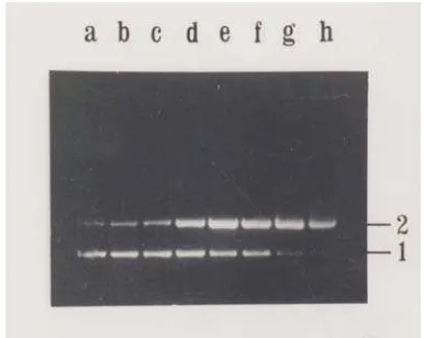
Fig. 1 Cleavage activity of protein extract of fresh C. mangga Val. (Lane a) untreated pUC18 were run as markers. (Lane b - h) treated pUC18 with protein extract at various concentrations, namely 0,1 ; 0,5 ; 1,0 ; 2,5 ; 5,0 ; 7,5 ; and 10,0 µg.
Cleaving activity was shown by disappearing of (1). supercoiled form of pUC18 , (3), and appearing of open circular form of pUC18 and (2). linear form of pUC18
Fig. 2 Cleavage activity of protein extract of 400C dried C. mangga Val. (Lane a) untreated pUC18 were run as markers. (Lane b - h) treated pUC18 with protein extract at various concentrations, namely 0,1 ; 0,5 ; 1,0 ; 2,5 ; 5,0 ; 7,5 ; and 10,0 µg. Cleaving activity was shown by disappearing of (1). supercoiled form of pUC18 , (2), and appearing of open circular form of pUC18.
When pUC18 was treated with protein of C. mangga Val. dried 400C, similar result was also obtained. At a concentration of 0,1 µg of protein extract exhibited apparent activity on supercoiled DNA in a fashion similar to fresh extract. On the other hand, at the highest concentration (10 µg), supercoiled DNA haven’t been cleaved at all to supercoiled DNA that shown by fade band
of
Fig. 3 Cleavage activity of protein extract of freezed dryC. mangga Val. (Lane a) untreated pUC18 were run as markers. (Lane b - h) treated pUC18 with protein extract at various concentrations, namely 0,1 ; 0,5 ; 1,0 ; 2,5 ; 5,0 ; 7,5 ; and 10,0 mg. Cleaving activity was shown by disappearing of (1). supercoiled form of pUC18 , (2), and appearing of open circular form of pUC18
supercoiled DNA (Line h). Besides, the linear DNA have not appeared yet by adding 10 µg protein
Tested protein extract of freezed dry C. mangga Val rhizomes on the ability of cleaving supercoiled double stranded DNA indicated that at the concentration of 0,1µg, the protein extract was able to cleave double stranded pUC18 into nick-circular form as indicated in Fig 3, Line b. The supercoiled band gradually became fade away and nick circular band became bolder than before, in line with increasing protein concentration. However, not all of supercoiled DNA have been cleaved to nick circular DNA at the highest concentration (10 µg) (Line h).
Since the cleavage of supercoiled DNA is one of the characteristic of RIPs beside the N-glycosidase activity [2, 3], so it was strongly suggest that this protein extract contained RIPs.
The protein extract of fresh C. mangga rhizome have the highest activity on DNA supercoiled cleaving. It can be seen from Figure 1, 2 and 3 that at the same concentration, fresh C. mangga was able to cleavege supercoiled DNA more extensively than 400C dried dan freezed dry, indicating that fresh was more active than 400C dried and freezed dry C. mangga rhizome, respectivelly.
0% 20% 40% 60% 80% 100% 120%
250 100 50 25 10 5 2.5 1 0.5 0.25
Concentration (ug/mL)
% of C
ytotox
ic
ity
Fresh 40oC dried Freezed dry
A2
B2
C2
Fig 4 Morphological appearance of HeLa, Raji, and mononuclear cells in the presence of protein extract. (A1) treated HeLa cells; (A2) untreated HeLa cells; (B1) treated Raji cells (B2) untreated Raji cells; (C1) treated mononuclear cells; (C2) untreated mononuclear cells.; (t) death cell
untreated cells as shown at Fig 4. On the treated cancer cells, densityof cells and percentage of cell death were higher compared to the untreated cells. The higher of the added extract the higher of the cancer cells death. For normal mononuclear cells, however, there were no differences on the cells viability between untreated and treated cells. Interestingly, this protein extract showed different level of cytotoxicity in between cancer cells.
Protein of fresh C. mangga Val gave the highest cytotoxic effect on Hela cell line, Raji cell line and normal mononuclear cells, followed by freeze drying and 400C drying respectively. It was demonstrated in Fig 5, 6, 7 and Table 1. The protein of fresh C. mangga Val still have native conformation, so it provided the highest on both cleaving of supercoiled DNA and cytotoxic activity. Similar activity was appeared on freeze dried C. mangga Val, because the rhizome was not dried by heating. The lowest activity was shown by 400C drying, since RIP underwent denaturation after heating at 400C.
0%
Fresh 40oC dried Freezed dry
0%
Fresh 40oC dried Freezed dry
Fig. 6 Cytotoxic activity of fresh, 400C dried and freezed dry C. mangga Val rhizome on HeLa cell line. The percent cytotoxicity was determined based on the absorbance at 550nm of the lysate of MTT-stained cells. Each value came from triplicate determinations.
Fig. 7 Cytotoxic activity of fresh, 400C dried and freezed dry C. mangga Val rhizome on normal mononuclear cells. The percent cytotoxicity was determined based on the absorbance at 550nm of the lysate of MTT-stained cells. Each value came from triplicate determinations.
Table 1 The LC50 of protein extract of fresh, 40OC dried and freeze dried C. mangga Val rhizome on HeLa cell line, Raji cell line and normal mononuclear cells.
LC50 (µg/mL)
Interestingly, this protein extract showed different level of cytotoxicity in between cancer cells. It was more cytotoxic to HeLa cell line with LC50 levels 18,2 µg/mL for fresh, 15,0 µg/mL for freeze dried and 105,1 µg/mL for 400C dried of C. mangga Val rhizome rather than Raji cell line with with LC50 levels 41,3 µg/mL for fresh, 80,4 µg/mL for freeze dried and 244,2 µg/mL for 400C dried of C. mangga Val. The cytotoxicity of C. mangga Val’s protein to normal mononuclear cells was similar to Raji cell line with LC50 levels 37,8 µg/mL for fresh, 81,0 µg/mL for freeze dried and 87,0 µg/mL for 400C dried of C. mangga Val rhizome.
These two cancer cells, HeLa, and Raji, represent different kind of cancer cell, which will enable us to describe the sensitivity of the protein extract on different cell types. HeLa cell line is an epithelial cells which are developed from human cervix carcinoma transformed by human papiloma virus 18 (HPV18) [14]. While Raji cell is lymphoblast-like cells which are developed from lymphoblast of burkitt lymphoma cancer patient. This differences on the cytotoxic level seemed to be caused by the differences on the mechanism of the cell death in these cancer cells. The mechanism by which the protein extract killed the
cells was not clearly understood yet. However, it has been reported that some type 1 RIP isolated from Aspergillus giganteus and RIPs-like protein isolated from Mirabilis jalapa had cytotoxic effect and induce the cells death via apoptosis, programmed of the cell death [15-17].
bcl-2 and c-myc which could affect synergistically on the inhibition of apoptosis [21]. Apoptotic resistance of Raji cell-line was demonstrated while this cell was treated by RIPs-like protein isolated from Mirabilis jalapa [16].
As indicated on Table 1, there were no different cytotoxic effects with Raji cell line when normal mononuclear were treated with the protein extract. The cytotoxic effect to normal mononuclear cells presumed due to glycosil residues of type 1 RIP could be bound to carbohydrat or by endocytosis [1]. Besides, some RIP have been founded, they have different structure with the exception of their homolog sequens. Base on the idea, some RIPs had different cytotoxic effect on normal mononuclear cells. For example, type 1 RIP of Mirabilis jalapa had cytotoxic effect to normal mononuclear cells, however, the protein of Annona squamosa had no effect [10]
CONCLUSION
1. The protein fraction of fresh Curcuma mangga Val. gave the highest activity on supercoiled DNA cleaving, followed by freeze drying and 40oC drying, respectively.
2. The protein fraction of fresh Curcuma mangga Val. gave the highest cytotoxic activity to cancer cell lines and normal cell, followed by freeze drying and 40oC drying, respectively
3. The protein fraction of Curcuma mangga Val. gave the highest cytotoxic activity to HeLa cell line, followed by Raji cell line and normal mononuclear cells with similar LC50 value.
REFERENCES Bourinbaiar, AS., Morell, JL., Brown, JH., Tsai, WP., Chen, AY., Huang, HI., and Chen, H.C., 1994, Proc. Natl. Acad. Sci. USA., 91. 12208 – 12212
5. Goldmacher, Y.S., Bourret, L.A., Levine, B.A., Rasmussen, R.A., Pourshadi, M., Lambert, J.M., and Andeson, K.C., 1994, Blood 84, 3017 - 3025.
6. Lestariana, W., Triandiyasih, H., Mubarika, S., and Sismindari, 2000, Buletin ISF, Vol.3, 25-30 7. Lestariana, W., Mubarika, S., and Sismindari,
1999, Laporan Penelitian, Proyek QUE Batch I UGM/KU/424/QUE/IV/99, Fakultas Kedokteran UGM, Yogyakarta
8. Stirpe, F., 1983, Biochem. J., 216, 617-625 9. Deutcher, M.P., 1990, Methods in Enzymology;
Guide to Protein Purification, Vol.182, Academic Press Inc., California
10. Sismindari, Husaana, A., and Mubarika S., 1998, Indon. J. Pharm., 9,4, 146-152.
11. Anonim,1997,http://numi.crc.nus.edu.sg/numiwe b/ numimcu/training/cytometriy/ficol.html, June 2001
12. Berridge, M.V., Tan, A.S., McKoy, K.D., and Wang, R., 1996, Biochemica, 4, 15-20
13. Riss, T. and Moravee, R., 1996, Promega Notes Magazine, 59, 19-25
14. Bosch, F.X., Schwart, E., Boukamp, P., Fusening, N.E., Bartsch, D., and zur Hausen, H., 1990, J.Virol, 64, 4743 – 4754
15. Sismindari, Mubarika, S., and Sudjadi, 2001, Indon.J.Biotech. June, 459 – 462.
16. Ikawati, Z., Sudjadi, Widyaningsih, E., Puspitasari, and D., Sismindari, 2003, Oriental Pharmacy and Experimental Medicine, Korea. Inpress.
17. Olmo, N., Turnay, J., Buitrago, G.H., Silanes, I.L., Gavilanes, J.G., and Lizarbe, M.A., 2001, Eur.J.Biochem., 268, 2113 – 2123.
18. Nishimura, A., Ono, T., Ishimoto, A., Dowhanic, J.J., Frizell, M.A., Howley, P.M., and Sakai, H., 2000, J.Virol, 74 (8), 3752-3760.
19. Houge, G., Robaye, B., Eikhom, T.S., Goldstein, J., Mellgren, G., Gjertsen, B.T., Lanotte, M., and Doskeland, S.O., 1995, Mol.Cell.Biol. 15, 2051-2062.
20. Kawabata, Y., Hirokawa, M., Kitabayashi, A., Horiuchi, T., Kuroki, J., and Miura, A.B., 1999, Blood, 94, 3523 – 3530.