Cytotoxic selectivity of MJC
0.3
and MJC
0.5
,
acidic ribosome-inactivating proteins isolated
from
Mirabilis jalapa
L. leaves against various
cancer cell-lines
Sismindari1*
, Mae Sri Hartati2
, Adhyatmika1
, Retno S. Sudibyo
1Faculty of Pharmacy, 2Faculty of Medicine, Universitas Gadjah Mada, Yogyakarta, Indonesia
ABSTRACT
Mirabilis jalapaL. contains basic (MJ30) and acidic (MJC) Ribosome-inactivating proteins (RIPs). Further purification of MJC has been found two RIPs, MJC0.3and MJC0.5. This study is aimed to prove the cytotoxic selectivity of MJC0.3and MJC0.5against many cancer cell-lines and normal cell line. The two RIPs, MJC0.3and MJC0.5were tested their cytotoxic effect on 8 human cancer cell lines and normal cell using MTT assay compared with MJC protein. The highest cytotoxic activities of MJC0.3and MJC0.5were against EVSA-T followed by T47D, HeLa, WiDR, SiHa, Raji, NS1, and MCF7, with the IC50of 59.3, 102.4, 162.9, 190.5, 249.5, and 304.5 µg/mL, respectively for MJC0.3 and 32.8, 75.5, 86.0, 108.3, 346.7, and 220.06 µg/mL, respectively for MJC0.5. Based on these IC50values, MJC0.3and MJC0.5were specific to EVSA-T and T47D, whereas they were not selective against Raji and SiHa (SI<10.0). It can be concluded that he acidic RIPs isolated fromM. jalapaL. leaves was potential to be developed as anticancer agents for breast cancer.
Key words:acidic RIPs – M. jalapaL.- cytotoxic selectivity - cancer cell lines.
INTRODUCTION
Mirabilis jalapaL. is one of the medicinal plat
that has been scientifically proven to contain ribosome-inactivating proteins (RIPs), a potential toxin isolated from plants that inhibits mamalian protein synthesis.1,2Seed and root of M. jalapaL.
contain MAP (mirabilis antiviral protein), which is actively strive against mechanical transmission from TMV (Tobacco Mozaic Virus) in tobacco, pepper, and tomato.3 Beside these activities, RIPs which
are isolated from some plants have an antioxidant activity in the same level as Fe-superoxide dismustase from Escherichia coli,4,5 and has a
potential to be developed as immunotoxin, when conjugated with antibody for acquiring selective anticancer agent.6
Ribosome-inactivating proteins fromAnnona
squamosaL. andM. jalapaL. have been identified,7
as demonstrated for their ability to cleave super-coiled DNA and the adenine glycoside bond on 4324-rRNA ofSaccharomyces cerevisae.8Total protein
fraction isolated from the leaves of M. jalapaL. has cytotoxic effect and has been proven can induce the apoptotic process of HeLa cell line.9
Ribosome-inactivating proteins isolated from theM. jalapaL. root also have cytotoxic activity against HeLa cell-line and relatively less cytotoxic against Raji cell line.10Based from the purification process, it was
found that leaves ofM. jalapaL. was demonstrated to contain more than one RIPs, basic and acidic RIPs respectively.5,11
Basic-RIP (MJ30) which was obtained from purification using CM-Sepharose CL-6B has cytotoxic activity against breast cancer T47D, HPV-18 induced uterine cervix cancer cell line HeLa, and blood myeloma cancer cell line NS1 with IC50as
0.36, 7.06 and 1.25 mg/mL respectively. This protein was able to induce apoptotic process as demonstrated by the appearance of DNA fragmentation.12The unbound fractions which are
negatively-charged, called MJC, has been known to cleave super-coiled DNA11 and has more
powerfull cytotoxic effects compare to the MJ30.13
The MJC inhibits carcinogenesis process as shown by its ability in decreasing the expression of oncogen RAS and COX-2 in HeLa cell-line.5Further
purification step of MJC has been developed and gaining two acidic RIPs, MJC0.3 and MJC0.5.5
However, the cytotoxic activity of these two acidic RIPs against cancer cell-lines has not been studied.
MATERIALS AND METHODS
Chemicals
Medium of RPMI 1640 (Sigma), DMEM (Sigma), M199 (Sigma), sodium bicarbonat (Sigma), and HEPES (Sigma), Fetal Bovine Serum (FBS) 0.5% v/v and 10% v/v (Gibco), penicillin-streptomycin 1% (v/v) (Gibco), and fungison 0.5% (v/v) (Gibco), sodium phosphat 5mM pH 6.5, sterile aquadest, trypsin 0.5%, Phosphat Buffer Saline solution, ethanol 70%, and MTT reagent 5 mg/mL were used in the study. The remaining chemicals were high purified grade (pro analysis) from Merck.
Cell Lines
Three kinds of cell lines (MCF7, EVSA-T, and WiDR) were gained from Erasmus Medical Center Netherlands, which were recultured by the Integrated Research and Testing Laboratory (IRTL), Universitas Gadjah Mada Yogyakarta. Other cell lines were the collections of the IRTL Universitas Gadjah Mada Yogyakarta. Cell lines were cultured from the stock which was stored at -60oC and freshly
used post-culture. Cells were routinely maintained in their appropriate media supplemented with 10% FBS in 37oC and 5% CO
2incubation.
MJC Proteins Preparation
MJC0.3and MJC0.5 were prepared from fresh
M. jalapaL. leaves using 5mM phosphate buffer
pH 7.2 containing 0.14 M NaCl and precipitated using aceton. Total protein was then purified using CM-Sepharose CL-6B resins. Followed by
DEAE-sepharose resins coloum for the unbound fraction. Two different active protein fraction (MJC0.3 and MJC0.5) were obtained from the purification processes.5
Cytotoxicity Studies
The study were started by preparing the appropriate media for each cell line and the 2x10⁴
cell/100µL was grown in the 96 well plates. These cells were then treated by a serial concentration of MJC’s. The citotoxicity tests were performed by MTT (3-[4,5-dimethylthiazol-2-yl]-2,5-diphenyl tetrazolium bromide)-assay method. The formed formazans formed can not penetrate the life cell’s membrane and accumulated in the live cells. This intracellular formazan can be dissolved with detergent, in this experiments we used sodium dodecyl sulphate (SDS). The intensity of the colour which appeared then can be measured with 96-well plate scanning spectrophotometer, where the percent of the colour intensity of the chambers treated over the control represents the amount of the live cells left by treatment over the cells before treatment, otherwise the percent of cells inactivated because of the treatment of the sample then can be assayed. Value of IC50was determined by regression correlation method, using protein concentration as the independent variable and percentage of inhibition as the dependent variable. Then using the regression equation, the IC50 value can be found as the concentration by putting 50% value as the y value at the regression. Value of p as the degree of significance was analyzed using SPSS statistical program version 16.0 on 95% confidence-interval.
RESULT AND DISCUSSION
Tested MJC, MJC0.3, and MJC0.5 proteins on the ability of cleaving supercoiled double stranded DNA indicated that the protein isolates were able to cleave double stranded pUC18 into nick-circular form as indicated in FIGURE 1. This result supported the previous study which using the crude extract.8Since the cleavage of supercoiled DNA is
one of the characteristic of RIPs besides their N-glycosidase activity,13-15hence it was strongly suggest
FIGURE 1. MJs cleavage activity study, shows that there were cleavage activities as the forming of linear and nick circular structure of the DNA
These MJC0.3, and MJC0.5proteins demonstrated cytotoxic activity against EVSA-T, T47D and MCF7 (breast cancer lines), WiDR (colon cancer cell-lines), SiHa and HeLa (uterine cervix cancer cell-lines induced by HPV-16 and HPV-18 respectively), Raji (naso-pharynx cancer cell-lines), NS1 (myeloma cancer cell-lines), but not on normal Vero cell-lines. The induction of the cells death was characterized by the changing of the cells viability that was quantified by the intensity of the color formed by the formazans deposition. On the treated cancer cell-lines, the percentage of the cell death was higher compared to the untreated cells. The higher addition of protein samples, the higher the cancer cells death. For normal Vero cells, there were no differences on the cells viability between untreated and treated cells in the low concentration, however cytotoxicity was observed in very high concentrations.
Interestingly, these proteins showed different level of cytotoxicity in between cancer cell-lines. From the data TABLE 1, MJC0.3and MJC0.5gave best activities for EVSA-T as 32.8 µg/mL and 59.3 µg/mL respectively. Unfortunately, the exact IC50 for MJC fraction was not determined. However, against other cell lines, T47D, we could get those all of the 3 MJs fractions gave best results, as 111.0 µg/mL for MJC, 102.4 µg/mL for MJC0.3, and 75.6 µg/mL for MJC0.5. These MJC0.3and MJ0.5showed various 2 or 3 times higher IC50values against another cell lines (TABLE 1).
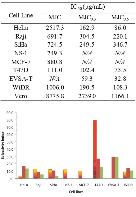
TABLE 1. IC50of protein MJC, MJC0.3, and MJC0.5 against various cell lines
Cell Line
IC50(µg/mL)
MJC MJC0.3 MJC0.5
HeLa 2517.3 162.9 86.0
Raji 691.7 304.5 220.1
SiHa 724.5 249.5 346.7
NS-1 749.3 N/A N/A
MCF-7 880.8 N/A N/A
T47D 111.0 102.4 75.5
EVSA-T N/A 59.3 32.8
WiDR 1006.0 190.5 108.3
Vero 8775.8 2739.0 1166.1
FIGURE 2. Selectivity index values show that MJC(orange), MJ0.3(brown) and MJC0.5(green) were majority selective compared with Vero normal cell-lines. Selectivity indexes were measured by dividing IC50value against each cancer cell lines by the IC50value against the normal cell line Vero
Selectivity index is an important factor in the development of anticancer agent, because this index is the factor that ensure the safety of the xenobiotic. Selectivity index is the ratio between cytotoxic
O C O 3
sc nc
lin
activity of the xenobiotic against certain cancer cell line by the activity against normal cell line. In this study, Vero cell line was used as normal cell line comparator. After comparison of IC50against cancer cell line with IC50 against normal cell line as a quantification for activity of protein MJC, MJC0.3, and MJC0.5, it was known that generally protein MJ was selective, except against some several cell lines (FIGURE 2). The value of a selectivity index can be accepted if it was higher than 10, which means that the xenobiotic is active to kill cancer cells 10 times stronger than to kill normal cells.
The selectivity index values of MJC were below the standard for HeLa and WiDR, but due to the activities of MJC against those two cell lines were very low, the IC50 values were very high. Same cases and discussion were applied for protein MJC0.3, and MJC0.5against Raji and SiHa. For unmeasured IC50values, selectivity index values also can not be measured.
Comparing this result with the result of the study performed by Ikawati et al.10 in which the RIP
total protein fraction which was isolated from the root ofM. jalapa L. was cytotoxic against HeLa cell line but less cytotoxic against Raji cell line, it could be concluded that RIPs which were isolated from the leaves were more potent, because they were relatively more toxic against both HeLa and Raji cell-lines. This result can be explained by two probable reasons, the first is because the RIPs in the root and the leaves itself were different, and second because the concentration of RIPs in the root were smaller than the amount of RIPs in the leaves.
Furthermore, if we compare with the cytotoxicty of MJ-30,12 against T47D, NS-1, and HeLa cell
lines, with IC50 as 0.36, 7.06 and 1.25 mg/mL respectively, the results of this study for MJC gave smaller IC50against T47D and NS-1. However, if this results are compared with the study by Sudjadi,
et al.13where citotoxicity study of the fraction in
the same level of MJC but purified by
ionenaustauscher type II, gave IC50values as 0.28
mg/mL (T47D), 0.007 mg/mL (NS1), and 0.014 mg/mL (HeLa), so that this study only gave smaller IC50value for T47D as 111.0 µg/mL, but for NS-1 and HeLa were higher, as 0.749 and 2.517 mg/mL respectively (TABLE 1).
Those differences can be caused by various conditions, such as the purification method, and also the method of the study itself. Sudjadi, et al.13
performed cytotoxicity analysis using tryphan-blue direct counting method, whereas this study was using MTT method. These can cause some perspective differences. With the result that, we can get actual values compilation which can be compared well just in this study where the method of isolation, purification, and also cytotoxicity analysis itself are in the same way, so that the results can show a valid comparison of the selectivity and the specificities of MJ proteins against each cancer cell line.
The next interesting discussion is that MJ proteins from the entire studies both for MJ-30 and MJC itself, showed a consistency of IC50 values that always relatively small against T47D cell line. For HeLa cell line, good result was shown by Sudjadi,et al.13and this study for MJC
0.3and MJC0.5
fractions. Contradictive result was shown for MJC fraction (TABLE 1). This fact strengthen the indication that MJ proteins were cytotoxic-specific against breast cancer, besides selective as shown by selectivity-index values (FIGURE 2).
CONCLUSION
From the study it can be concluded that MJC protein was relatively potent for T47D cells, MJC0.3 for T47D and EVSA-T cells, and MJC0.5was potent for T47D and EVSA-T cells. Since both of T47D and EVSA-T cells are breast cancer cell-lines, the study showed that these protein fractions were potential against the breast cancer. Further study for other types of cancer cell-lines is needed. For the follow up of this study, further development of these isolates to be formulated as a potential anticancer agent against breast cancer will actuate research on anticancer therapy.
ACKNOWLEDGMENT
REFERENCES
1. Barbieri L, Batelli MG, Stirpe F. Ribosome-inactivating proteins from plants. Biochim Biophys Acta 1993;1154: 237-82.
2. Stirpe F. Ribosome-inactivating proteins: review.Toxicon 2004;44:371-83.
3. Kubo S, Ikeda T, Imaizumi S, Takanami Y, Mikami Y. A potent plant virus inhibitor found inMirabilis jalapaL. Ann. Phytopathol Soc Jpn 1990;56:481-7.
4. Barbieri L, Polito L, Bolognesi A, Ciani M, Pelosi E, Farini V,et al.Ribosome-inactivating proteins in edible plants and purification and characterization of a new ribosome-inactivating protein fromCucurbita moschata. Biochim Biophys Acta 2006;170:783-92.
5. Sismindari, Sudjadi, Ikawati Z, Wijayanti N. Uji aktivitas dan kloning MJ-C, ribosome- inactivating protein (RIP) asam dari Mirabilis jalapa L. Laporan Kemajuan Program Insentif Riset Dasar, Ristek 2008. Universitas Gadjah Mada, Yogyakarta.
6. Stirpe F, Battelli MG. Riobosome-inactivating proteins: progress and problems. Cell Mol Life Sci 2006;63:1850-66.
7. Sismindari, Husaana A, Mubarika S.In vitrocleavage of supercoiled DNA byAnnona squamosaextract. Majalah Farmasi Indonesia 1998;(9)4:146-52.
8. Sismindari, Lord JM. 2000, RNA-N-glikosidase activity of leaves crude extract fromCarica papaya, Morinda citrifolia, Mirabilis jalapa. (Special issue) Indon J Biotech 2000;342-5.
9. Sudjadi, Ikawati Z, Sismindari, Fajar Arini K. Efek fraksi protein bijiMirabilis jalapapada proses kematian kultur sel HeLa. Biologi 2002;2:743 -53.
10. Ikawati Z, Sudjadi, Sismindari, Sari RP, Maulani N. Efek fraksi protein sejenis RIP (ribosome inactivating protein) yang diisolasi dari akar Mirabilis jalapaL. terhadap proses kematian Sel Raji. J Biologi 2002;2:769-83. 11. Sudjadi, Sismindari, Herawati T, Prasetyowati AT.
Pemurnian ribosome inactivating protein (RIP) dari daun
Mirabilis jalapaL. dengan kolom CM-sepharose CL-6B dan Sephacryl S-300HR. Majalah Farmasi Indonesia 2003;14(2):316-21.
12. Ikawati Z, Sudjadi, Widyaningsih E, Puspitasari D, Sismindari. Induction of apoptosis by protein fraction isolated from the leaves of Mirabilis jalapaL. on HeLa and Raji cell-lines, OPEM 2003;3:151-6.
13. Sudjadi, Sadarum MT, Nastiti N, Witasari LD, Sismindari, 2007, Cytotoxic effects of acidic RIPs isolated from
Mirabilis jalapaL. leaves on cancer cell-lines, Indon J Pharm 2007;18 (1):8–14
14. Li MX, Yeung HW, Pan LP, Chan SI. Trichosanthin, a potent HIV-1 inhibitor, can cleave supercoiled DNAin vitro. Nucleic Acids Res 1991;19: 6309-12.
15. Ling J, Lui W, Wang TP. Cleavage of supercoiled double stranded DNA by several ribosome inactivating proteins
in vitro. FEBS Letters 1994;345:143-6.