RETROSPECTIVE VALIDATION OF A STRUCTURE-BASED VIRTUAL SCREENING
PROTOCOL TO IDENTIFY LIGANDS FOR ESTROGEN RECEPTOR ALPHA AND ITS
APPLICATION TO IDENTIFY THE ALPHA-MANGOSTIN BINDING POSE
Agustina Setiawati
1, Florentinus Dika Octa Riswanto
2, Sri Hartati Yuliani
3,
and Enade Perdana Istyastono
2,3,4,*
1
Laboratory of Pharmacognosy-Phytochemistry, Faculty of Pharmacy, Sanata Dharma University, Paingan, Maguwoharjo, Depok, Yogyakarta 55284, Indonesia
2
Laboratory of Pharmaceutical Chemistry, Faculty of Pharmacy, Sanata Dharma University, Paingan, Maguwoharjo, Depok, Yogyakarta 55284, Indonesia
3
Laboratory of Pharmaceutical Technology, Faculty of Pharmacy, Sanata Dharma University, Paingan, Maguwoharjo, Depok, Yogyakarta 55284, Indonesia
4
Center for Environmental Studies Sanata Dharma University (CESSDU), Soropadan, Condongcatur, Depok, Yogyakarta 55283, Indonesia
Received April 23, 2014; Accepted June 6, 2014
ABSTRACT
The publicly available enhanced data of ligands and decoys for estrogen receptor alpha (ERα) which were
recently published has made the retrospective validation of a structure-based virtual screening (SBVS) protocol to
identify ligands for ERα possible. In this article, we present the retrospective validation of an SBVS protocol using
PLANTS molecular docking software version 1.2 (PLANTS1.2) as the backbone software. The protocol shows better enrichment factor at 1% false positives (EF1%) value and the Area Under Curve (AUC) value of the Receiver
Operator Characteristic (ROC) compared to the original published protocol. Moreover, in all 1000 iterative attempts
the protocol could reproduce the co-crystal pose of 4-hydroxitamoxifen in ERα binding pocket. It shows that the protocol is not only able to identify potent ligands for ERα but also able to be employed in examining binding pose of known ligand. Hence, the protocol was successfully employed to examine the binding poses of α-mangostin, an ERα
ligand found in the Garcinia mangostana, L. pericarp.
Keywords: Structure-based virtual screening (SBVS); molecular docking; estrogen receptor alpha (ERα); α-mangostin
ABSTRAK
Keberadaan data termutakhir ligan-ligan reseptor estrogen alfa (ERα) beserta pengecohnya memungkinkan
dilakukan validasi retrospektif pada protokol-protokol Penapisan Virtual Berbasis Struktur (PVBS) yang
dikembangkan untuk identifikasi ligan-ligan ERα. Artikel ini membahas validasi retrospektif protokol PVBS yang
menggunakan aplikasi penambatan molekuler PLANTS versi 1,2 (PLANTS1.2) sebagai tulang punggung protokol
tersebut dalam identifikasi ligan-ligan ERα. Hasil validasi retrospektif menunjukkan bahwa protokol yang
dikembangkan memiliki nilai faktor pengayaan pada 1% false positives (EF1%) dan nilai di bawah kurva dari Receiver
Operator Characteristic (ROC) yang lebih baik daripada protokol original yang dipublikasikan bersama data ligan dan pengecoh. Protokol tersebut juga divalidasi untuk melihat kemampuannya dalam menambatkan senyawa aktif
dengan penambatan ulang 1000 kali ligan ko-kristal 4-hidroksitamoksifen pada kantung ikatan ERα. Dalam 1000 kali
iterasi, keseluruhan simulasi menunjukkan kemampuan protokol dalam mereproduksi pose ko-kristal. Hal ini menunjukkan bahwa protokol yang dikembangkan memiliki kemampuan untuk identifikasi ligan-ligan poten pada
ERα dan menambatkan ligan ERα dengan tepat di kantung ikatan. Oleh karena itu, protokol tervalidasi ini selanjutnya digunakan untuk meneliti pose-pose ikatan α-mangostin, senyawa aktif yang terdapat pada kulit
manggis (Garcinia mangostana, L.).
2004), breast cancer was the highest prevalence cancer with 26% of the patients were less than 40 years old [3]. Among other molecular determinants in breast cancer
development, estrogen receptor α (ERα) is one of
molecular targets in the therapy [4]. Tamoxifen, one of drug of choice in breast cancer therapy [5], is targeting
ERα. Tamoxifen itself is a ligand with high affinity for ERα, which is metabolized to 4-hydroxytamoxifen and
N-des-methyl-4-hydroxo-tamoxifen with affinities circa 30
to 100 times stronger than tamoxifen for ERα [6]. Fortunately, the ERα crystal structure with
4-hydroxytamoxifen as its co-crystal ligand is publicly available in the Protein Data Bank (PDB) with the PDB code of 3ERT (Fig. 1) [7], which could be employed to
be a virtual target to identify potential ERα fragments [8].
On the other hand, a public database of enhanced useful decoys (A database of useful decoys: Enhanced (DUD-e)) has recently published for 102 molecular drug
targets, including ERα [9]. The article presenting DUD-e shows that employing ERα as the molecular target in a
structure-based virtual screening (SBVS) campaign gave enrichment factor at 1% false positives (EF1%) value and
the Area Under Curve (AUC) value of the Receiver Operator Characteristic (ROC) of 15.4 and 67.48%, respectively [9]. Some improvements in the virtual screening protocol are therefore required to have more convincing SBVS tools [10]. Therefore, development of SBVS protocols to discover drugs in order to cure or even to prevent the development of breast cancer by
targeting ERα is of considerable and timely interest.
In this post genomics era, the development of computer technology is remarkably boosting and assisting the drug discovery and development [11]. One of developing technique in the field is structure-based drug design and discovery [10,12–15], which uses the availability of the three-dimensional (3D) structures of the protein targets [7,9,16-17] to identify or even to
design novel ligands [14]. The availability of ERα
structure and its ligands and decoys (which has been publicly available since 2006 in a database of useful decoys (DUD) [17]) has led some attempts to construct
valid SBVS protocols to identify novel ligands for ERα
[8-9,17-18]. By employing PLANTS docking software [19], Anita et al. [8] has developed and retrospectively
validated SBVS protocol to identify ERα ligands. The
protocol showed a better VS quality compared to the original protocol accompanying the publication of the
Fig 1. The co-crystal ligand 4-hydroxytamoxifen
(carbon atoms are in cyan) in the ERα (carbon atoms are in green) binding pocket [7]. The ERα is presented
in the cartoon mode, while the crystal structure pose is presented in the sticks mode. Only polar hydrogens (presented in white), residues (presented in sticks mode, carbon atoms are in green) with hydrogen bond interaction (presented in black dashes) and ionic interaction (presented in red dashes) to the ligand, and a conserved water molecule [8] are presented for the sake of clarity. Nitrogen and oxygen atoms are presented in blue and red, respectively
Fig 2.Structure α-mangostin [21]
ligands and decoys by Huang et al. [17]. Subsequently, in order to test the applicability of a software to identify protein-ligand interaction fingerprints PyPLIF in a SBVS campaign, Radifar et al. [18] re-scored and re-validated the results from the SBVS protocol constructed by Anita et al. [8]. PyPLIF-aided SBVS protocol showed a significant increase in quality by filtering on the hydrogen bond interactions of the ligands to the
ASP351 of the ERα [18]. Interestingly, the co-crystal
ligand 4-hydroxytamoxifen in the crystal structure of the
ERα (3ERT.pdb) does not have the hydrogen bond interactions to the ASP351 of the ERα (Fig. 1) [7]. The
SBVS protocol developed by Radifar et al. [18] is therefore not able to reproduce the pose of the
co-crystal ligand in the crystal structure of the ERα.
Fig 3.The representatives of the identified binding poses of α-mangostin (carbon atoms are in magenta) in the ERα (carbon atoms are in green) binding pocket [7] from cluster 1(a)and cluster 2(b). The rendering is similar to Fig. 1
Fig 4.The ROC curves of the SBVS protocol developed in this research (solid line) and the random sampling (dashed line)
decoys DUD-e was published for 102 molecular drug
targets, including ERα [9]. This database has more
ligands and decoys compared to DUD [9,17].
This research aimed to develop a robust computational medicinal chemistry tool in order to
discover novel ERα ligands and to examine the binding poses of known ERα ligands. In this article, the
retrospective validation of a modified SBVS protocol developed by Anita et al. [8] using the DUD-e database is presented. The validated protocol was subsequently examined to see its ability to reproduce the pose of the
co-crystal ligand in the ERα binding pocket and to examine the binding pose of α-mangostin (Fig. 2), an ERα ligand found in the Garcinia mangostana, L. pericarp [20-21]. The modified SBVS protocol developed in this research showed better VS quality compared to the original SBVS protocol accompanying the publication of DUD-e [9] and was able to reproduce the pose of the co-crystal ligand [7]. Two distinct binding poses of
α-mangostin in the ERα binding pocket (Fig. 3) were
identified using the retrospectively validated protocol in this research.
METHODS
Materials
A dataset of ERα ligands (383 compounds) and
their decoys (20,685 compounds) in file type of .mol2
obtained from DUD-e [9]. The ERα crystal structure
and its co-crystal ligand 4-hydroxytamoxifen (3ERT.pdb) downloaded from Protein Data Bank (PDB) submitted by Shiau et al. [7]. Configuration files to perform molecular docking simulations in order to
perform SBVS to identify ligands for ERα using
PLANTS docking software prepared by Anita et al. [8]: (i) The virtual target protein.mol2, (ii) the conserved water molecule water.mol2, and (iii) the configuration files to run PLANTS docking softwareplants.config.
Computation Details
Computational medicinal chemistry applications used in this research were: PLANTS docking software version 1.2 (PLANTS1.2) [19,22] to perform molecular docking simulations, R computational statistics software to calculate EF1% and AUC and to perform
statistical tests [23] and PyMOL [24, 25] to calculate the root mean square distances (RMSD) values and to generate molecular pictures. All calculations and computational simulations were performed on a Linux (Ubuntu 10.04 LTS Lucid Lynx) machine with Intel(R) Xeon(R) CPU E31220 (@ 3.10 GHz) as the processors and 8.00 GB of RAM.
Procedure
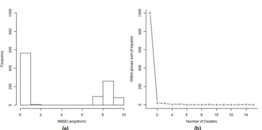
Fig 5.The histogram(a) and the Scree plot(b) of the RMSD values of α-mangostin docking poses compared to its binding pose with the best ChemPLP value
pocket of ERα by using configuration files from Anita et
al. [8]. Every run resulted in 50 docked poses. Therefore, every compound had 150 poses. The best pose of each compound was selected as the pose with the best ChemPLP score [19]. The compounds were then ranked based on their ChemPLP score [10]. The EF1% and the
AUC values were then calculated [26] by using R computational statistics software [23].
The similar procedure was performed to dock
co-crystal ligand 4-hidroxytamoxifen in the ERα binding
pocket 1000 times to see the ability of the protocol to reproduce the pose of the co-crystal ligand. The best poses collected in every run were compared to the pose of the co-crystal ligand and the RMSD values were calculated by using PyMOL [24-25,27]. The protocol was considered as acceptable in reproducing the co-crystal ligand pose if resulted in the RMSD value of less than 2.0 Å [28].
The same procedure to dock co-crystal ligand
4-hidroxytamoxifen in the ERα binding pocket was performed to dock α-mangostin which resulted in 1000
poses. Those poses were compared to the pose with the best ChemPLP score and the RMSD values were calculated [27]. Based on the RMSD values, the poses were clustered by employing k-means clustering [29] in R computational statistics software [27].
RESULT AND DISCUSSION
This research aimed to retrospectively validate an
SBVS protocol in order to develop a tool to identify ERα
ligands and to examine the binding poses of identified
ERα ligands. The SBVS protocol used in the research
was initially developed by Anita et al. [8] and has been
modified in this research. The modification in the SBVS protocol was using three independent molecular docking simulations for each compound instead of one as performed in the initial SBVS protocol [8]. One of the advantages of using some more independent simulations is that we can sample more converged docking poses for every compounds although it increases the computing cost [27,30-31].
Fig. 4 presents the ROC of the % true positives (%TP) and % false positives (%FP) results from the
retrospective validations to identify ERα ligands by
employing the DUD-e dataset [9,26]. The retrospective validation showed that the EF1% value of the modified
protocol was 18.54 (Fig. 4). This EF1% value is higher
than the EF1% value of original SBVS protocol (15.4)
[9]. The EF1% represents the early enrichment results
from the protocol. The higher the EF1%value, the better the protocol in the identification of known ERα ligands
is [9,26]. It means that in the first 1% of the ranked database the protocol can identify known ligands and put them in the higher rank compared to their decoys [9,26]. Based on Fig. 4, the AUC value could be calculated by using pROC package in R computational statistics software [23]. The AUC value resulted from the retrospective validations was 76.41% with 95% confidence interval of 74.05%-78.78%. This value is also better than the AUC value of the original protocol (67.48%) [9]. The ideal value of the AUC is 100% [26], which indicates that all known ligands are ranked higher than their decoys. In random sampling, the AUC value is 50% [32]. The EF1%value represents the early
this research are better than the original protocol [9], we are confident that the protocol is more robust to identify
ERα ligands.
The developed protocol was intended to be employed also in the examination of the binding pose of
known ERα ligands. The protocol was then challenged to
redock the co-crystal ligands 4-hydroxytamoxifen in the
ERα binding pocket [7] for 1000 times [27,31].
Remarkably, in all redocking simulations the protocol showed its ability to reproduce the co-crystal pose with RMSD values of < 2.00 Å. The developed protocol in this
research is therefore able to identify ERα ligands and to examine their binding pose in the ERα binding pocket.
The protocol was then employed to examine the
binding pose of α-mangostin (Fig. 2) resulted in 1000
selected poses from 1000 different iterations of the
protocol. The compound α-mangostin, which can be
found in the pericarp ofGarcinia mangostana, L. [21,33]
is a known ligand for ERα [21,33]. Garcinia mangostana, L. has recently gained its popularity [34] due to its applications as herbal medicines to treat inflammation and bacterial infections [33] as well as its application in cancer chemoprevention [35]. Therefore, it is of interest
to select α-mangostin as the lead compound in the
structure-based drug design in this research. By examining the RMSD values of the poses compared to the pose with the best ChemPLP value presented in a histogram and a Scree plot in Fig. 5, two distinct poses
of α-mangostin were identified (Fig. 3). Interestingly, both poses shows that α-mangostin located in the ERα
binding pocket (Fig. 3) only in the subpocket where the co-crystal ligand 4-hydroxytamoxifen interacts to
THR347 and ASP351 (Fig. 1). α-mangostin could not go
to the subpocket where 4-hydroxytamoxifen interacts to GLU353 and ARG394 (Fig. 1). This indicates that referring to 4-hydroxytamoxifen as the co-crystal ligand,
α-mangostin interacts to ERα as an allosteric ligand.
CONCLUSION
The SBVS protocol developed in this research is a
robust computational tool to identify ERα ligands and to examine their poses in the binding pocket ERα. The
application of the SBVS protocol to examine the binding
poses of a known ERα ligand α-mangostin resulted in
two distinct binding poses. The binding poses of
α-mangostin indicate that it interacts in the allosteric site of ERα.
ACKNOWLEDGEMENT
This work was supported by Sanata Dharma University Special Internal Research Grant 2013 (No. 083/Panel/LPPM USD/SP/X/2013).
REFERENCES
1. Siegel, R., Naishadham, D., and Jemal, A., 2012, CA Cancer J. Clin., 62 (1), 10–29.
2. Jemal, A., Bray, F., and Ferlay, J., 2011, CA Cancer J. Clin., 61 (2), 69–90.
3. Purnomosari, D., 2006,PhD Thesis, VU University, Amsterdam.
4. Harvey, J.M., Clark, G.M., Osborne, C.K., and Allred, D.C., 1999, J. Clin. Oncol., 17 (5), 1474–1481.
5. Mouridsen, H., Giobbie-Hurder, A., Goldhirsch, A., Thürlimann, B., Paridaens, R., Smith, I., Mauriac, L., Forbes, J.F., Price, K.N., Regan, M.M., Gelber, R.D., and Coates, A.S., 2009, N. Engl. J. Med., 361 (8), 766–776.
6. Desta, Z., Ward, B.A., Soukhova, N.V., and Flockhart, D.A., 2004, J. Pharmacol. Exp. Ther., 310 (3), 1062–1075.
7. Shiau, A.K., Barstad, D., Loria, P.M., Cheng, L., Kushner, P.J., Agard, D.A., and Greene, G.L., 1998,Cell, 95 (7), 927–937.
8. Anita, Y., Radifar, M., Kardono, L., Hanafi, M., and Istyastono, E.P., 2012, Bioinformation, 8 (19), 901–906.
9. Mysinger, M.M., Carchia, M., Irwin, J.J., and Shoichet, B.K., 2012, J. Med. Chem., 55 (14), 6582–6594.
10. De Graaf, C., Kooistra, A.J., Vischer, H.F., Katritch, V., Kuijer, M., Shiroishi, M., Iwata, S., Shimamura, T., Stevens, R.C., de Esch, I.J.P., and Leurs, R., 2011,J. Med. Chem., 54 (23), 8195–8206.
11. Van Gunsteren, W.F., Bakowies, D., Baron, R., Chandrasekhar, I., Christen, M., Daura, X., Gee, P., Geerke, D.P., Glättli, A., Hünenberger, P.H., Kastenholz, M.A., Oostenbrink, C., Schenk, M., Trzesniak, D., van der Vegt, N.F.A., and Yu, H.B., 2006,Angew. Chem. Int. Ed., 45 (25), 4064–4092. 12. Kooistra, A.J., Leurs, R., de Esch, I.J.P., and de
Graaf, C., 2014, “From Three-Dimensional GPCR Structure to Rational Ligand Discovery” in G Protein-Coupled Receptors - Modeling and Simulation., Ed. Filizola, M., Springer Netherlands, 129–157.
13. De Graaf, C., Oostenbrink, C., Keizers, P.H.J., van der Wijst, T., Jongejan, A., and Vermeulen, N.P.E., 2006,J. Med. Chem., 49 (8), 2417–2430.
14. Istyastono, E.P., Nijmeijer, S., Lim, H.D., van de Stolpe, A., Roumen, L., Kooistra, A.J., Vischer, H.F., de Esch, I.J.P., Leurs, R., and de Graaf, C, 2011,J. Med. Chem., 54 (23), 8136–8147.
Bioinformation, 9 (6), 325–328.
19. Korb, O., Stützle, T., and Exner, T.E., 2009, J. Chem. Inf. Model., 49, 1, 84–96.
20. Hopert, A.C., Beyer, A., Frank, K., Strunck, E., Wünsche, W., and Vollmer, G., 1998, Environ. Health Perspect., 106 (9), 581–586.
21. Shibata, M.A., Iinuma, M., Morimoto, J., Kurose, H., Akamatsu, K., Okuno, Y., Akao, Y., and Otsuki, Y., 2011,BMC Med., 9 (69), 1–18.
22. Korb, O., Stützle, T., and Exner, T.E., 2007,Swarm Intell., 1 (2), 115–134.
23. Robin, X., Turck, N., Hainard, A., Tiberti, N., Lisacek, F., Sanchez, J.C., and Müller, M., 2011, BMC Bioinf., 12 (77), 1–8.
24. Lill, M.A., and Danielson, M.L., 2011, J. Comput. Aided Mol. Des., 25 (1), 13–19.
25. The PyMOL Molecular Graphics System, Version 1.2r1, Schrödinger, LLC.
30. Wijtmans, M., de Graaf, C., de Kloe, G., Istyastono, E.P., Smit, J., Lim, H., Boonnak, R., Nijmeijer, S., Smits, R.A., Jongejan, A., Zuiderveld, O., de Esch, I.J.P., and Leurs, R., 2011,J. Med. Chem., 54 (6), 1693–1703.
31. Radifar, M., Yuniarti, N., and Istyastono, E.P., 2013,Indo. J. Chem., 13 (3), 283–286.
32. Jain, A.N., and Nicholls, A., 2008, J. Comput. Aided Mol. Des., 22 (3-4), 133–139.
33. Yodhnu, S., Sirikatitham, A., and Wattanapiromsakul, C., 2009,J. Chromatogr. Sci., 47 (3), 185–189.
34. Purwanto, A., 2008, Skripsi, Universitas Sebelas Maret Surakarta
![Fig 2. Structure α-mangostin [21]](https://thumb-ap.123doks.com/thumbv2/123dok/2204056.1619791/2.595.319.561.112.295/fig-structure-a-mangostin.webp)
![Fig 3. The representatives of the identified binding poses of α-mangostin (carbon atoms are in magenta) in the ERα (carbon atoms are in green) binding pocket [7] from cluster 1 (a) and cluster 2 (b)](https://thumb-ap.123doks.com/thumbv2/123dok/2204056.1619791/3.595.79.268.294.472/representatives-identified-binding-mangostin-magenta-binding-cluster-cluster.webp)