VOL. 7, NO. 1, pp. 23 – 29, January, 2017 Submitted May 2015; Revised August 2015; Accepted April 2016
Effect of Tetrodotoxin from Crude Puffer Fish (Tetraodon fluviatilis) Liver Extract on Intracellular
Calcium Level and Apoptosis of HeLa Cell Culture
Natanael Untario1
*, Titik Cinthia Dewi1
, M Aris Widodo2
, Pudji Rahaju3
1
Biomedical Science, Medical Faculty, Brawijaya University, Malang, Indonesia 2
Pharmacology Department, Brawijaya University, Malang, Indonesia 3
Department Otolaryngology Head and Neck Surgery , Saiful Anwar Public Hospital, Malang, Indonesia
ABSTRACT
Cervical cancer is the third most commonly diagnosed cancer and fourth leading cause of women death with 8% of total death caused by cancer in women in 2008. Tetrodotoxin (TTX) is a potent neurotoxin found in inner or-gans puffer fish, with the specific mechanism of sodium channel blocking, and widely used for research purposes. Previous reports claimed that TTX has the capability of inhibiting metastatic process of cancer and apoptotic effect. Studies also show that apoptosis is a process involving increase of intracellular calcium level, yet the connec -tion between TTX and increase of intracellular calcium level, therefore triggering apoptosis, has not been estab-lished. This is an experimental study with post-test only control group design, carried out by exposing HeLa cell culture to crude liver extract of a puffer fish species, Tetraodon fluviatilis. Crude puffer fish liver extract is admin-istered into HeLa cell culture well in different concentrations 10-4
, 10-2
, and 10-1
. Intracellular calcium level and apoptosis were then measured after 18 hours of incubation. Measurements of intracellular calcium level were done by using CLSM with Fura-2AM staining, and apoptosis by using flowcytometry with Annexin V/PI. The result shows that there is a significant differences between samples both in intracellular calcium (p < 0.05) and apoptosis (p < 0,05). Both intracellular calcium and apoptosis levels are proportional to liver fish extract concentration. Pear-son’s correlation test shows correlation between treatment and intracellular calcium levels (p = 0.000), between treatment and apoptosis (p = 0.002), but not between intracellular calcium and apoptosis (p = 0.05). These results suggest that TTX induces an increase in intracellular calcium level and apoptosis, but calcium pathway is not the sole cause of the apoptosis.
Keywords: Apoptosis, HeLa cell culture, intracellular calcium, Tetraodon fluviatilis, tetrodotoxin
Cervical cancer is the third most commonly diagnosed cancer and fourth leading cause of women death, contributing to 9% (528,000) from total new cancer cases per year and 8% (257,100) total cancer death for women in 2008 [1]. Conventional strategy for cancer management including surgery, radiotherapy, and chemotherapy with both synthetic agents and natural agents have been established for years [2], yet cancer is still the leading cause of death. Side effects of chemotherapy includes headache, hair loss, nausea and vomiting, sore throat, diarrhea, and many more [3]. This clearly calls for other effective therapeutic agents
with less intolerable side effects.
Tetrodotoxin (TTX) is a potent neurotoxin found in the inner organs of puffer fish, including Tetraodon fluviatilis. Found in deep water, these fishes are known to be toxic, hence they are are usually returned to the sea by fisherman. TTX is a highly selective voltage-gated sodium channel (VGSC) blocker [4]. TTX have been used for anaesthetic agents [5], reducing opioid withdrawal symptoms [6], preventing ischemia in post stroke patients [7]. Recent studies also show that TTX has anti-metastatic [8] and anti tumor properties [9]. Study by Fouda et al. (2005) shows that TTX extracted from masked puffer fish (Arothron diedematus) can
JTLS | J. Trop. Life. Science 23 Volume 7 | Number 1 | January | 2017
INTRODUCTION
*Corresponding author: Natanael Untario
Biomedical Science, Medical Faculty, Brawijaya University Jalan Veteran, Malang 65145, Indonesia
E-mail: [email protected]
How to cite:
induce apoptosis [9]. Connection between apoptosis and increased intracellular calcium level is already well-known [10], but on how TTX may induce apoptosis is yet to be explained. Therefore, this study investigates the effects of TTX to intracellular calcium level and whether this pathyway is the main contributing factor to TTX-induced apoptosis in tumor cells.
This is an experimental study with post-test only control group design. The subject was HeLa cell line culture from cervical cancer. There are 4 groups with 5 repeating samples each, which are 5 samples of HeLa cell culture, 5 samples each of HeLa cell culture ex-posed to crude T. fluviatilis liver extract with concen-trations of 10-4, 10-2, and 10-1. This research took time from July 2013 until October 2013 at Biomedical Labo-ratory, Medical Faculty of Brawijaya University, In-donesia.
HeLa cell culture
HeLa cell line was procured from Badan Pengkajian dan Penerapan Teknologi (BPPT) Serpong, Jakarta, In-donesia. HeLa cells were grown in F12 medium supple-mented with L-glutamine and 7% FBS on tissue-cul-ture plastic in 5% CO2 incubator at 37ºC, 95% atmos-phere, 100% humidity. After the culture is in mono-layer state, crude T. fluviatilis liver extract were then added to each well in different concentrations like mentioned above. After the period of 18h incubation, cells were then removed and examined.
Extraction of Tetraodon fluviatilis liver
This method is a modification of TTX extraction method introduced by Zhou et al. In 2003 [11]. The liver was washed then mashed using mortar. Mush were then weighed and added into an Erlenmeyer glass with aquadest equal to 1.5× tissue weight. Acetic acid was added with concentration 0.2% tissue weight and then stirred for 1.5 hours in room temperature. Solu-tion was then filtered. Remaining soluSolu-tion was then heated in 80ºC using water bath for 10 min, and cooled until room temperature. It was then centrifuged at 600 × g for 10 min. Supernatan then filtered using microfilter 0.2 µm, and stored at -4ºC.
Identification of TTX
(1) Nanodrop spectrophotometry. Solution was pre-pared by dissolving it in 2 mL of 2M NaOH and heated in boiling water bath for 45 min. After cooling to room temperature, TTX was identified by the UV
spectrum for characteristic absorptions associated with C9-base, 2-amino-6-hydroxymethyl-8-hydroxyquinazo-line, which are possibly formed from TTX. UV spec-trum of alkali-decomposed TTX compounds appears as a shoulder at near 276 nm [12]. (2) Liquid Chromatog-raphy Mass Spectrometry (LC-MSMS). LC-MSMS was performed using TSQ Access Max Type Triple Quadrupole by Thermofisher for the MSMS and Ac-cella type LC and autosampler. 0.1% Formic acid with aquabidest and 0.1% formic acid in acetonitrile (90%, injection volume 2 µL) used as mobile phase. Sample was prepared my diluting 25 µL in 1 ml methanol. Ion peak (M+H+) appeared at m/z = 320, showing a molec-ular weight of the toxin (319) [12].
Intracellular Calcium Level Measurement
HeLa cells were washed 2 times using HBSS, and loaded Fura-2AM by incubation in standard medium containing 5 µm acetoxymethyl ester form of fura-2-(fura-2-AM) for 30 min at room temperature (37ºC). Cells were then washed using HBSS twice and resus-pensed with density of 2 × 10⁹ cells/L. Cells were then incubated for 45 min at room temperature and mounted in a cell chamber on the stage of Zeiss Ax-iovert 200 microscope under continuous perfusion. Single-cell fluorescence was excited at 340 nm and 380 nm using a Cairn monochromator (100 ms excitation at each wavelength every 2 s, 10 nm bandwidth) and images of the emitted fluorescence obtained with a 40 × Fluar objective were collected using a 400 DCLP dichroic mirror and a D510/80 emission filter (both from Chroma Technology) and recorded with a Hama-matsu ORCA-ER camera. Single-cell fluorescence was recorded as 340/380 nm fluorescence ratio and
cali-Apoptosis was measured using flowcytometry An-nexin V-FITC kit (Trevigen, Gaithersburg, MD, cata-log# 4830-01-K) staining. Adherent cells released from their substrate using 0.25 trypsin. Cells were collected by centrifugation approximately 300 × g for 5 to 10 min at room temperature, then wash in cold (4ºC) phosphate-buffered saline (PBS). Cells were then incu-bated with 1 µL of Annexin-FITC and PI for 15 min at room temperature, according to manufacturer’s in-structions. Annexin-FITC/PI-stained samples were di-luted in 300 annexin binding buffer and examined im-mediately using a BD FACSort flow cytometer with
CellQuest software. Five thousand cells were excited at 488 nm and examined at 530 and 585 nm for Annexin-FITC and PI fluorescence, respectively.
Data Analysis
The data was analyzed using a test of variance (ANOVA) technique with p < 0.05. All tests employed SPSS 16 for Windows.
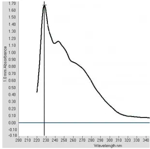
Identification of TTX in T. fluviatilis crude liver extract Spectrophotometry analysis of T. fluviatilis crude liver extract shows a peak around 246 nm and a shoul-der around 276 nm. The shoulshoul-der at near 276 nm is in accordance to the study by Asakawa et al., indicating the formation of C9-base, 2-amino-6-hydroxymethyl-8-hydroxyquinazoline specific to TTX or related sub-stances [12]. However, the peak around 246 nm is not specific to TTX or related substances, and might be due to other substances, considering this is a crude ex-tract (Figure 1).
Spectrophotometry of T. fluviatilis shows a shoul-der at near 276 nm indicating the presence of TTX. Other peak at around 246 nm shows the presence of other substances other than TTX.
Identification of TTX using LC-MSMS showed pre-dominant peak at m/z 320, in accordance to TTX molecular weight. Ion transition was found at 302 m/z and 257 m/z (Figure 2), as assigned to [M+H2H2O]+, [M+H3H2O]+.12,25 There are other peaks other than those mentioned, indicating that there are other sub-stances other than TTX in the extract solution.
Effect of T.fluviatilis Crude Liver Extract On Intracellu-lar Calcium and Apoptosis Level of HeLa Cells Culture
This study showed that intracellular calcium level is in proportion to the concentration of liver extract (sig < 0.05, ANOVA) (Figure 3, Figure 4). In addition, Pearson’s correlation test showed that there was a posi-tive correlation between liver extract concentration and intracellular calcium (p = 0.000, Pearson). This means that the higher concentration of liver extract, in which contained TTX, the higher intracellular calcium will be. Regression analysis showed that 79.6% of intracel-lular calcium is related to treatment.
Study for apoptosis showed that there was a signifi-cant difference between treatment group in terms of apoptosis (p < 0.05, ANOVA) and positive correlation between treatment and apoptosis (p = 0.002, Pearson) (Figure 3, Figure 5). Again, the higher concentration of liver extract, the higher apoptosis level of HeLa cells culture become. In this case, regression analysis
showed that 41.6% of apoptosis level is related to treat-ment. In addition, we analyzed whether there was a correlation of intracellular calcium of apoptosis, and the result was none (p = 0.05, Pearson). There was no correlation between intracellular calcium with apopto-sis level of HeLa cells culture.
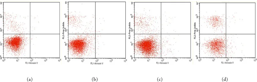
Culture exposed by different concentration of T. fluvi-atilis crude liver extract
There is an increase in HeLa cells only (A); HeLa cells + liver extract with 10-4
concentration (B); HeLa cells + liver extract with 10-2
concentration (C); HeLa cells + liver extract with 10-1 concentration (D).
This result confirmed our hypothesis that T.fluvi-atilis liver extract which contains TTX will trigger an increase in intracellular calcium and apoptosis level. We conclude that calcium increase did contribute to the apoptosis level, but not exclusively, in which many other pathway might played role in the apoptosis process of HeLa cells culture. Hence, apoptosis is a complex, multifactorial, and multi pathway process. This result is in accordance to the study from Fouda saying that TTX has an anti tumor effect, in which he was using crude extract from a different species of puffer fish A. diedematus. Fouda confirmed that TTX exposure increase the number of apoptotic cells, using Annexin V staining [9].
Our results also confirmed that T. fluviatilis liver extract containing TTX increased intracellular calcium level. This probes to the question as for where those calcium ion came from. Previous study have shown that calcium ion might originated from intracellular and extracellular pool [10].
Figure 1. Spectrums of UV absorption of alkaline hydrolyzates of TTX fraction from T. fluviatilis liver extract and authentic TTX with UV spectroscopy nanodrop is around 240 nm
JTLS | J. Trop. Life. Science 24 Volume 7 | Number 1 | January | 2017
RESULTS AND DISCUSSION
Extracellular calcium pool
Concentration of calcium is higher on the extracel-lular side than intracelextracel-lular. Calcium ion influx from extracellular side might come from different channels, which are voltage-gated calcium channels (VGCC), re-ceptor-operated channels (ROC), or store-operated channels (SOC). VGCC is usually activated after depo-larization of plasma membrane because of sodium channel influx from extracellular to intracellular side. This activated channels will allow calcium influx, re-garding the high concentration gradient of calcium be-tween two sides of plasma membrane [10]. Voltage-gated channels conventionally were thought to be exist in excitable cells, but recent study by Chakrabarti in 2006 showed that these channels also exist in
non-ex-citable cells, despite the working mechanism in these type of cells has yet to be established [13].
TTX is known for its ability to blocks sodium chan-nel, preventing sodium influx. This action will have 2 effect: (1) depolarization will not happen hence VGCC will not open, preventing calcium influx; (2) low intra-cellular sodium level, debilitating cancer cells need of sodium ion to function properly, such as the capability to maintain normal cell membrane distribution, a process to hold cells’ integrity, hence lowering the ca-pability of cancer cells’ proliferation and invasion [14, 15]. Related to the fact that influx of sodium is needed to open VGCC allowing calcium influx, which is sup-posedly inhibited by TTX treatment, intracellular cal-cium should be low in the presence of TTX. On the
(a) (b)
Figure 2. Product ion mass spectra of with LC-MSMS of T. fluviatilis liver extract (A); Authentic TTX (B) [16]. Predominant peaks showed at m/z 320, in accordance to TTX molecular weight, 302 m/z and 257 m/z for TTX-related substances. Other peaks indicating presence of other non TTX-related substances.
(a) (b)
contrary, our study shows that there is an increase of calcium level. This phenomenon suggests that there are other pathway of VGCC activation which are indepen-dent to sodium ion depolarization pathway, that is from cellular potassium efflux [17].
Extracellular calcium influx might also come from activation of ROC, which are mainly mediated by cell membrane receptor, especially glutamate receptor. Binding of glutamate to its receptor will activate and open ROC, allowing calcium influx.10 Another path-way might be from activation of SOC, which are af-fected by intracellular calcium storage level, specificly by reticulum endoplasma (RE). Decrease of intracellu-lar calcium storage in RE is mediated by Inositol-1,4,5 triphosphate receptor (Ins(1,4,5)P3Rs), and ryanodine receptor (RYR). Activation of these two receptor will deplete RE calcium storage to cytoplasm. This relation between calcium storage depletion with calcium influx
is known as Ca2+
release-activated Ca2+
current or I(CRAC). Despite all the efforts, signal connecting cal-cium storage and CRAC channel in plasma membran has yet to be established. Some reports showed that SOC activity is related to transient receptor potential (TRP) channel. In addition, I(CRAC) regulation is thought to be affected by mitochondrial activity. Hence, I(CRAC) is thought to be a dynamic and mul-tifactorial work between RE, mitochondria, and plasma membrane [18].
Intracellular calcium pool
Intracellular pool source of calcium ion are RE and mitochondria. Depletion of calcium ion storage to cy-toplasma is mediated by Ins(1,4,5)P3Rs and RYR as ex-plained before. These two receptors were found to be up-regulated in plasma membrane of apoptotic lym-phocyte. In cells with deficiency of these receptor,
re-JTLS | J. Trop. Life. Science 24 Volume 7 | Number 1 | January | 2017
(a) (b) (c) (d)
Figure 4. Identification of intracellular calcium level by confocal laser scanning microscope with Fura-2AM Staining. Green fluorescence shows the color of Fura-2AM. HeLa cells only (A); HeLa cells + liver extract with 10-4 concentration (B); HeLa cells + liver extract with 10-2 concentration (C); HeLa cells + liver extract with 10-1 concentration (D)
(a) (b) (c) (d)
Figure 5. Identification of apoptosis using flowcytometry with Annexin V/PI Staining. Lower left (Annexin -/PI -): viable cells; upper left (Annexin -/PI +): necrotic cells; lower right (Annexin +/PI -): early apoptotic cells; upper right (Annexin +/PI +): late apoptotic cells. HeLa cells only (A); HeLa cells + liver extract with 10-4 concentration (B); HeLa cells + liver extract with 10-2 concentration (C); HeLa cells + liver extract with 10-1 concentration (D).
sistancy to apoptosis were found, which can be re-verted pharmacologically by increasing cytoplasmic cal-cium level [10]. Mitochondria is involved in intracellu-lar calcium compartementalization. Mitochondrial up-take of calcium from cytoplasma is done via uniport transporter, and can be released from multiple route such as uniporter reversion, Na+
H+
-dependent-Ca2+ ex-change, or formation of permeable transition pore (PTP).
Mitochondria is a calcium buffer, but overload of calcium intake by mitochondria will trigger apoptosis via intrinsic pathway. Calcium ion play role in activat-ing Bcl-2 protein via dephosphorilation, catalized by calcineurin. Calcium also trigger an increase in outer mitochondrial membrane (OMM) and pro-apoptotic mitochondrial protein release. This permeability transi-tion requires a pore complex, known as PTP between two mitochondrial membrane. This mitochondrial in-nermembrane space (IMS) contains pro-apoptotic fac-tors such as cytochrome C, apoptosis inducing factor (AIF), procaspase-9, Smac/DIABLO, and endonuclease G [19, 20, 21, 22].
PTP is an important point of apoptosis which can be activated not only by calcium overload, but also other mitochondrial stress, such as reactive oxygen species (ROS), and high cellular pH. These factors will activate PTP and pro-apoptotic protein to cytoplasma, altogether with apoptotic protease activating factor 1, cleaves procaspase-9 to become caspase-9, and so the apoptosis cascade begins [23]. This theory is supported by study showing that TTX is able to trigger ROS for-mation [24]. Both ROS and calcium are mayor inducer of PTP mitochondrial formation [25].
T. fluviatilis crude liver extract containing TTX triggered an increase in intracellular calcium and apoptosis level of HeLa cells culture, but apoptosis is not exclusively caused by increased intracellular calcium. As for the origin of intracellular calcium increase level still needs further research to define whether it came from extracellular or intracellular pool.
This work was supported by Research Development Foundation of Medical Faculty, Brawijaya University, Malang, Indonesia.
1. Ferlay J, Shin HR, Bray F et al. (2010) Estimates of world-wide burden of cancer in 2008: GLOBOCAN 2008.
Inter-national Journal of Cancer 127: 2893–2917.
2. Gupta A, Mazumder UK, Kumar RS, Kumar TS (2004) Anti-tumor activity and anti-oxident role of Bauhinia racemosa against Ehrlich ascites carcinoma in Swiss albino mice. Acta Pharmacologica Sinica 25 (8): 1070–1076. 3. X-Plain (2011) Patient education: Chemoterapy.
http://online.x-plain.com/. Accessed: March 2015. 4. Benzer TI (2007) Toxicity, Tetrodotoxin.
http://emedicine.medscape.com/. Accessed: March 2015. 5. Hagen NA, Fisher KM, Lapointe B et al. (2007) An
open-label, multi-dose efficacy and safety study of intramuscular tetrodotoxin in patients with severe cancer-related pain. Journal of Pain Symptom Manage 34 (2): 171-182. 6. Madhavan A, He L, Stuber GD et al. (2010) Micro-Opioid
receptor endocytosis prevents adaptations in ventral tegmental area GABA transmission induced during nalox-one-precipitated morphine withdrawal. The Journal of Neuroscience 30 (9): 3276–3286.
7. Zhao X, Yeh JZ, Narahashi T (2001) Post-stroke demen-tia. Nootropic drug modulation of neuronal nicotinic acetylcholine receptors. Annals of the New York Academy of Sciences 939: 179-186
8. Brackenbury W, Djamgoz M (2007) Nerve growth factor enhances voltage-gated Na+
channel activity and Transwell migration in Mat-LyLu rat prostate cancer cell line. Jour-nal of Cell Physiology 210 (3): 602–608.
9. Fouda FM (2005) Anti-tumor activity of tetrodotoxin ex-tracted from the Masked Puffer fish Arothron diadematus. Egyptian Journal of Biology 7:1-13.
10. Orrenius S, Zhivotovsky B, Nicotera P (2003) Regulation of cell death: The calcium-apoptosis link. Nature Publisi-hing 4 (7): 552–565.
11. Zhou M, Shum FHK (2003) Method of extracting tetrodotoxin. https://www.google.com/patents/US6552191 Accessed: January 2015.
12. Asakawa M, Shida Y, Miyazawa K, Naguchi T (2012) Chromatography - The most versatile method of chemical analysis, Dr. Leonardo Calderon (Ed.), InTech. http://www.intechopen.com/books/chromatography-the- most-versatile-method-of-chemical-analysis/instrumental-analysis-of-tetrodotoxin. Accessed: February 2015. doi: 10.5772/48668.
13. Chakrabarti R, Chakrabarti R (2006) Calcium signaling in non-excitable cells: Ca2+ release and influx are indepen-dent events linked to two plasma membrane Ca2+
entry channels. Journal of Cellular Biochemistry 99 (6): 1503– 1516.
14. Bragadeeswaran S, Therasa D, Prabhu K, Kathiresan K (2010) Biomedical and pharmacological potential of tetrodotoxin-producing bacteria isolated from marine pufferfish Arothron hispidus (Muller, 1841). The Journal
ACKNOWLEDGMENT
of Venomous Animals and Toxins including Tropical Diseases 16 (3): 421–431. doi: 10.1590/S1678-91992010000300008.
15. Abd El-Dayem SM, Fouda FM, Ali EH, Abd El Motelp BA (2012) The antitumor effects of tetrodotoxin and/or doxorubicin on Ehrlich Ascites carcinoma-bearing female mice. Toxicology and Industrial Health 29 (5): 404–417. doi: 10.1177/0748233711434955
16. Rusnak M, Tóth ZE, House SB, Gainer H (2007) Depo-larization and neurotransmitter regulation of vasopressin gene expression in the rat suprachiasmatic nucleus in vitro. The Journal of Neuroscience 27 (1): 141–151. doi: 10.1523/JNEUROSCI.3739-06.2007
17. Ferri KF, Kroemer G (2001) Organelle-specific initiation of cell death pathways. Nature Cell Biology 3 (11): E255– E263.
18. Martinou JC, Desagher S, Antonsson B (2000) Cy-tochrome c release from mitochondria: all or nothing. Na-ture Cell Biology 2 (3): E41-E43.
19. Joza N, Susin SA, Daugas E et al. (2001) Essential role of the mitochondrial apoptosis-inducing factor in
pro-grammed cell death. Nature 410 (6828): 549–554. 20. Verhagen AM, Ekert PG, Pakusch M et al. (2000)
Identifi-cation of DIABLO, a mammalian protein that promotes apoptosis by binding to and antagonizing IAP proteins. Cell 102 (1): 43–53.
21. Parrish J, Li L, Klotz K et al. (2001) Mitochondrial en-donuclease G is important for apoptosis in C. elegans. Na-ture 412 (6842): 90–94. doi: 10.1038/35083608.
22. Hengartner MO (1998) Apoptosis: Death cycle and Swiss army knives. Nature 391: 441–442. doi:10.1038/35036 23. Abd El-Motelp BA, Zaazaa AM (2013) Potential
antitu-mor effect of tetrodotoxin and/or quercetin against ehrlich carcinoma in mice. World Journal of Pharmacy and Phar-maceutical Sciences 2 (6): 6653–6667.
24. Tanaka T, Nangaku M, Miyata T et al. (2004) Blockade of calcium influx through L-type calcium channels attenuates mitochondrial injury and apoptosis in hypoxic renal tubu-lar cells. Journal of the American Society of Nephrology 15 (9): 2320–2333. doi: 10.1097/01.ASN.0000138287.468 49.82
![Figure 2. Product ion mass spectra of with LC-MSMS of T. fluviatilis liver extract (A); Authentic TTX (B) [16]](https://thumb-ap.123doks.com/thumbv2/123dok/3598172.1788742/4.595.108.496.478.682/figure-product-spectra-msms-fluviatilis-liver-extract-authentic.webp)