S
ymbiogenesis (the merger of two sym-bionts to form a novel chimeric organism1) was crucial for creating photosynthetic eukaryotes and eukaryotes in general. Although mitochondria do not define eukaryotes2, recent evidence indicates that the common ancestor of all extant eukaryotes had mitochondria3 and that their symbiotic a-proteobacterial ancestor contributed various non-mitochondrial proteins to the eukaryotic chimera4. Symbiogenesis is one of the rarest evolutionary phenomena, hav-ing occurred only about six times over 4 billion years. Only four of these events were deeply influential: the origins of mitochondria and chloroplasts, and two major secondary transfers of different chloroplasts from one eukaryote toanother. Thus, three momentous symbiogenetic events involved the origin or acquisition of chloroplasts to form eukaryotic algae. Ad-ditional weaker evidence suggests at least two tertiary symbiogenetic replacements of endoge-nous dinoflagellate chloroplasts by foreign ones. Symbiogenesis is rare because it requires not just the addition of an extra genome to a pre-existing cell, but also the integration of host and symbiont genetic membranes by the evolution of novel protein-targeting systems5,6. The diver-sity and origins of chloroplast genomes have been reviewed elsewhere7,8 – the focus of this article is the role of membrane heredity2and membrane mutation9in chloroplast origins and early diversification6.
Origins of chloroplast and plastid diversity: stasis and novelty
It was first proposed1that there were five inde-pendent origins of chloroplasts from different photosynthetic bacteria, but it is now widely agreed that chloroplasts originated only once4–7. In spite of their diversity (Table 1), chloroplasts of green plants, red algae and the entirely uni-cellular glaucophyte algae (e.g. the flagellate Cyanophoraand coccoid Glaucocystis), which together constitute the kingdom Plantae10, almost certainly diverged directly from a single ancestral cyanobacterium incorporated into the ancestral plant in one symbiogenetic event11 (Fig. 1). Red algae and glaucophytes (collec-tively termed biliphytes because they retained the ancestral cyanobacterial phycobilisomes and unstacked thylakoids10,11) have chloroplasts that closely resemble entire cyanobacteria, whereas the ancestor of green plants (subkingdom Viridaeplantae, i.e. green algae, bryophytes and vascular plants) lost phycobilisomes and evolved thylakoid stacking11. The discovery that the chlorophyll b-synthesizing enzyme of green plants is homologous to that of the prokaryotic prochlorophytes12makes it likely that the cyanobacterium ancestral to chloroplasts was aberrantly pigmented, with both phycobili-somes and chlorophyllb. However, green plant chlorophylla/b-binding (cab) proteins evolved convergently with those of prochlorophytes from an unrelated protein13. Chloroplast mono-phyly is strongly supported by the presence of cognate-derived proteins14in red algae and in chromophytes [a collective term for algae with
Membrane heredity and early
chloroplast evolution
Thomas Cavalier-Smith
Membrane heredity was central to the unique symbiogenetic origin from cyanobacteria of chloroplasts in the ancestor of Plantae (green plants, red algae, glaucophytes) and to subsequent lateral transfers of plastids to form even more complex photosynthetic chimeras. Each symbiogenesis integrated disparate genomes and several radically different genetic membranes into a more complex cell. The common ancestor of Plantae evolved transit machinery for plastid protein import. In later secondary symbiogeneses, signal sequences were added to target proteins across host perialgal membranes: independently into green algal plastids (euglenoids, chlorarachneans) and red algal plastids (alveolates, chromists). Conservatism and innovation during early plastid diversification are discussed.
Table 1. The nine types of chloroplasts or plastids
Kingdom Plastid location Protein-targeting Thylakoids/ RuBisCo type Pigments Special features mechanism (peripheral stack
membrane no.)
Plantae Cytosol Transit (2)
Viridaeplantae Many CB I Chl a, b Plastid starch
(green plants)
Biliphyta 1 Chl a, PB Cytosolic starch
Red algae P I
Glaucophytes CB I Peptidoglycan
Chromista RER lumen Signal/transit (4) P I
Cryptomonads 2 Chl a, c2 Nucleomorph
iPE/C Periplastid starch
Chromobiotes 3 Chl a, c1, c2, c3 No starch
(heterokonts, Fucoxanthin
haptophytes)
Protozoa Cytosol Signal/Golgi/transit
Cabozoa Many CB I Chl a, b Paramylum
Euglenoids 3
Chlorarachneans 4
Alveolata Cytosolic starch
Dinoflagellates 3 3 P II Chl a, c2 peridinin
Sporozoa (5 4 None None None
apicomplexans)
chlorophyllc(plus eustigmatophytes that sec-ondarily lost it), i.e. photosynthetic dinoflagel-lates and chromists, sometimes restricted to the chromists or more narrowly to chromobiotes or, even to heterokont algae alone].
Thus chloroplast evolution involves a fasci-nating mixture of conservatism – retention of ancestral characters – and innovation. Usually novelties arose by recruiting and modifying host proteins, but occasionally foreign proteins were implanted by horizontal gene transfers entirely distinct from the primary endosymbi-otic acquisition of the chloroplast genome and membranes. For example, the non-cyanobacterial rubiscos obtained from pro-teobacteria by red algae7and dinoflagellates7,15. The cyanobacterial rubisco was retained only by glaucophytes and green plants7.
Distinct from such lateral gene or operon transfer involving only DNA is the lateral trans-fer of whole chloroplasts, plasma membranes and many nuclear genes from a symbiotic eukaryotic alga into a foreign protozoan host to form a more complex cell chimera or ‘meta-alga’2,5–7,9,11,16. Such lateral organelle transfer, also called secondary symbiogenesis, occurred with a red algal endosymbiont to produce the chromophytes and, independently, with a green algal endosymbiont to generate euglenoid and chlorarachnean algae17. Some researchers pos-tulate as many as seven secondary transfers7, but a single secondary symbiogenesis generating the common ancestor of all chromophytes plus a second major green algal transfer is more likely17. This is because for each symbiogen-esis, whether primary or secondary, organelle-specific protein-targeting mechanisms had to evolve for .1000genes to acquire the requisite topogenic signals for transfer to the host nucleus (by duplication, illegitimate recombination and deletion from organelle genomes)5,11,17,18. In-voking more independent symbiogenetic events than the minimum required by phylogenetic analysis is unparsimonious.
With regard to membrane topology and pro-tein-import mechanisms, chloroplasts fall into three groups, reflected in their classification in separate kingdoms in the six-kingdom system of life10. First, those of the plant kingdom (bili-phytes and Viridaeplantae)10are located in the cytosol and have a double-membraned enve-lope; these arose directly from a cyanobac-terium11and need only transit sequences19–21to specify import of nuclear-encoded proteins. Second, those of kingdom Chromista9(Box 1), which arose by secondary symbiogenesis from a red alga are surrounded additionally by its for-mer plasma membrane – the periplastid mem-brane – and are located in the rough endo-plasmic reticulum (RER) lumen, and thus need both signal and transit sequences on imported proteins6,17,18. Third are the more disparate chloroplasts of kingdom Protozoa, also acquired by secondary symbiogenesis but never located
within the RER. Protozoan chloroplasts are sep-arated from the cytosol by three or four smooth membranes and probably import proteins more indirectly via both RER and Golgi17,18. Two independent membrane losses, probably of the endosymbiont’s plasma membrane17, generated the triple envelopes of dinoflagellates and euglenoids. The three kingdoms’ different membrane topologies are frozen relics of their chequered symbiogenetic history, not purely functional engineering designs6,17,18.
Differential losses in various lineages of pig-ments, losses of photosynthesis that led to the conversion of chloroplasts into non-photosyn-thetic plastids (typically retained for fatty acid synthesis22as in most sporozoan protozoa23), and of the entire plastid (in chromists and pro-tozoa)22, also increased diversity11. Molecular sequence phylogenetics documents the loss of the secondary plastid within euglenoids24, heterokont chromists25 and dinoflagellates26. Therefore, the closer relationship of some eukaryotic algae to heterotrophs than to other
algae results from multiple losses22as well as multiple plastid gains, contrary to widespread assumptions. Many more losses than gains probably occurred because they are genetically simpler, but their precise number remains to be determined – hence the controversy over the number of lateral transfers of plastids7,17.
Underlying these rare transitions was a remarkable stasis in membrane topology and pigment types within the eight major algal taxa (Table 1). A key role for membrane heredity in cell evolution and stasis has become apparent over the past two decades6,9.
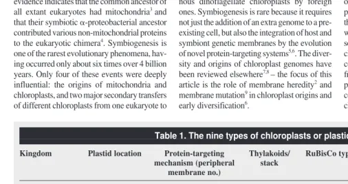
Membrane heredity and cell evolution Two universal constituents of cells never form de novo: chromosomes and membranes2,27. Unlike ribosomes and microtubules, which form by self assembly, cell membranes always form by growth and division or fusion of pre-existing membranes2,27,28The diverse membranes of the millions of extant species are all lineal descen-dants of those of the first bacterial cell2,27,28. A Fig. 1.The symbiogenetic origin of chloroplasts. Chloroplasts arose monophyletically from a cyanobacterium with phycobilins and chlorophyllsaand bthat was phagocytosed by a bicili-ate protozoan host6,11
that converted it to an organelle35
. In the ancestral plant, the food vacuole membrane was lost. Therefore plant chloroplasts have only a double envelope, which evolved a protein-import mechanism5,11,18–21
distinct from that of endoplasmic reticulum, mitochondria and peroxisomes. Green plants lost phycobilisomes and evolved thylakoid stacking instead; whereas red algae and glaucophytes lost chlorophyllb, possibly independently.
Trends in Plant Science
Biciliate protozoan
Glaucophytes
Peptidoglycan Food vacuole
membrane
Cyanobacterium
Loss of peptidoglycan Proteobacterial RuBisCo;
loss of chlorophyll b Loss of phycobilisomes;thylakoid stacking
Green plants Red algae
Triose phosphate translocator transit machinery
eukaryotic cell contains numerous distinct mem-brane types. Just as DNA replication requires information from a pre-existing DNA template, membrane growth requires information from pre-existing membranes – their polarity, type and topological location relative to other mem-branes. During these processes, polarity and type-specificity are continuously maintained. Some membranes, such as the nuclear enve-lope/RER membranes or mitochondrial inner or outer membranes, were called genetic mem-branes2. This is because they always arise by growth and division of membranes of the same type, and therefore have a permanent genetic and evolutionary continuity. Others, such as lysosomal membranes, lack direct genetic con-tinuity, forming instead by differentiation from dissimilar membranes (‘derived membranes’). Genetic membranes are as much a part of an organism’s germ line as DNA genomes; they could not be replaced if accidentally lost, even if all the genes remained. For some membranes this distinction is less clear cut: Golgi and per-oxisomal membranes should perhaps be referred to as semi-genetic. Although normally arising by division (or in the Golgi of higher eukaryotes arising by fragmentation and reassembly) of pre-existing membranes of the same type, Golgi and
peroxisomal membranes might be slowly re-generated from other endomembranes if lost28,29 – this argues against a symbiogenetic origin for peroxisomes5.
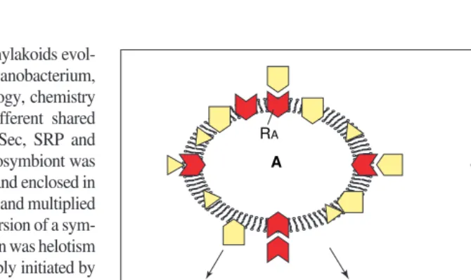
Inheritance of the architecture of membranes (by membrane heredity) and of the manufacture of their component molecular bricks (by gene heredity and protein and lipid biosynthesis) are independent genetic processes that combine constructively through a rapid and spontaneous perpetual Brownian motion of molecules, whose chemical affinities govern their insertion into continuously pre-existing supramolecular structures. Each genetic membrane type has a unique composition of membrane proteins, and usually also lipids; this individuality is typically maintained by the type-specific insertion of new proteins and lipids during growth (Fig. 2). Some genetic membranes (e.g. RER and the bacterial cytoplasmic membrane) are primary genetic membranes, growing by the direct insertion of individual protein and lipid molecules. Others (e.g. the outer membrane of gram-negative bacteria and eukaryote plasma membrane) are secondary genetic membranes, growing instead by intracellular transfer of molecules (individu-ally in bacteria; as membrane vesicles in eukaryotes) from a primary genetic membrane.
Self-targeting of specific membrane receptors for protein insertion (Fig. 2) and the differential targeting of specific lipid-synthesizing or -bind-ing proteins are the essential molecular bases for the specificity of membrane heredity. Mem-brane heredity depends on DNA heredity in that genes for the receptors and the targeted proteins encode their properties, but although necessary this is insufficient. Preformed cell structure30 also is essential – for every distinct kind of genetic membrane a cell must perpetually main-tain a topologically distinct membrane with spe-cific receptors in the correct polarity2,6,27. This is because a cell cannot create them de novoin spite of possessing all the correct genes28.
This central fact of cell biology makes an increase in the number of distinct genetic mem-branes one of the rarest evolutionary steps in the history of life2,17. A few increases have occurred autogenously, such as the origin of the endomembrane system2, but more have come about by symbiogenesis2,5,6. Their growth mech-anism ensures that the relative locations of topo-logically distinct membranes are conserved over hundreds of millions of years, for example:
(1) The cyanobacterial and homologous11,31 plant and chromist plastid outer membrane outside the former cyanobacterial cyto-plasmic membrane and the thylakoids inside it.
(2) The location of plastids in the cytosol of Plantae, but within the RER lumen in Chromista6.
(3) The periplastid membrane between the plastid envelope and the cytosol-facing membrane in chromists, sporozoans and chlorarachneans6,17.
The origin of chloroplasts: plastids and nuclei as chimeras
Most chloroplasts have three genetic membranes, all acquired in a single symbiogenetic event from a cyanobacterium7,8,11. All cyanobacteria except Gloeobacter are more complex than other Gram-negative bacteria in having thylakoids, and therefore also have three genetic membranes. Thylakoids are a third type of genetic membrane topologically separate from the cytoplasmic membrane (Fig. 1), bearing phycobilisomes with blue and red antennal pigments on their cytoso-lic surfaces. Glaucophytes still retain the cyano-bacterial peptidoglycan between their two chloroplast envelope membranes. This was the first conclusive evidence for the symbiogenetic origin of chloroplasts (Fig. 1). Peptidoglycan was lost by green plants and red algae, possibly once only in their common ancestor11, as implied by trees indicating an earlier divergence for glauco-phytes32,33, but independent losses cannot be ruled out because all three groups diverged so closely (probably soon after the origin of plastids) that tree topology remains uncertain.
Chloroplast and cyanobacterial thylakoid lipids are predominantly glycolipids and sulpholipids, Box 1. Chromista, the third botanical kingdom
The kingdom Chromista9,10is fundamentally chimeric like the plant kingdom, but arose by merging two eukaryotic cells. Chromista include all chlorophyll-c-containing ‘chromophyte’ algae except dinoflagellates, which belong in kingdom Protozoa (infrakingdom Alveolata)10. Chromista typically have chlorophyllc2and some have retained both phagotrophy and photosynthesis; most are coloured brown or yellow by carotenoids. Chromistan plastids uniquely lie in the rough endoplasmic reticu-lum reticu-lumen, not in the cytosol, and are also surrounded by a periplastid membrane, a relic of the plasma membrane of the red algal symbiont that provided their chloroplasts. Secondarily aplastidic chromists (e.g. the soil zooflagellates Oikomonas andGoniomonas; the human pathogen Blastocys-tis; oomycetes such as the plant-pathogen Phytophthora) are distinguishable from members of king-doms Fungi and Protozoa by molecular phylogeny and by mostly having rigid bi- or tri-partite tubular ciliary hairs.
Outline classification10 Subkingdom Cryptista
• Usually bipartite tubular hairs on both cilia; extrusive ejectisomes; cristae flat. Phylum Cryptophyta
• Cryptomonads with nucleomorph, starch and 80S ribosomes within periplastid space; usually with an intrathylakoid phycobiliprotein.
• Goniomonas, without nucleomorph or plastids. Subkingdom Chromobiota
• Nucleomorph, periplastid ribosomes, starch, phycobilins all absent; brown carotenoid fucoxanthin widespread; one cilium typically autofluorescent; cristae tubular.
Infrakingdom 1. Heterokonta
Tripartite, rarely bipartite, tubular hairs on anterior cilium only; rarely on cell body or absent). Three phyla:
1. Ochrophyta For example, brown algae, diatoms, chrysophytes, Oikomonas, xantho-phytes, eustigmatoxantho-phytes, raphidoxantho-phytes, silicoflagellates, pedinellids and pelagophytes; chloroplasts or colourless plastids typically present.
2. Bigyra For example, oomycetes, hyphochytrids, opalinates (no ciliary hairs); no plastids. 3. SagenistaFor example, labyrinthulids, thraustochytrids, bicoecid and anoecid flagellates; no plastids.
Infrakingdom 2. Haptophyta
Haptonema; no rigid tubular ciliary hairs. One phylum:
not phospholipids. Chloroplast thylakoids evol-ved directly from those of a cyanobacterium, with little or no change in topology, chemistry or function, including three different shared protein insertion mechanisms (Sec, SRP and TAT)34. The photosynthetic endosymbiont was probably initially phagocytosed and enclosed in a vacuole, from which it escaped and multiplied freely in the cytosol11. The conversion of a sym-biont to a plastid or mitochondrion was helotism (slavery) not mutualism – probably initiated by inserting host protein translocators into its en-velope to tap photosynthesate11. The key differ-ence between an obligate intracellular symbiont and an organelle is the presence of a generalized organelle-specific protein-import mechanism35. This was important in allowing integration directly by inserting host proteins, and as a pre-requisite for effective gene transfer from the symbiont to the host nucleus11. Early on, the cyanobacterial double envelope was radically altered by losing bacterial lipoprotein and lipolysaccharide11, replacing the lipolysaccha-ride with phosphatidylcholine shuttled by lipid transfer proteins from the ER to the outer leaflet of the former cyanobacterial outer membrane22. This replacement probably preceded perfection of the protein-import mechanism, because pre-cursor transfer into the main protein-channel (Toc75) depends on phospholipids in the outer leaflet21 (of putative host origin22), whereas galactolipids of cyanobacterial origin are essen-tial for correctly folding the transit peptide18,20.
The chimeric theory of the origin of the plant plastid (and mitochondrial) outer membrane5 predicted that the proteins are also of dual ori-gin: from host and symbiont. Proteins from the symbiont either retained their original polarity or acquired an inverted polarity through inser-tion directly from the cytosol after their genes moved to the nucleus. Although such inversion is a theoretically simple way of converting a receptor for bacterial protein secretion to one for plastid import5, a receptor originating from a host protein is even simpler as no gene transfer is involved5. Recent studies of protein-targeting substantiate these predictions. The major recep-tor, Toc159 GTPase, is of host origin in green plants. It is partly cognate with and possibly derived from the a-subunit of the host-signal-recognition-particle receptor19–21. The fact that glaucophyte and red algal transit sequences tar-get proteins to green algal plastids implies that both groups have a similar receptor; verifying this would further strengthen arguments for plastid monophyly. The channel protein Toc75 is clearly of cyanobacterial origin36,37, support-ing the homology of the outer plastid membrane with that of cyanobacteria11,31. However, the dual mode of Toc75 import across both membranes via a cleaved transit sequence and back across the inner membrane by a signal sequence21implies that it has probably retained its ancestral polarity and now works in reverse.
Therefore it is not an example of a receptor hav-ing undergone polarity reversal, as suggested36. The third translocon protein (Toc34), which interacts with it and is self inserting via a C-ter-minal sequence21, might even be a chimera of host and symbiont. Toc34 has weak similarity to a cyanobacterial protein21as well as an N-ter-minal GTPase domain related to that of Toc159 (Refs 19,20). The first transit sequences might have evolved from bacterial export signals using the Toc75-related channel36 or from modified host signal sequences or mitochondrial pre-sequences. Three inner membrane translocon
proteins (Tic20; Tic22; Tic55) are of cyanobac-terial ancestry19,21, as are most chaperones that help with import19,21.
However, the ADP–ATP exchange protein that imports ATP to allow for more efficient CO2fixation is not38 of cyanobacterial ancestry. The ADP–ATP exchange protein is unrelated to the mitochondrial ADP–ATP translocator37, the insertion of which probably initiated the mito-chondrial enslavement5. Related translocators are known only from the obligate parasites, Chlamydiaand Rickettsia38. Contrary to the pos-sibly misleading rRNA trees, these parasitic Fig. 2.Maintenance of genetic membrane individuality. Membrane proteins reach their specific locations because their genes encode sequences27 that interact with type-specific insertion, trans-fer or retention machineries on membranes. These types of machinery consist of integral mem-brane proteins with receptors for the topogenic sequences of other memmem-brane proteins, and must themselves be correctly targeted exclusively to their own membrane type to maintain individu-ality. Two different genetic membranes (A and B) within the same cell must have two different receptors RA(red) and RB (blue), symbolized with angular (A) or curved (B) receptor pockets. Each receptor must have a topogenic sequence complementary to its own receptor site to ensure its own targeting, shown as a triangular projection in A and curved in B. This self-targeting and molec-ular self-complementarity of the protein-targeting receptors of genetic membranes is a key basis of membrane heredity. By sharing similar topogenic sequences many diverse proteins (symbol-ized by different shapes: yellow in A, green in B) are correctly targeted to each membrane. Thus molecular complementarity is the central requirement for both nucleic acid and membrane heredity; one transmits inherited molecular structure and the other inherited supramolecular structure. Theoretically, if a genetic membrane has more than one receptor, as thylakoids34do, they could be either self-targeting (i.e. self-complementary) or mutually targeting (i.e. mutually complementary). However, if a genetic membrane has a single receptor complex, as higher plant chloroplast surfaces appear to, it must be self-targeting and self-complementary. Correct target-ing might also involve bindtarget-ing to specific lipids, maktarget-ing co-evolution of protein strucure, lipid structure and composition central to the specificity of membrane heredity. Membrane protein polarity (orientation within the membrane) is strongly conserved. It depends not only on the structure of the inserted protein (and therefore its gene) but also on the insertion machinery and upon whether the protein is inside or outside the membrane before insertion. Polarity usually has to be conserved to maintain function; this is automatically achieved if the site of synthesis is always on the same side of the membrane – another aspect of the importance of pre-existing structure30. The same principle is the fundamental explanation for lipid polarity – the fact that the two leaflets of the bilayer typically have different lipids. This arises partly through polarity of insertion (e.g. in plastid outer membranes phosphatidylcholine from the cytosol and glycol-ipids from the plastid) coupled with the slow rate of flip/flop (spontaneous polarity inversion), and partly from differential binding to polarized membrane proteins. If the location of synthesis changes (e.g. following gene transfer from the chloroplast into the nucleus where proteins for-merly made inside the plastid are now made outside it) a protein might need to be translocated back across the membrane(s) before insertion to retain the correct polarity5.
Trends in Plant Science
RA
RA A
A
B
RB
A
RA RB
B B
bacteria might actually be related, with a com-mon ancestor possessing the gene, possibly acquired from their eukaryotic hosts (probably early algae as it is vastly more similar to the chloroplast than to the mitochondrial protein) and not donated to chloroplasts as others39have suggested. No convincing ancestor of this gene family is known, possibly because it was too drastically modified at its origin; algal translo-cators need sequencing to clarify its history. Some outer membrane components are self-inserting (e.g. an amino acid channel)21; some of these might have played an integrative role before transit machinery evolved.
The import machinery, whose origin con-verted the modified cyanobacterium into the first chloroplast11,35, probably arose to insert host proteins5such as the triose phosphate transloca-tor and the related glucose-6-phosphate translo-cator. These lack prokaryotic relatives and probably evolved from the host phosphate/ phosphoenol pyruvate translocator (T. Cavalier-Smith, unpublished). The ability to tap photo-synthesate in this way would confer a selective advantage to the host; initially there would have been much less advantage in inserting proteins of cyanobacterial origin because each gene would normally remain for a while in the chloroplast after a copy entered the nucleus. The probable priority of host-protein insertion from a mutationist–selectionist view point has been overlooked by some studies19. Although acci-dental transfers of gene copies might have occurred before transit machinery originated, only when they acquired an effective transit sequence could cells with deletions of the plas-tid version survive. Plasplas-tid to nucleus gene transfer required three steps:
• Escape of one copy from a plastid and its insertion into nuclear DNA (several genes could go together in a single piece of DNA). • Mutation(s) to form a transit sequence. • Deletion of the plastid copy.
Mutations adapting the promoters to RNA polymerase II and eukaryotic (rather than prokaryotic) transcription factors, and deletions removing some genes from transferred operons, were probably essential for reasonable levels of gene expression and thus effective transfer. Thousands of cyanobacterial genes that were useless to the host cell were simply deleted; this reduction of the cyanobacterial genome some-times involved shifts of function. For example, a cyanobacterial peptidoglycan synthesis en-zyme (MurG), when no longer needed for pep-tidoglycan, became a monogalactolipid synthase replacing the original galactolipid synthase18. Once import became efficient, transit sequences could be acquired (probably by transposition5, e.g. exon-shuffling4rather than point mutations) by other host proteins or former cyanobacterial proteins encoded by gene copies accidentally incorporated into the nucleus. Even if only 1500 genes were transferred (not 2000–5000 as some
suggest4) the total number of mutations in-volved in the origin of plastids cannot be less than 10 000.
Thus, the origin of the transit machinery allowed two cells to become one35, enabling thousands of symbiont genes to be transferred to the nucleus. When these genes duplicated host functions, host genes were sometimes lost and symbiont genes retained; their proteins were either retargeted to the plastid (e.g. those for fatty acid synthetase) or remained in the cytosol as well or instead (e.g. phospho-glycerate kinase4). Sometimes, cyanobacterial proteins were retargeted to the plastid in one plant lineage (e.g. green plant starch-making enzymes) but became cytosolic in the others. The vast majority of transfers probably pre-ceded divergence of the three plant lineages, but many happened later in parallel32.
This raises the question of why genomes were retained by most chloroplasts and mito-chondria (although most mitomito-chondria that con-verted to hydrogenosomes5lost them40). The idiosyncratic distribution of the organelle genes across taxa8,32throws doubt on any simple func-tional explanation41. Much of the variation prob-ably arises by historical chance, because effective gene transfer requires several rare mutations in each gene – some lineages might not have had the right combination. Because most widely-conserved organelle genes in the mitochondria and in the plastids are relatively large membrane proteins, a difficulty in effec-tively retargeting large hydrophobic proteins across multiple membranes5is probably a major selective factor. The fact that the large rubisco subunit and the largest RNA polymerase sub-units have never been naturally retargeted sug-gests similar difficulties for large multi-subunit soluble proteins that must be synthesized and assembled by chaperones in the same compart-ment. This interpretation is strongly favoured by the fact that both genes can be lost if their func-tions are replaced by smaller, more easily tar-geted, single-subunit analogues: for example, eubacterial RNA polymerase genes replaced by a phage-type polymerase in both plastids and mitochondria42, and multi-subunit type-I rubisco replaced by a small single subunit type-II molecule in dinoflagellate chloroplasts15. This generalized ‘retargeting difficulty’ argument is not invalidated as others argue41by the ex-perimental retargeting of rbcL, because such mutants, although viable in the laboratory, have so little useful rubisco that natural selection would eliminate them.
Important genes transferred to the nucleus include ftsZ(encoding a GTPase that proba-bly mediates division of all plastids, as it did their cyanobacterial ancestors43), ftsH and minicell (involved in plastid growth or division)18. In addition, plastid and mitochon-drial divisions require host proteins such as dynamin18. Thus the division and the import
machinery underlying plastid growth are both evolutionary chimeras of host and symbiont molecules. Even though many host lipids and proteins have been individually inserted into the former outer membrane of the cyanobac-terium, thereby greatly modifying its proper-ties, we may still regard it as being homologous with the cyanobacterial outer membrane5,11,31. Without massive recruitment of pre-existing symbiont and host proteins, such a complex evo-lutionary innovation as the origin of chloroplasts could never have happened. But the scale of the transformation was so vast, with shared-derived features of plastid genomes7,8,44, chlorophyll-a/b or c-binding proteins14, transit protein-targeting machinery, and the integration of thousands of prokaryotic genes into the different transcrip-tional environment of the nucleus, that it appears unlikely that it occurred convergently in green plants, red algae and glaucophytes. Phylogenies of many concatenated nuclear protein sequences suggest that red algae and green plants are sis-ters33(as do mitochondrial sequences8) and there is weaker evidence that glaucophytes are their closest relatives33. This supports monophyly of an ancestrally photosynthetic kingdom Plantae embracing these three groups10, which the equiv-ocal nuclear 18S rRNA trees only sometimes weakly show.
Intensive study of biliphyte plastid envelopes and their biogenesis and division should also help to resolve plastid and plant monophyly. The primary synthesis of fatty acids in green plants is by enzymes in the plastid stroma, of cyanobacterial origin, but which are nuclear-encoded22. If biliphytes also have the cyanobac-terial instead of the original host cytosolic enzymes, their loss and the evolution of machin-ery for exporting C-16 fatty acids, to allow mem-brane growth in the rest of the cell, probably occurred in the common ancestor of all Plantae – another potentially homologous novelty in the origin of plastid envelopes. The site of fatty acid synthesis in meta-algae that arose by secondary symbiogenesis is also unknown, but their origin must have involved major innovations in trans-envelope fatty acid transport that, like those in protein import, caution against postulating numerous independent symbiogeneses.
Secondary symbiogeneses
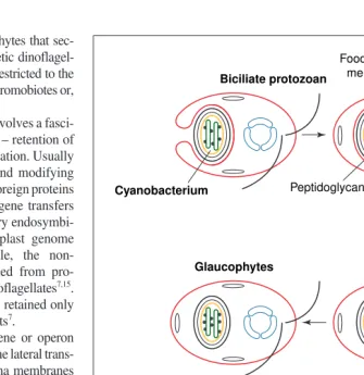
membrane was retained as a fourth membrane – the periplastid membrane – which is sandwiched between the double plastid en-velope and the former phagosomal membrane. Nuclear-coded plastid proteins are imported across four peripheral membranes, not two as in plants. The import mechanism across the periplastid membrane is a major unknown for chloroplast biology (for possibilities see Ref. 17). Euglenoids and dinoflagellates independently lost the periplastid membrane and attached the peri-algal vacuole locally to the plastid envelope, yielding a novel triple-membraned envelope; Golgi-based protein-import and phylogenetic evidence make alternative scenarios relatively implausible17.
Lateral transfer of green algal chloroplasts by secondary symbiogeneses
Photosynthetic euglenoids and chlo-rarachnean algae have plastids with chlorophyllsaand b, cab proteins, stacked thylakoids and cyanobac-terial rubisco, obtained from green algae by lateral organelle trans-fer16,17. However, unlike all green plant plastids these two phyla lack starch. Because the protozoan phyla to which they belong (Euglenozoa and Cercozoa10,45) are ultrastruc-turally rather different, two separate endosymbiotic events are usually assumed7. However, in both phyla, nuclear-coded plastid proteins are targeted via RER and Golgi17,46, and polysaccharide stores are cytosolic b-glucans (paramylum), not starch as in all Plantae17. The long branches of Euglenozoa on 18S rRNA trees that place them well below, rather than near Cercozoa, are suspected to be misleading6,17,47. Therefore,
to minimize the number of independent symbio-geneses that need to be postulated, these phyla, plus a third non-photosynthetic phylum (Perco-lozoa), were suggested to be related17. The com-mon ancestor of this putative cabozoan clade possibly acquired green algal plastids once only, in which case all non-photosynthetic cabozoans evolved by secondary plastid losses17.
Unlike those of green plants, chlorarachnean plastids are surrounded by two additional smooth membranes (Fig. 3). The innermost is the erstwhile plasma membrane of a eukaryotic endosymbiont; between it and the chloroplast lies a minute relic of the symbiont’s nucleus – the nucleomorph. The nucleomorph, which has three tiny chromosomes, 90–135 kb in size, is
the smallest eukaryotic genome48. The fourth, outermost membrane is the former phagosomal membrane of the host, now a permanent genetic membrane. In this symbiogenetic event the ancestral chlorarachnean acquired five novel genetic membranes directly from an endosym-biotic green alga, and a sixth novel type by modifying the host phagosomal membrane, making chlorarachnean membrane topogenesis far more complex than in any animal or plant. Photosynthetic euglenoids also obtained plas-tids from a green alga but entirely lost the green algal nucleus and its plasma membrane (Fig. 3). Genes for proteins formerly encoded by the green algal nucleus were transferred to the host nucleus. Curiously, euglenoid nuclear genes for
plastid proteins have numerous introns without canonical splice junctions, whether derived from the green algal nucleus (e.g. cab pro-teins49) or from the plastid [e.g. RbcS (Ref. 50)]. Perhaps these unique in-trons originated from typical spliceo-somal introns in a miniaturized version of the green algal nucleus (such as the chlorarachnean nucleo-morph)48before their transfer to the host nucleus. One euglenoid gene of host origin (fibrillarin) has typical spliceosomal introns51, but as some such introns are mobile52, both types might now be present in genes of symbiont or host origin. The aberrant type is found in a Euglenacytosolic GAPDH, which is related to the gly-cosomal GAPDH of the non-photo-synthetic kinetoplastid Euglenozoa rather than to those of green plant plastids53. This GAPDH subfamily is weakly related to another of the three cyanobacterial isozymes – if the ancestral euglenozoan was indeed photosynthetic17it could have come from the green algal part of the chimera if (unlike angiosperms) it still had that isozyme. In either case, the ancestral euglenozoan must have evolved differential retargeting between euglenoids and kinetoplas-tids. Chlorarachniophyte nucleo-morph introns, which are recog-nisably spliceosomal, are the tiniest known48. On the cabozoan theory, which postulates a common photo-synthetic ancestor for euglenoids and chlorarachneans17, both aberrant in-tron types might have diverged from a common ancestor. Possibly the aberrant euglenozoan introns were also minute initially, but expanded secondarily after transfer to the host nucleus, where there is far less selection for small genome size54.
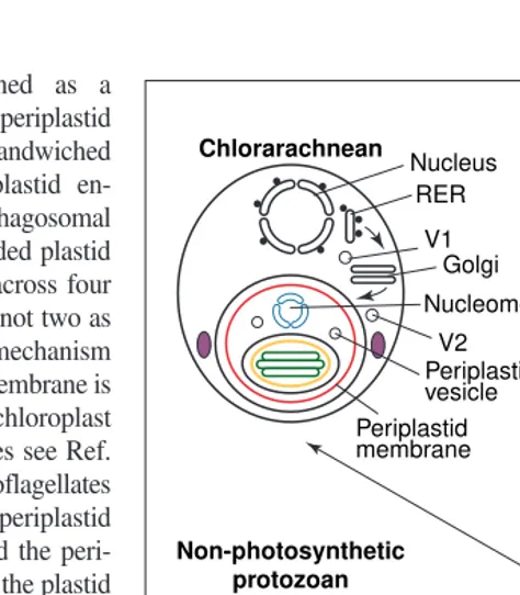
Secondary origin of the Chromista Another secondary symbiogenesis created chromistan algae (e.g. brown algae, diatoms, haptophytes, cryptomonads; Box 1). Un-like all other eukaryotes their chloroplasts lie within the RER lumen5,6,9,18, usually the perinuclear cisterna (Fig. 4). Chromists and plastid-bearing alveolates (dinoflagellates and sporozoans) obtained their plastids from red algae6,7,55, but lost the red algal phyco-bilisomes, evolving chlorophyll c2 as a replacement secondary pigment, and stacked thylakoids in pairs (cryptomonads) or triplets (the others). In both groups, there were multi-ple subsequent losses of the photosynthetic machinery or of the entire plastid. Because of Fig. 3.The cabozoan theory of the common origin of euglenoid
and chlorarachnean chloroplasts suggests that both were acquired by secondary symbiogenesis from the same green alga in a common biciliate host. Following engulfment of the green alga by phago-cytosis, the hypothetical cabozoan common ancestor evolved (1) Golgi vesicle targeting to the perialgal vacuole by fusion of Golgi vesicles (V2 ) with the former perialgal vacuole and (2) periplastid vesicle shuttling between the periplastid membrane and the outer membrane of the plastid envelope; it lost the green algal mitochon-dria, starch and all other organelles. Paramylum evolved as a cytoso-lic carbohydrate storage compound. Following divergence of the two groups, the green algal plasma membrane was retained in chlo-rarachneans, but was lost in an early euglenoid that integrated the food vacuole membrane with its plastid envelope to make a triple envelope. Most green algal nuclear-encoded genes for plastid pro-teins were transferred to the host nucleus and signal sequences added to their pre-existing transit sequences before the divergence of the two groups. The signal sequences inserted the nascent pro-teins into the rough endoplasmic reticulum (RER) lumen, from where vesicles (V1) carried them to the Golgi and then the perialgal vacuole. This gene transfer was completed in euglenoids only, enabling the nucleomorph to disappear. Adapted from Ref. 17.
Trends in Plant Science
these and other characters, chromists and alve-olates, are informally grouped as ‘chromalve-olates’17, and might be a clade whose common ancestor acquired a red algal plastid and evolved chlorophyll c2 once only; other researchers assume that there were up to five independent symbiogeneses for chromalveolates7. It is con-troversial whether chlorophyllc2evolved after the uptake of the red alga17, or was already present [as is now accepted for chlorophyllb
(Ref. 12)] in the bacterium ancestral to all plas-tids56. The closer relationship of chlorophylla/c -binding proteins to cabs than to any bacterial proteins, supports a purely eukaryotic origin14. The ‘chlorophyllc-like’ pigments of prochloro-phyte cyanobacteria56and of a few green algae are not chlorophyllc2(or c1and c3widespread in chromobiote chromists), but Mg-protopor-phyrins, which could easily have evolved in-dependently from chlorophyll a precursors.
Sequence data for enzymes synthesizing these pigments are needed to resolve this. Unlike dinoflagellates, chromists retained the red algal plastid-coded proteobacterial form I rubisco7.
After the red alga55was phagocytosed by a biciliate host11the phagosome membrane fused with the nuclear envelope9,55, thereby placing the chloroplast in the RER lumen. The first step in targeting nuclear-encoded proteins into the chromist chloroplast across four membranes is by ER-type signal sequences17,18. The ancestral chromist lost phycobilisomes, but one chromist group, cryptomonads, retained a single phyco-erythrin pigment (a subset of cryptomonads later converted this to a blue protein57), but retargeted it into the thylakoid lumen. The plas-tid-coded bsubunit of this phycoerythrin pig-ment is clearly of red-algal origin, but the affinities of its nuclear-coded a-polypeptide, which is imported successively across five membranes, are unclear. Cryptomonads retain the former red algal nucleus (but drastically reduced in genome size54) as a nucleomorph6,11 with three linear chromosomes densely packed with largely intron-free genes encoding many proteins for nucleomorph replication, division and gene expression, and a few imported into the plastid, notably FtsZ and rubredoxin58. Most genes for chloroplast proteins were transferred from the nucleomorph to the host nucleus and their proteins retargeted to the periplastid space. Other chromists (chromobiotes) lost the nucleo-morph entirely after transferring the remaining essential genes for plastid proteins into the host nucleus. The retention of the chromobiote periplastid membrane hundreds of millions of years after the loss of the genome of the cell that it once enclosed, emphasizes the immense stability of membrane heredity.
Alveolate plastids
Most sporozoan parasites (e.g. malaria) have plastids with relict genomes, probably retained from photosynthetic ancestors because they con-tain the cells’ only fatty acid synthesis enzymes (of cyanobacterial origin)23; Cryptosporidium appears to have lost the plastid59 because it retained the host mechanism of fatty acid syn-thesis instead60. Four membranes bound these plastids, at least in Toxoplasma. Their gene com-plement and order implies an origin from a red44,61not a green alga, as TufA trees62 uncon-vincingly suggested. In the chromalveolate theory, the merger with a red alga was initially the same symbiogenesis as for chromists but the two groups diverged before the chromistan fusion of perialgal vacuole and RER (Ref. 17).
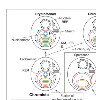
The sisters of Sporozoa are dinoflagellates; their plastid proteins are related to those of chromists and red algae63. This is consistent with the common ancestor of all alveolates (sporo-zoa, dinoflagellates, protalveolates, ciliates) being a photosynthetic sister to chromists17. According to this theory, the dinoflagellate Fig. 4.The chromalveolate theory of the common secondary symbiogenetic origin of chromist
and alveolate plastids. Following phagocytosis of a red alga into a host food vacuole, chloro-phyllc2evolved in the ancestral chromalveolate plastid, and the phagosomal membrane became a stable perialgal vacuole by the specifically targeted fusion of Golgi-derived vesicles (V1). After evolving an unknown mechanism17
for protein import across the periplastid membrane (PPM), plastid proteins coded by any genes accidentally transferred from the red algal to the host nucleus could be correctly retargeted to the plastid as soon as they acquired signal sequences distal to their pre-existing transit sequences. This enabled the loss of these genes from the symbiont nucleus. Eventually the red algal nucleus disappeared entirely in alveolates and independently in chromo-biotes [which independently lost phycobilin pigments (PB)] but remained in miniaturized form as the cryptomonad nucleomorph (N). Mitochondria and all other organelles were lost. The sym-biont starch (S)-making machinery was retained by cryptomonads in the periplastid space (its location in the progenitor red alga), relocated to the host cytosol in dinoflagellates (simply by the transfer of their genes to the host nucleus), and lost by the chromobiotes. The periplastid mem-brane was lost and a triple envelope evolved in the ancestral dinoflagellate, which also evolved the carotenoid peridinin. Thylakoids (T) and photosynthesis were lost in the progenitor of Sporozoa. Chlorophyllsc1and c3and fucoxanthin (f) evolved in the chromobiote ancestor alone, which evolved thylakoid stacking in threes independently of dinoflagellates. The divergence of chromobiotes into haptophytes and heterokonts (not shown) probably occurred soon after their origin, but the divergence of sporozoans from dinoflagellates occurred well after the origin of alveolates. Adapted from Ref. 17.
Trends in Plant Science
Cryptomonad Chromobiote
Ancestral chromalveolate alga
Nucleus RER
RER Eochromist
Chromista
Protozoa Nucleomorph
–NM, –PB –S
+ f, chl c1, c3
Golgi Sporozoan
Golgi Dinoflagellate
Non-photosynthetic protozoan
–T
–PPM –N, –PB Fusion of
nuclear envelope and perialgal membrane
Starch
Red alga
Host nucleus Endosymbiont
nucleus
Chloroplast + chl c2 Perialgal vacuole
ancestor lost the red algal plasma membrane after its divergence from sporozoa, and a triple plastid envelope was generated analogously to but independently from euglenoids17. The number of sporozoan plastid membranes is con-troversial62 – if, as is sometimes suggested, some actually have only three, such reduction would be independent of that in dinoflagellates. Protein-targeting to alveolate plastids is prob-ably via the ER and Golgi17,18,23. As in eugle-noids46, imported dinoflagellate light-harvesting proteins are made as polyproteins that are later proteolytically cleaved. Because proteolytic cleavage is common in Golgi-secreted proteins, the convergent import via the Golgi in both groups might have predisposed them to evolve similar proteolytic processing, differently from plant and chromist chloroplast proteins.
Dinoflagellate plastid genomes have frag-mented into single gene circles63, reversing the concatenation of genes that created the first chromosomes2. Dinoflagellates appear to have transferred to the nucleus or lost far more plas-tid genes than other eukaryotes63, primarily retaining a handful of hydrophobic thylakoid protein genes. This possibly facilitated acci-dental transposition of replicon origins beside every gene to fragment their genome. As cyanobacterial RNA polymerase genes have not been found in dinoflagellate plastid DNA (Ref. 63), they might (like most mitochondria8) have lost them and instead use the unigenic phage-type polymerase that transcribes gene expression genes in higher plant plastids42.
Tertiary chloroplast replacements Typical dinoflagellate peridinin-containing chloroplasts have probably been replaced sym-biogenetically at least twice by differently pig-mented chloroplasts from other eukaryotic algae. As their plastids must lack enough genes for self-reproduction and neither endosymbiont nuclei nor mitochondria remain, the host must have evolved a protein-insertion mechanism, making them true tertiary organelles, but direct evidence is lacking. One aberrant dinoflagel-late, Lepidodinium, has a green plastid with chlorophyllaand bplus a membrane topology suggesting a tertiary symbiogenetic incorpora-tion of a green alga. By contrast, a few species, such as Gymnodinium breve, have hexanoyl-fucoxanthin and plastid DNA indicative of a haptophyte endosymbiont7. A third possible case is Dinophysis: several species have cryp-tomonad chloroplasts – because Dinophysis cannot be cultured we cannot rule out the pos-sibility that they do not multiply and are sim-ply kleptochloroplasts (‘stolen’ and maintained temporarily for photosynthesis as many het-erotrophic protozoa do). The nesting of all three cases within the peridinin-containing dino-flagellates on nuclear rRNA trees26shows that they are tertiary chloroplast replacements and not secondary uptakes by a primitively
non-photosynthetic host. By contrast with primary and secondary symbiogenesis, which created whole kingdoms, phyla and classes, ter-tiary symbiogenesis merely replaced existing chloroplasts and made a few obscure aberrant species within a single class – the peridinean dinoflagellates – which is unusually prone to plastid loss and replacement.
We must firmly distinguish symbiogenesis, which is exceedingly rare, from symbiosis, which is frequent and evolutionarily easy. In addition to a non-photosynthetic relic (retained as an eyespot) of the typical triple-enveloped plastid, the fucoxanthin-containing dinofla-gellates Peridinium balticumand P. foliaceum contain a photosynthetic diatom cell64 that is greatly reduced by loss of mitochondria and frustule. However, the diatom retains a nucleus and therefore does not need to import any host proteins – unless it does it would be an obligate symbiont not a symbiogenetically acquired replacement organelle.
The plastid big bang and snowball earth From the branching patterns of the major groups of eukaryotic algae on molecular trees6,45,47it appears that there was a rapid, almost simulta-neous, diversification of the three major plant and three chromist groups, alveolates and cabo-zoan algae. This explosive radiation was part of the eukaryotic ‘big bang’, in which single gene trees cannot confidently resolve the relative branching order of the major lineages6,45,47, sug-gesting that the cabozoan and chromalveolate symbioses occurred shortly after the origin of plastids when host and symbiont were still closely related. If this is the case, the merger of two different genomes might have been easier than if it occurred long after their divergence. Comparisons with the fossil record suggest that the origin of plastids might have been in the late Proterozoic, around 600 My ago shortly after (perhaps even stimulated by) the melting of the icecap that enclosed earth for millions of years65. Protein trees45,47and the fossil record are consis-tent with an explosive early radiation of all eukaryotic algal phyla, implying a relatively short timescale for the major transitions dis-cussed here. This interpretation might surprise some people because it assumes that the scale of early eukaryotic divergences on 18S rRNA trees is inflated6,47, and brings into question the identification of any fossils as eukaryotic before ~700 My ago. Although the new body plans that distinguish kingdoms and phyla probably all originated by explosive quantum evolution, its speed appears less than was theoretically con-ceivable11. Therefore the view that the big bang was so rapid that we cannot establish the relative branching order of kingdoms and phyla is prob-ably over-pessimistic. Concatenated protein trees already support monophyly of kingdom Plantae and their primary chloroplasts33; in con-junction with other molecular evidence, such as
shared introns and gene order44, such studies might eventually confirm or refute the monophyly of cabozoa, Chromista and chromalveolates. Elucidating the machinery for protein-targeting into plastids and the division mechanisms of their extra membranes in each group will provide even more direct evidence.
Acknowledgements
I thank NSERC (Canada) for a research grant and the Canadian Institute for Advanced Re-search for Fellowship support. Space limitations and this article’s wide scope prevented the cita-tion of hundreds of important papers. I apolo-gise to those whose work had to be referred to obliquely via other reviews rather than directly.
References
1Mereschkowsky, C. (1910) Theorie der zwei Plasmaarten als Grundlage der Symbiogenesis, einer neuen Lehre von der Entstehung der Organismen. Biologisches Centralblatt30, 278–303, 321–347, 353–367
2Cavalier-Smith, T. (1991) The evolution of prokaryotic and eukaryotic cells. In Fundamentals of Medical Cell Biology(Vol. I) (Bittar, G.E., ed.), pp. 217–272, JAI Press, Greenwich, CT, USA.
3Roger, A.J. (1999) Reconstructing early events in eukaryotic evolution. Am. Nat. 154, S146–S163
4Martin, W. and Herrmann, R.G. (1999) Gene transfer from organelles to the nucleus: how much, what happens and why? Plant Physiol. 118, 9–17
5Cavalier-Smith, T. (1987) The simultaneous symbiotic origin of mitochondria, chloroplasts, and microbodies. Ann. New York Acad. Sci.503, 55–71
6Cavalier-Smith, T. (1995) Membrane heredity, symbiogenesis, and the multiple origins of algae. In Biodiversity and Evolution(Arai, R. et al., eds), pp. 75–114, The National Science Museum Foundation, Tokyo, Japan
7Delwiche, C.F. and Palmer, J.D. (1997) The origin of plastids and their spread via secondary symbiosis. Plant Syst. Evol. (Suppl.) 11, 53–86
8Gray, M.W. (1999) Evolution of organellar genomes. Curr. Opin. Genet. Dev.9, 678–687
9Cavalier-Smith, T. (1986) The kingdom Chromista: origin and systematics. In Progress in Phycological Research(Vol. 4) (Round, F.E. and Chapman, D.J., eds), pp. 309–347, Biopress, Bristol, UK
10Cavalier-Smith, T. (1998) A revised six-kingdom system of life. Biol. Rev. 73, 203–266
11Cavalier-Smith, T. (1982) The origins of plastids.
Biol. J. Linn. Soc.17, 289–306
12Tomitami, A. et al. (1999) Chlorophyll b and phycobilins in the common ancestor of cyanobacteria and chloroplasts. Nature400, 159–162
13La Roche, J. et al. (1996) Independent evolution of the prochlorophyte and green plant chlorophyll a/b light-harvesting proteins. Proc. Natl. Acad. Sci. U. S. A.93, 15244–15248
15 Morse, D. et al. (1995) A nuclear encoded form II RuBisCo in dinoflagellates.Science268, 1622–1624
16 Gibbs, S.P. (1978) The chloroplasts of Euglena
may have evolved from symbiotic green algae.
Can. J. Bot.56, 2883–2889
17 Cavalier-Smith, T. (1999) Principles of protein and lipid targeting in secondary symbiogenesis: euglenoid, dinoflagellate, and sporozoan plastid origins and the eukaryote family tree. J. Euk. Microbiol. 46, 347–366
18 McFadden, G.I. (1999) Endosymbiosis and the evolution of the plant cell. Curr. Opin. Plant Biol. 2, 513–519
19 Keegstra, K. and Froehlich, J.E. (1999) Protein import into chloroplasts. Curr. Opin. Plant Biol.
2, 471–476
20 Chen, X. and Schnell, D.J. (1999) Protein import into chloroplasts. Trends Cell Biol.9, 222–227
21 Heins, L. et al. (1998) The protein translocation apparatus of chloroplast envelopes. Trends Plant Sci.3, 56–61
22 Cavalier-Smith, T. (1993) The origin, losses and gains of chloroplasts. In Origin of Plastids: Symbiogenesis, Prochlorophytes and the Origins of Chloroplasts(Lewin, R.A., ed.), pp. 291–348, Chapman & Hall
23 Waller, R. et al. (1998) Nuclear-encoded proteins target to the plastid in Toxoplasma gondii and
Plasmodium falciparum. Proc. Natl. Acad. Sci. U. S. A.98, 12352–12357
24 Linton, E. et al. (1999) A molecular study of euglenoid phylogeny using small subunit rDNA.
J. Euk. Microbiol. 46, 217–223
25 Cavalier-Smith, T. et al. (1996) Oikomonas, a distinctive zooflagellate related to chrysomonads.
Archiv f. Protistenkunde146, 273–279
26 Saunders, G.W. et al. (1997) Small-subunit ribosomal RNA sequences from selected dinoflagellates: testing classical evolutionary hypotheses with molecular systematic methods.
Plant Syst. Evol.(Suppl.) 11, 237–259
27 Blobel, G. (1980) Intracellular membrane topogenesis. Proc. Natl Acad. Sci. U. S. A.77, 1496–1500
28 Warren, G. and Wickner, W. (1996) Organelle inheritance. Cell 84, 395–400
29 South, S.T. and Gould, S.J. (1999) Peroxisome synthesis in the absence of preexisting peroxisomes. J. Cell Biol. 144, 255–266
30 Sonneborn, T.M. (1963) Does preformed cell structure play an essential role in cell heredity? In
The Nature of Biological Diversity(Allen, J.M., ed.), pp. 165–221, McGraw-Hill
31 Douce, R. and Joyard, J. (1981) Does the plastid envelope derive from the endoplasmic reticulum?
Trends Biochem. Sci.6, 237–239
32 Martin, W. et al. (1998) Gene transfer to the nucleus and the evolution of chloroplasts. Nature393, 162–165
33 Moreira, D. et al. The origin of red algae: implications for the evolution of chloroplasts.
Nature (in press)
34Dalbey, R.E. and Robinson, C. (1999) Protein translocation into and across the bacterial plasma membrane and the plant thylakoid membrane.
Trends Biochem. Sci. 24, 17–22
35Cavalier-Smith, T. and Lee, J.J. (1985) Protozoa as hosts for endosymbioses and the conversion of symbionts into organelles.J. Protozool. 32, 376–379
36Reumann, S. et al.(1999) The evolutionary origin of the protein-translocating channel of chloroplastic envelope membranes: identification of a cyanobacterial homolog. Proc. Natl. Acad. Sci. U. S. A.96, 784–789
37Bölter, B. et al. (1998) Origin of a chloroplast protein importer. Proc. Natl. Acad. Sci. U. S. A.
95, 15831–15836
38Winkler, H.H. and Neuhaus, H.E. (1999) Non-mitochondrial ATP transport. Trends Biochem. Sci. 24, 64–68
39Wolf, Y.I. et al. (1999) Rickettsiae and Chlamydiae: evidence of horizontal gene transfer and gene exchange. Trends Biochem. Sci. 15, 173–175
40Palmer, J.D. (1997) Organelle genomes: going, going, gone! Science275, 790–791
41Race, H.L. et al. (1999) Why have organelles retained genomes? Trends Genet. 15, 364–370
42Hedtke, B. et al. (1997) Mitochondrial and phage type RNA polmerase in Arabidopsis. Science277, 809–811
43Erickson, H.P. (1997) FtsZ, a tubulin homologue in prokaryote cell division. Trends Cell Biol. 7, 362–367
44Stoebe, B. and Kowallik, K.V. (1999) Gene cluster analysis in chloroplast genomics. Trends Genet.15, 344–347
45Keeling, P. et al.(1998) The phylogenetic position of a- and b-tubulins from the
Chlorarachnionhost and Cercomonas
(Cercozoa). J. Euk. Microbiol.45, 561–570
46Sulli, C. and Schwartzbach, S.D. (1995) The polyprotein precursor to the Euglena light-harvesting chlorophyll a/b-binding protein is transported to the Golgi apparatus prior to chloroplast import and polyprotein processing.
J. Biol. Chem. 270, 13084–13090
47Philippe, H. and Adoutte, A. (1998) The molecular phylogeny of Eukaryota: solid facts and uncertainties. In Evolutionary Relationships Among Protozoa
(Coombs, G.H. et al., eds), pp. 25–56, Kluwer
48Gilson, P.R. and McFadden, G.I. (1996) The miniaturized nuclear genome of a eukaryotic endosymbiont contains genes that overlap, genes that are cotranscribed, and the smallest known spliceosomal introns. Proc. Natl. Acad. Sci. U. S. A. 93, 7737–7742
49Muchal, U.S. and Schwartzbach, S.D. (1994) Characterization of the unique intron–exon junctions of Euglena gene(s) encoding the polyprotein precursor to the light-harvesting chlorophyll a/b-binding protein of photosystem II.Nucleic Acids Res.22, 5737–5744
50Tessier, L.H. et al. (1995) Structure and expression of Euglena gracilisnuclear rbcS
genes encoding the small subunits of the ribulose 1,5-bisphosphate carboxylase/oxygenase: a novel splicing process for unusual intervening sequences? J. Mol. Biol.245, 22–33
51 Breckenridge, D.G. et al. (1998) U1 small nuclear RNA and spliceosomal introns in Euglena gracilis.
Proc. Natl. Acad. Sci. U. S. A. 96, 852–856
52 Tarrío, R. et al. (1998) New Drosophila introns originate by duplication. Proc. Natl. Acad. Sci. U. S. A.95, 1658–1662
53 Henze, K. et al. (1995) A nuclear gene of eubacterial origin in Euglena gracilisreflects cryptic endosymbioses during protist evolution.
Proc. Natl. Acad. Sci. U. S. A.92, 9122–9126
54 Beaton, M.J. and Cavalier-Smith, T. (1999) Eukaryotic non-coding DNA is functional: evidence from the differential scaling of cryptomonad genomes.
Proc. R. Soc. London Ser. B266, 2053–2059
55 Whatley, J.M. et al. (1979) From extracellular to intracellular: the establishment of mitochondria and chloroplasts. Proc. R. Soc. London Ser. B
204, 165–187
56 Larkum, A.W.D. et al. (1994) Light-harvesting chlorophyll-c like proteins in Prochloron. Proc. Natl. Acad. Sci. U. S. A.91, 679–683
57 Marin, B. et al. (1998) Phylogenetic relationships among the Cryptophyta: analyses of nuclear encoded SSU rRNA sequences support the monophyly of extant plastid containing lineages.
Protist149, 265–276
58 Zauner, S. et al. (2000) Chloroplast protein and centrosomal genes, a tRNA intron, and odd telomeres in an unusually compact eukaryotic genome, the cryptomonad nucleomorph.Proc. Natl. Acad. Sci. U. S. A.97, 200–205
59 Zhu, G. et al.(2000) Cryptosporidium parvum
appears to lack a plastid genome. Microbiology
146, 315–321
60 Zhu, G. et al.(2000) Molecular analysis of a type I fatty acid synthase in Cryptosporidium parvum.
Mol. Biochem. Parasitol.105, 253–260
61 Blanchard, J.L. and Hicks, J. (1999) The non-photosynthetic plastid in malarial parasites and other apicomplexans is derived from outside the green plastid lineage. J. Euk. Microbiol. 46, 367–375
62 Köhler, S. et al. (1997) A plastid of probable green algal origin in apicomplexan parasites.
Science275, 336–342
63 Zhang, Z. et al. (1999) Single gene circles in dinoflagellate chloroplast genomes. Nature400, 155–159
64 Chesnick, J. et al. (1997) Ribosomal RNA analysis indicates a benthic diatom ancestry for the endosymbionts of the dinoflagellates
Peridinium foliaceumand Peridinium balticum
(Pyrrhophyta). J. Euk. Microbiol. 44, 314–320
65 Hoffmann, P.F. et al. (1998) A neoproterozoic snowball earth. Science281, 1342–1346
Tom Cavalier-Smith is at the Dept of Zoology, University of Oxford, South Parks Road, Oxford, UK OX1 3PS
(tel 144 1865 281065; fax 144 1865 281310;