With Advanced Cirrhosis and Ascites
Rub´en Franc´es,
1Susana Benlloch,
2Pedro Zapater,
3Jos´e M. Gonz´alez,
1Beatriz Lozano,
1Carlos Mu ˜noz,
1Sonia Pascual,
2Juan A. Casellas,
2Francisco Uceda,
2Jos´e M Palaz ´on,
2Fernando Carnicer,
2Miguel P´erez-Mateo,
2and Jos´e Such
2Bacterial translocation is currently considered the main pathogenic mechanism leading to spontaneous bacterial peritonitis in patients with advanced cirrhosis and ascites. However, to the authors’ knowledge there is no information regarding the characteristics of this process in humans. The goals of the current study were to pursue partially identified bacte-rial DNA in blood (what the authors consider molecular evidence of bactebacte-rial translocation) through its relative quantification in a 72-hour study period by using real-time polymerase chain reaction (PCR). A consecutive series of 17 patients with advanced cirrhosis and cul-ture-negative, nonneutrocytic ascites were studied. Therapeutic paracentesis was performed at the time of admission, and blood samples were obtained at baseline and every 8 hours in a 3-day period. Bacterial DNA was detected by a PCR-based method, relatively quantified by real-time PCR, and identified by automated nucleotide sequencing. Seven of 17 patients demonstrated the simultaneous presence of bacterial DNA in blood and ascitic fluid at the time of admission. After therapeutic paracentesis was performed, bacterial DNA persisted in the blood for a minimum of 24 hours, and was reported to last as long as 72 hours in some patients. In addition, different patterns of bacterial DNA appearance and clearance from the blood were identified. The nucleotide sequencing process demonstrated that bacteria de-tected in the first sample were identical to those noted in subsequent detections over time. In conclusion, bacterial translocation is a single-species, dynamic process that appears to de-velop in a subgroup of patients with advanced cirrhosis.(HEPATOLOGY2004;39:484 – 491.)
S
pontaneous bacterial peritonitis (SBP) is a severe infection developing in patients with advanced cir-rhosis, in the absence of any intraabdominal, surgi-cally treatable source of infection.1It is considered to bethe final consequence of repeated episodes of bacterial translocation (BT) from the intestinal lumen and even-tual arrival of bacteria in the ascitic fluid (AF). However, the predisposition to develop a SBP episode is related to its intrinsic bactericidal properties.2– 4
BT is an incompletely understood process by which in-testinal bacteria can cross the epithelial wall, thereby reaching
mesenteric lymph nodes and other organs.5BT has been
studied extensively in cirrhotic rats,6,7but for obvious
rea-sons it is difficult to study its incidence in patients with cir-rhosis.8We recently reported the presence of bacterial DNA
(BactDNA) in blood and AF in roughly 40% of patients with cirrhosis and culture-negative, nonneutrocytic ascites9
and, although more experimental work is needed to confirm our hypothesis, the data available to date may represent mo-lecular evidence of BT. This method allows the study of BT in patients without clinical evidence of infection, thus be-coming a useful tool with which to investigate the steps pre-ceding a fully developed infection.
To our knowledge, to date it is not known whether bacteria translocate as the result of a “single pulse” event or, conversely, bacteria continuously are crossing the in-testinal wall, and what is the rate of bacterial clearance from the systemic circulation. Although we previously reported thatEscherichia coliis the most prevalent bacteria found to cause episodes of BT at the time of admission,9
we do not know whether this finding may be different in the following hours or days.
Therefore, the objectives of the current investigation were to explore the temporal pattern of BactDNA clear-Abbreviations: SBP, spontaneous bacterial peritonitis; BT, bacterial
transloca-tion; AF, ascitic fluid; S, serum; PCR, polymerase chain reaction.
From the Unidad Hepa´tica1Seccio´n de Inmunologı´a,2Servicio de Farmacologı´a
Clı´nica,3Liver Unit, Hospital General Universitario, Alicante, Spain.
Received July 25, 2003; accepted November 17, 2003.
Supported, in part, with grants from Fondo de Investigaciones Sanitarias (FIS PI02/1291), Instituto de Salud Carlos III (03/02), and an unrestricted grant from Schering-Plough.
Address reprint requests to: Jose´ Such, M.D., Unidad Hepa´tica, Hospital General Universitario, Alicante, Spain. Fax:⫹34-965-93-85-20.
Copyright © 2004 by the American Association for the Study of Liver Diseases. Published online in Wiley InterScience (www.interscience.wiley.com). DOI 10.1002/hep.20055
ance in patients with cirrhosis and culture-negative, non-neutrocytic ascites, the identification of their sequences, and their comparison with the BactDNA sequences de-tected in the AF obtained from these patients. Because of ethical reasons and because to our knowledge this is the first study undertaken in this setting, we decided to limit the current study to a 3-day period.
Materials and Methods
Between May 2001 and May 2002, 17 consecutively admitted patients with cirrhosis and ascites were included in the current study. Cirrhosis was diagnosed by histology or by clinical, laboratory, and/or ultrasonographic find-ings. Exclusion criteria were the presence of culture-pos-itive blood or AF, neutrocytic AF (ⱖ 250 polymorphonuclear leukocytes [PMN]/L), signs or symptoms of a systemic inflammatory response syndrome (ⱖ2 of the following: temperature⬎38°C or⬍36°C, heart rate⬎90 beats per minute, respiratory rate⬎20 breaths per minute, a leukocyte count⬎12.0⫻109/L, or the presence of⬎0.10 immature neutrophils) according to previously published criteria,10upper gastrointestinal
bleeding or intake of antibiotics in the preceding 2 weeks including selective intestinal decontamination with nor-floxacin, hepatocellular carcinoma and/or portal throm-bosis, alcoholic hepatitis, and refusal to participate in the study. The Ethics Committee of the Hospital General Universitario approved the study protocol, and all pa-tients provided informed consent for inclusion into the study. None of the patients had presented previously with an episode of SBP.
Blood was obtained for routine hematologic, biochem-ical, and coagulation studies. Simultaneously, therapeutic paracentesis was performed in all patients at the time of admission in aseptic conditions following the usual pro-cedures,11and samples for routine biochemical study and
PMN count were obtained. Total protein, albumin, leu-kocyte count, and PMN count were performed in all AF specimens. Both blood and AF were inoculated at the bedside in aerobic and anaerobic blood culture bottles (10 mL each),12 and in rubber-sealed pyrogen-free tubes (5
mL each) (Endo Tube ET威; Chromogenix AB, Vienna, Austria). In addition, subsequent blood samples were col-lected in aseptic conditions every 8 hours in a 3-day pe-riod. All blood samples were obtained by the same physician (J.S.) under sterile conditions. Urine was col-lected for culture in all patients at the time of admission, and cultures were performed during hospitalization when clinically indicated. Patients did not receive antibiotics during the study period (3 days), and were followed dur-ing the hospitalization period to assess the incidence of bacterial infections.
DNA Isolation, Amplification, and Sequencing
DNA extraction and polymerase chain reaction (PCR) amplification of the complete bacterial 16S rRNA were performed in all serum and AF samples as previously described.9 The primers used were: 5⬘
TTCCGGTT-GATCCTGCCGGA 3⬘ as forward, and 5⬘ GGTTAC-CTTGTTACGACTT 3⬘ as reverse.13 PCR amplicons
were partially sequenced using the ABI PRISM Termina-tor Cycle Sequencing Ready Reaction Kit (Version 3.1) and an ABI PRISM 310 automated sequencer (Perkin-Elmer, Foster City, CA), according to the manufacturer’s directions. The same forward primer used for PCR am-plification was used for sequencing.
Sequences obtained were compared with 16S rRNA sequences available both in the Ribosomal Database Project14and the GenBank and European Molecular
Bi-ology Laboratory (EMBL) obtained from the National Center for Biotechnology Information (NCBI) Database by the advanced BLAST search.15
Real-Time PCR Assays
Design of Primers. The 16S rRNA gene sequences from a variety of bacterial species were obtained from GenBank. Using the Clustal W program from the Euro-pean Bioinformatics Institute (EBI) (available from URL: http://www.ebi.ac.uk/clustalw.htm) aligned sequences were found to demonstrate two highly conserved regions as universal primer annealing sites. Primers were designed according to the guidelines presented in the ABI Primer Express software program (PE Applied Biosystems, Foster City, CA). The forward (5⬘ AGAGGGTGATCGGC-CACA 3⬘) and reverse primers (5⬘ TGCTGCCTCCCG-TAGGAGT 3⬘) amplify a fragment of 59 base pairs (bp). As an endogenous control for relative quantitation, a 65-bp fragment of the locus exon 3 of the factor VIII gene with the forward primer (5⬘ TGGCTTCCCATCCT-GTCAGT 3⬘) and the reverse primer (5⬘ CTCACCCT-CAGAAGCTTTCCA 3⬘) was amplified.
1⫻SYBR Green PCR mix. All PCRs were performed in triplicate and visualized on 4% NuSieve威 agarose gels (BMA, Rockland, ME) that were stained with ethidium bromide.
Post-PCR Analysis. Amplification data were analyzed using SDS software (PE Applied Biosystems), which cal-culates⌬Rn using the equation Rn (⫹) – Rn (-), in which Rn (⫹) is the emission intensity of the reporter dye di-vided by the fluorescence emission intensity of the passive reference dye, whereas Rn (-) is the value of Rn (⫹) prior to PCR amplification. Thus,⌬Rn indicates the magni-tude of the signal generated. The threshold cycle (Ct) is the cycle at which a statistically significant increase in
⌬Rn first is detected. The Ct is inversely proportional to the starting amount of target DNA. Amplification plots were generated by plotting⌬Rn versus Ct.
Comparative Ct Method. The amount of target, nor-malized to an endogenous reference and relative to a cal-ibrator, is determined by the arithmetic formula 2-⌬⌬Ct, in which ⌬⌬Ct is the difference between the ⌬Ct of any sample and the⌬Ct of the baseline sample. A validation experiment was performed to demonstrate that efficien-cies of target (16S rRNA) and endogenous reference (fac-tor VIII) were approximately equal. The plot of log input amount versus⌬Ct had a slope of approximately zero in which⌬Ct is the difference in threshold cycles for target and endogenous genes. Once this was proven, we used the
⌬⌬Ct calculation for the relative quantitation of target without running standards curves on the same plate.
Melting Curve and Visualization of PCR Products.
Melting curve analysis was performed to measure the specificity of quantitative PCR. After PCR, samples were heated to 95°C for 30 seconds and 63°C for 20 seconds, and then slowly heated to 95°C at a ramp rate of 0.2°C/ second for 19 minutes and 59 seconds. The results were analyzed using the melting curve analysis software of the 7700 Sequence Detector. The melting temperature (Tm) of the PCR products was calculated at the same time. The correct size of the PCR product from each assay was ver-ified by running an amplver-ified sample from each reaction tube on 4% NuSieve agarose gels stained with ethidium bromide.
Statistical Analysis. The total detected BactDNA was calculated as the area under the curve (AUC) from 0 – 64 hours of the 2-⌬⌬Ct values, in which⌬⌬Ct is the difference between the⌬Ct of any sample and the⌬Ct of the corresponding sample at baseline. The AUCs were calculated by the trapezoidal method. Data were ex-pressed as arbitrary units. Bivariate correlations among total detected BactDNA and clinical variables of patients with the presence of BactDNA were evaluated using the Pearson test. Observations were reported as the mean⫾
the standard deviation (SD) or the median and ranges when appropriate. Statistical differences were analyzed using the chi-square test for categoric data applying the Yates correction when required, or the Mann–WhitneyU
test for quantitative data. AllPvalues were two-tailed. A
P⬍0.05 indicated statistical significance. Analyses were performed using the SPSS statistical software package (Version 8.0; SPSS Inc., Chicago, IL).
Results
Patient Characteristics and Laboratory Data
Seventeen consecutively admitted patients with cirrho-sis and ascites who fulfilled the inclusion and exclusion criteria as defined earlier were included in the study co-hort. BactDNA was not detected in the serum or AF in 10 patients at the time of admission and these patients com-prised a group that was termed BactDNA-neg, whereas BactDNA was detected simultaneously in the blood and AF in 7 patients, who comprised a group termed BactDNA-pos. Urine cultures obtained at the time of admission and during the hospitalization period were found to be negative in all patients.
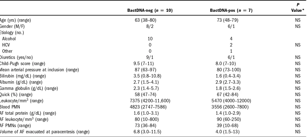
The baseline clinical, basic hemodynamic, and serum and AF analytical characteristics of the cohort distributed according to the absence (BactDNA-neg) or presence (BactDNA-pos) of BactDNA in the serum and AF are shown in Table 1. Although a trend toward more ad-vanced liver disease (lower serum albumin and prothrom-bin time, higher total biliruprothrom-bin, and median Child-Pugh score) was observed in BactDNA-pos patients, these dif-ferences did not reach statistical significance. The volume of AF evacuated was similar in both groups. The compar-ison of the rest of the parameters analyzed did not reach statistical significance (Table 1).
An upper gastrointestinal endoscopy was performed at the time of index admission or in the preceding 3 months in 7 BactDNA-neg patients and 6 BactDNA-pos patients. One BactDNA-neg patient was found to have Grade 1 esophageal varices and four patients had Grade 2 esopha-geal varices. Gastric varices were detected in one patient and portal hypertensive gastropathy was detected in two patients. Four patients demonstrated more than one find-ing. Two BactDNA-neg patients were found to have Grade 1 esophageal varices, one patient was found to have Grade 2 esophageal varices, and two of the studied pa-tients also presented with portal hypertensive gastropathy. Overall, no differences were observed between both groups of patients with regard to the severity of portal hypertension-related endoscopic findings.
whomCitrobacter freundiiwas identified during the study period developed an episode of SBP because ofEscherichia coli, and 1 BactDNA-pos patient developed a vertebral abscess resulting fromStreptococcusspecies 6 months and 3 months, respectively, after the conclusion of the study. One BactDNA-pos patient (in whomE. coliwas detected during the study period) and one BactDNA-neg patient
developed a urinary tract infection resulting fromE. coli
in subsequent admissions.
Detection and Identification of BactDNA. All de-tections of BactDNA in the serum and AF at the time of admission and in the serum during the study period are detailed in Table 2. As can be observed, the intermittent presence of BactDNA was observed in Patients 7 and 11, Table 1. Basic Clinical and Analytical Characteristics of the Cohort Distributed According
to the Absence or Presence of BactDNA in the Serum and AF
BactDNA-neg (nⴝ10) BactDNA-pos (nⴝ7)
P Value*
Age (yrs) (range) 63 (38–80) 73 (48–79) NS
Gender (M/F) 8/2 6/1 NS
Etiology (no.)
Alcohol 10 4
HCV 0 2 NS
Other 0 1
Diuretics (yes/no) 9/1 6/1 NS
Child-Pugh score (range) 9.5 (7–11) 8.0 (7–10) NS
Mean arterial pressure at inclusion (range) 87 (63–97) 80 (73–100) NS Bilirubin (mg/dL) (range) 3.5 (0.8–10.8) 1.6 (0.4–3.4) NS Albumin (g/dL) (range) 2.7 (1.5–4.1) 2.9 (2.7–3.3) NS Gamma globulin (g/dL) (range) 2.3 (1.4–5.7) 1.8 (1.5–2.6) NS
Quick (%) (range) 58 (47–74) 67 (42–84) NS
Leukocyte/mm3(range) 7375 (4200–11,600) 5470 (4000–12000) NS
Blood PMN 4823 (2747–7586) 3556 (2600–7800) NS
AF total protein (g/dL) (range) 1.6 (1.0–3.1) 1.4 (1.0–2.9) NS AF leukocyte/mm3(range) 80 (10–800) 90 (60–250) NS
AF PMNs (range) 73 (36–84) 39 (10–68) NS
Volume of AF evacuated at paracentesis (range) 6.8 (3.0–11.5) 4.0 (1.5–13) NS
BactDNA-negative, negative for the presence of bacterial DNA; BactDNA-pos, positive for the presence of bacterial DNA; AF, ascitic fluid; NS, not significant; M/F, male/female; HCV, hepatitis C virus; PMN, polymorphonuclear leukocytes.
Data are shown as the median and ranges.
Mean arterial pressure was estimated before paracentesis was performed. *A nonsignificantPvalue was aPvalue⬎.05.
Table 2. Sequential BactDNA Detections in the Blood and Ascitic Fluid During the Study Period
Patient No. AF S S8 S16 S24 S32 S40 S48 S56 S64 S72
1 ⫺ ⫺ ⫺ ⫺ ⫺ ⫺ ⫺ ⫺ ⫺ ⫺ ⫺
2 ⫺ ⫺ ⫺ ⫺ ⫺ ⫺ ⫺ ⫺ ⫺ ⫺ ⫺
3 ⫹ ⫹ ⫹ ⫹ ⫹ ⫹ ⫺ ⫺ ⫺ ⫺ ⫺
4 ⫺ ⫺ ⫺ ⫺ ⫺ ⫺ ⫺ ⫺ ⫺ ⫺ ⫺
5 ⫺ ⫺ ⫺ ⫺ ⫺ ⫺ ⫺ ⫺ ⫺ ⫺ ⫺
6 ⫺ ⫺ ⫺ ⫺ ⫺ ⫺ ⫺ ⫺ ⫺ ⫺ ⫺
7 ⫹ ⫹ ⫹ ⫹ ⫹ ⫹ ⫹ ⫺ ⫹ ⫺ ⫺
8 ⫺ ⫺ ⫺ ⫺ ⫺ ⫺ ⫺ ⫺ ⫺ ⫺ ⫺
9 ⫹ ⫹ ⫹ ⫹ ⫹ ⫹ ⫹ ⫺ ⫺ ⫺ ⫺
10 ⫹ ⫹ ⫹ ⫹ ⫹ ⫺ ⫺ ⫺ ⫺ ⫺ ⫺
11 ⫹ ⫹ ⫹ ⫹ ⫹ ⫹ ⫺ ⫹ ⫹ ⫹ ⫺
12 ⫺ ⫺ ⫺ ⫺ ⫺ ⫺ ⫺ ⫺ ⫺ ⫺ ⫺
13 ⫺ ⫺ ⫺ ⫺ ⫺ ⫺ ⫺ ⫺ ⫺ ⫺ ⫺
14 ⫹ ⫹ ⫹ ⫹ ⫹ ⫹ ⫹ ⫺ ⫺ ⫺ ⫺
15 ⫹ ⫹ ⫹ ⫹ ⫹ ⫹ ⫺ ⫺ ⫺ ⫺ ⫺
16 ⫺ ⫺ ⫺ ⫺ ⫺ ⫺ ⫺ ⫺ ⫺ ⫺ ⫺
17 ⫺ ⫺ ⫺ ⫺ ⫺ ⫺ ⫺ ⫺ ⫺ ⫺ ⫺
BactDNA, bacterial DNA; AF, ascitic fluid; S, serum;⫺, negative;⫹, positive.
whereas the disappearance of BactDNA was definitive in the remainder of the BactDNA-pos patients. BactDNA was not detected in any of the sequential samples of serum obtained from BactDNA-neg patients during the study period.
All PCR Fragments Were Sequenced for Bacterial Identification.E. coliwas identified in Patients 3, 9, 14, and 15;C. freundiiwas identified in Patients 10 and 11; andKlebsiella pneumoniaewas detected in Patient 7. The similarity between the isolated BactDNA and a 16s rRNA strain sequence present in the database in most cases was⬎99%, which is high enough to warrant identifica-tion of the species. In all cases the same bacterial species were found simultaneously in the blood and AF for each positive patient at the time of admission, with a similarity of nearly 100%. Also, the similarity between the basal sequence and that obtained at every time point during the study period was always⬎99.5%.
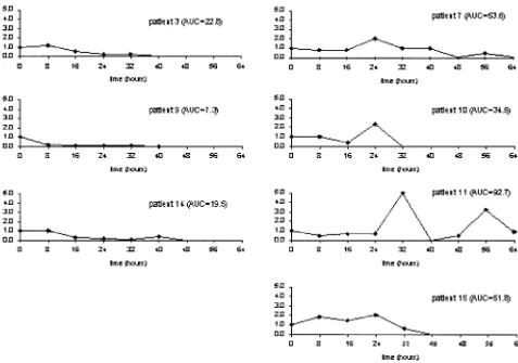
Temporal Evolution of Quantitative BactDNA. A relative quantification of BactDNA was performed in ev-ery specimen obtained at the time of admission and dur-ing an 8-hour period. Data obtained are shown in Fig 1. The mean⫾the standard deviation (SD) of⌬Ct values corresponding to the baseline BactDNA number of
pa-tients was –9.55⫾0.23 (a variation coefficient of 2.4%). Similarly, the mean⫾ SD of the average BactDNA Ct and factor VIII Ct were 22.1⫾0.4 and 31.6⫾0.4, respec-tively. The intersubject variability was considered small, and no significant variations were attained if the quantity of baseline BactDNA was assumed as the unit.
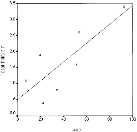
Figure 1 shows the temporal sequence of the relative BactDNA quantification in all BactDNA-pos patients. In 3 cases (Patients 3, 9, and 14), total paracentesis was followed by BactDNA clearance from the blood in a short period of time and the total detected BactDNA, calculated as the AUC from 0 – 64 hours of the 2-⌬⌬Ct values was 22.8, 7.3, and 19.5, respectively. In the remaining 4 patients (Patients 7, 10, 11, and 15), the total detected BactDNA quantity was 3– 4 times higher (53.6, 34.5, 92.7, and 51.8) and in some cases it com-pletely disappeared and reappeared. The AUC values in BactDNA-pos patients was found to be significantly correlated with the total bilirubin (P⫽0.031) (Fig. 2). No significant correlations were detected between total detected BactDNA and age, volume of AF evacuated, Child–Pugh score, albumin, Quick, AF WBC, and to-tal protein.
Discussion
In the current study we report the temporal evolution of the presence of BactDNA in the blood in patients with advanced cirrhosis and ascites, which we consider molec-ular evidence of BT, together with the relative quantifica-tion of circulating BactDNA and its species identificaquantifica-tion by real-time PCR and nucleotide sequencing, respec-tively. To our knowledge, this is the first study proving the continued presence of BactDNA in afebrile, asymptom-atic patients with advanced cirrhosis, which likely sug-gests the existence of repeated episodes of BT.
As discussed earlier, we obtained serum samples every 8 hours from all patients included in the current study over a 3 day-period using therapeutic paracentesis. In all sam-ples, we searched for molecular evidence of BT (i.e., de-tection of BactDNA by 16s rRNA), followed by relative quantification of PCR products and species identifica-tion. This methodologic approach allows us to not only identify those patients in whom BactDNA is present but also its temporal pattern and the amount of BactDNA detectable at every time point, which gives us indirect clues for the estimation of bacterial clearance.
SBP is a frequent and severe complication arising in patients with advanced cirrhosis and ascites1and is
con-sidered to be the consequence of the access of bacteria from the intestinal lumen to AF. However, most of the knowledge we currently possess regarding BT comes from experimental models because, for obvious reasons, it is difficult to obtain the adequate tissue specimens in
hu-mans.8 We have previously reported the detection of
BactDNA in the blood and AF in a subgroup of patients with advanced cirrhosis, which we consider to represent molecular evidences of BT.9
The results of the current study confirm that BT is a common event in patients with advanced and decompen-sated cirrhosis,9because 7 of 17 consecutively admitted
patients who fulfilled the strict inclusion and exclusion criteria detailed earlier showed the asymptomatic pres-ence of BactDNA in both the blood and AF. This per-centage is similar to that reported previously by our group,9and also to the percentage of patients
demonstrat-ing increased serum levels of lipopolysaccharide-binddemonstrat-ing protein (which has been considered to be indirect evi-dence of BT in patients with advanced cirrhosis and as-cites) reported in a previous study by Albillos et al.16The
origin of the detected BactDNA is that typical of bacteria that usually are reported to cause episodes of SBP.1
Although it has been shown that BT traveling from the intestinal lumen to the mesenteric lymph nodes is a pre-requisite for the development of SBP in experimental an-imals6,17,18 to our knowledge to date it is not known
whether BT is the result of single-pulse episodes of bacte-rial movement through the intestinal wall or, conversely, BT represents a continuous or repeated process leading to the development of infections once the immune system becomes unable to control the translocating bacteria.2,3In
fact, it has been previously shown that patients with ad-vanced cirrhosis demonstrate an impairment of the reticu-loendothelial system and that this may lead to the development of bacterial infections.19
The qualitative and sequential studies detailed in Table 2 show molecular evidence of BT in the blood during at least 24 hours after the time of admission and after ther-apeutic paracentesis was performed, and that may persist much longer in some patients. The clinical characteristics of BactDNA-neg and BactDNA-pos patients are similar (Table 1), a situation similar to what has been reported for cirrhotic patients with or without increased levels of li-popolysaccharide-binding protein.16 Therefore, we
can-not pinpoint the reasons leading to first, the development of BT, and second, the time until BactDNA disappears from the blood. Reasons not investigated in the current study, such as intestinal transit time, intestinal bacterial overgrowth, the degree of portal hypertension, and/or the efficacy of the reticuloendothelial system may influence the passage of bacteria from the intestinal lumen to other territories or its clearance from blood.
Figure 1 shows the temporal sequence of the relative BactDNA quantification in all samples. According to a visual evaluation of data, we arbitrarily separated two pat-terns of BactDNA clearance from the blood in all
BactDNA-pos patients. In 3 cases (Patients 3, 9, and 14), BactDNA was cleared from the blood in a short period of time, whereas in the remaining 4 patients the amount of circulating BactDNA changed over time, and in some cases it completely disappeared and reappeared until a total removal occurred at 64 hours, suggesting an inter-mittent process of BT and not a preponderance of the reduced ability of the reticuloendothelial system19to
re-move BactDNA. We have observed a significant and di-rect correlation between AUC and total bilirubin in BactDNA-pos patients (Fig. 2), suggesting that the degree of liver function is somehow related to the persistence of BactDNA in blood. However, patients with advanced cir-rhosis frequently demonstrate high levels of endotox-emia,20 and it has been observed that this factor may
down-regulate bilirubin transporters.21Although we did
not measured endotoxin in the current study, it is likely that the presence of BactDNA might be associated with endotoxemia, and therefore we cannot exclude the possi-bility of a endotoxin-induced hyperbilirubinemia in BactDNA-pos patients.
The fact that we have found an identical sequence of nucleotides in all instances in which BactDNA was de-tected points to a process of repeated episodes of BT. One may speculate regarding the reason for these findings, and two possible explanations likely arise: the existence of an increased or abnormal intestinal permeability, or the ex-istence of intestinal overgrowth of a certain bacterial clone. An increased permeability to macromolecules has been reported in patients with advanced stages of liver disease,22,23 and endotoxemia is a common event in
pa-tients with advanced cirrhosis,20 perhaps in relation to
ultrastructural changes in the intestinal mucosa.24
How-ever, if we assume that an abnormal intestinal permeabil-ity is the main factor responsible for BT, one would expect to find different bacterial species in the same patient that we would have likely identified with the methodology used in the current study. Indeed, only one bacteria spe-cies was found in each patient and in all subsequent sam-ples from the same patient, suggesting an alternative pathogenic explanation. Intestinal bacterial overgrowth is a common phenomenon in both experimental models and patients in cirrhosis,6,25and it is considered to be a
predisposing factor for the development of SBP.26In fact,
previous studies have reported a genetic identity of bacte-ria isolated in the AF and mesenteric lymph nodes and/or ileum in rats with cirrhosis and ascites,18and the findings
reported herein point to this pathogenic mechanism. To our knowledge, the clinical consequences of the presence of BactDNA in a subset of patients with ad-vanced cirrhosis are still unknown because the BactDNA-pos patients in the current study did not developed SBP or
episodes of bacteremia during hospitalization. However, we have observed a marked immune response in the peri-toneal macrophages obtained from AF during the course of paracentesis in BactDNA-pos patients, as represented by significantly higher levels of interleukin (IL)-2, IL-12, interferon-␥, and tumor necrosis factor-␣compared with those observed in BactDNA-neg patients.27This
activa-tion might, in turn, protect patients from developing a complete bacterial infection.28,29 A multicenter Spanish
study currently is underway to assess whether the detec-tion of BactDNA in patients with advanced cirrhosis is an indicator of the development of SBP.
In the current study, we determined the BactDNA clearance pattern through its relative quantification in pa-tients with in whom BactDNA was present in both the blood and AF at the time of admission. Although we consider that serum BactDNA may indicate bacterial translocation, further experimental evidence is necessary to confirm this contention. Bacteria persists in the blood during variable periods of time after the completion of therapeutic paracentesis, disappearing or peaking in se-quential blood samples, and therefore suggesting that this phenomenon is not only related to an impairment of the patient’s bacterial clearance ability, but to the existence of repeated episodes of BT from the intestinal lumen. The fact that we have found identical sequences of nucleotides in all BactDNA PCR fragments detected in every patient in the current study over time strongly supports the exis-tence of repeated episodes of BT due to the same bacteria species.
References
1. Such J, Runyon BA. Spontaneous bacterial peritonitis. Clin Infect Dis 1998;27:669 – 676.
2. Such J, Guarner C, Enriquez J, Rodriguez JL, Seres I, Vilardell F. Low C3 in cirrhotic ascites predisposes to spontaneous bacterial peritonitis. J Hepa-tol 1988;6:80 – 84.
3. Runyon BA. Patients with deficient ascitic fluid opsonic activity are pre-disposed to spontaneous bacterial peritonitis. HEPATOLOGY1988;8:632– 635.
4. Such J, Guarner C, Soriano G, Teixido M, Barrios J, Tena F, Mendez C, et al. Selective intestinal decontamination increases serum and ascitic fluid C3 levels in cirrhosis. HEPATOLOGY1990;12:1175–1178.
5. Berg RD. Bacterial translocation from the gastrointestinal tract. J Med 1992;23:217–244.
6. Guarner C, Runyon BA, Young S, Heck M, Sheikh MY. Intestinal bacte-rial overgrowth and bactebacte-rial translocation in cirrhotic rats with ascites. J Hepatol 1997;26:1372–1378.
7. Llovet JM, Bartoli R, Planas R, Cabre E, Jimenez M, Urban A, Ojanguren I, et al. Bacterial translocation in cirrhotic rats. Its role in the development of spontaneous bacterial peritonitis. Gut 1994;35:1648 –1652. 8. Cirera I, Bauer TM, Navasa M, Vila J, Grande L, Taura P, Fuster J, et al.
Bacterial translocation of enteric organisms in patients with cirrhosis. J Hepatol 2001;34:32–37.
9. Such J, Frances R, Munoz C, Zapater P, Casellas JA, Cifuentes A, Rodri-guez-Valera F, et al. Detection and identification of bacterial DNA in patients with cirrhosis and culture-negative, nonneutrocytic ascites. HEPA
10. Rangel-Frausto MS, Pittet D, Costigan M, Hwang T, Davis CS, Wenzel RP. The natural history of the systemic inflammatory response syndrome (SIRS). A prospective study. JAMA 1995;273:117–123.
11. Runyon BA. Paracentesis of ascitic fluid: a safe procedure. Arch Intern Med 1986;146:2259 –2261.
12. Runyon BA, Canawati HN, Akriviadis EA. Optimization of ascitic fluid culture technique. Gastroenterology 1988;95:1351–1355.
13. Lane DJ. 16S/23S rRNA sequencing. In: Stackebrandt E, Goodfellow M, editors. Nucleic acid techniques in bacterial systematics. New York: John Wiley and Sons, 1991:115–175.
14. Maidak BL, Cole JR, Lilburn TG, Parker CT Jr., Saxman PR, Farris RJ, Garrity GM, et al. The RDP-II (Ribosomal Database Project). Nucleic Acids Res 2001;29:173–174.
15. Altschul SF, Madden TL, Schaffer AA, Zhang J, Zhang Z, Miller W, Lipman DJ. Gapped BLAST and PSI-BLAST: a new generation of protein database search programs. Nucleic Acids Res 1997;25:3389 –3402. 16. Albillos A, de La HA, Gonzalez M, Moya JL, Calleja JL, Monserrat J,
Ruiz-del-Arbol L, et al. Increased lipopolysaccharide binding protein in cirrhotic patients with marked immune and hemodynamic derangement. HEPATOLOGY2003;37:208 –217.
17. Runyon BA, Squier S, Borzio M. Translocation of gut bacteria in rats with cirrhosis to mesenteric lymph nodes partially explains the pathogenesis of spontaneous bacterial peritonitis. J Hepatol 1994;21:792–796. 18. Llovet JM, Bartoli R, March F, Planas R, Vin˜ado B, Cabre E, Arnal J, et al.
Translocated intestinal bacteria cause spontaneous bacterial peritonitis in cirrhotic rats: molecular epidemiologic evidence. J Hepatol 1998;28:307– 313.
19. Rimola A, Soto R, Bory F, Arroyo V, Piera C, Rodes J. Reticuloendothelial system phagocytic activity in cirrhosis and its relation to bacterial infec-tions and prognosis. HEPATOLOGY1984;4:53–58.
20. Guarner C, Soriano G, Tomas A, Bulbena O, Novella MT, Balanzo J, Vilardell F, et al. Increased serum nitrite and nitrate levels in patients with cirrhosis: relationship to endotoxemia. HEPATOLOGY1993;18:1139 –1143. 21. Vos TA, Hooiveld GJ, Koning H, Childs S, Meijer DK, Moshage H,
Jansen PL, et al. Up-regulation of the multidrug resistance genes, Mrp1 and Mdr1b, and down-regulation of the organic anion transporter, Mrp2, and the bile salt transporter, Spgp, in endotoxemic rat liver. HEPATOLOGY
1998;28:1637–1644.
22. Pascual S, Such J, Esteban A, Zapater P, Casellas JA, Aparicio JR, Girona E, et al. Intestinal permeability is increased in patients with advanced cirrhosis. Hepatogastroenterology 2003;50(53):1482–1486.
23. Ersoz G, Aydin A, Erdem S, Yuksel D, Akarca U, Kumanlioglu K. Intes-tinal permeability in liver cirrhosis. Eur J Gastroenterol Hepatol 1999;11: 409 – 412.
24. Such J, Guardiola JV, de Juan J, Casellas JA, Pascual S, Aparicio JR, Sola-Vera J, et al. Ultrastructural characteristics of distal duodenum mu-cosa in patients with cirrhosis. Eur J Gastroenterol Hepatol 2002;14:371– 376.
25. Pardo A, Bartoli R, Lorenzo-Zuniga V, Planas R, Vinado B, Riba J, Cabre E, et al. Effect of cisapride on intestinal bacterial overgrowth and bacterial translocation in cirrhosis. HEPATOLOGY2000;31:858 – 863.
26. Guarner C, Soriano G. Spontaneous bacterial peritonitis. Semin Liver Dis 1997;17:203–217.
27. Frances R, Mun˜oz C, Zapater P, Pascual S, Pe´rez-Mateo M, Such J. Bac-terial DNA activates cell-mediated immune response in peritoneal macro-phages from patients with cirrhosis and ascites [abstract]. HEPATOLOGY
2003;38(Suppl 1):572A.
28. Krieg AM. CpG motifs: the active ingredient in bacterial extracts? Nat Med 2003;9:831– 835.