THE JOURNAL OF TROPICAL LIFE SCIENCE OPEN ACCESS Freely available online
VOL. 6, NO. 2, pp. 131 - 135, May 2016 Submitted March 2016; Revised May 2016; Accepted May 2016
Human and Animal Pentastomiasis in Malaysia: Review
Bahaa M A Latif*, Azdayanti Muslim, Heo Chong Chin
Faculty of Medicine, Universiti Teknologi MARA, Selangor, Malaysia
ABSTRACT
Pentastomiasis is a zoonotic parasitic disease induced by the larval stages of pentasomes. The disease has been re -ported in Africa, the Middle East, and Southeast Asia and caused by the nymphs of the two genera: Linguatula and Armillifer and the two species L. serrata and A. armillatus regard for more than 90% of human cases. The definitive hosts of Armillifer spp. are snakes, lizards, and other reptiles. The parasites live in the upper respiratory tracts and lay eggs that are passed out through respiratory secretions, saliva or feces. Intermediate hosts are hu -mans, rodents, and other mammals. Humans incidentally acquire the infestation by the consumption of uncooked infected snake meat or by drinking water contaminated with ova of the pentastomes. In the intestinal tract, the larvae hatch from the ova, penetrate the intestinal wall and migrate to organs in which the liver is the most com -mon site. In Malaysia, human pentastomiasis was reported a-mong aborigines in Peninsular and East Malaysia. Armillifer moniliformis was identified in wild animals and carnivores with infection rate 1.8% and 20.7% respect-ively. In addition to that, a previous study has discovered the adults of pentastomes of A. moniliformis in two out of six snakes from species Python reticulates. Recently a case of human pentastomiasis was reported in Sabah, East Malaysia, caused by the nymph of A. moniliformis. Therefore, the aim of this review is to provide the latest up-dates on human and animal pentastomiasis especially in Malaysia.
Keywords: Pentastomiasis, human, animal, Malaysia
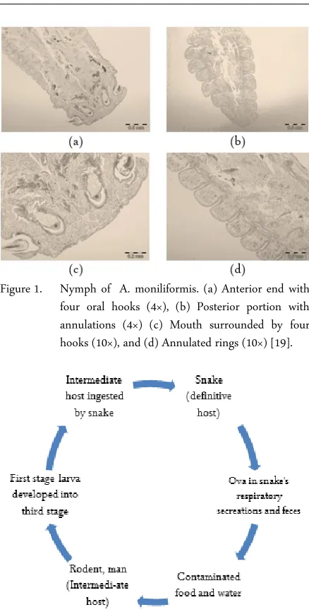
Pentastomes are wormlike parasites (“tongue worm”), 3 - 13 cm in length. They have centrally lo-cated mouth surrounded by four hooks making they seem like they have five mouths hence the name “pen-tastomes". Their body is segmented, forming annuli and covered in a chitinous cuticle [1] (Figure 1).
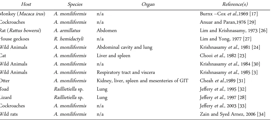
Adult parasites dwell in the upper respiratory tracts of the definitive hosts (snakes, lizards, and other reptiles), lay eggs that are passed out through respiratory secretions, saliva or feces. Humans, rodents, and other mammals act as intermediate hosts which acquired the infection by; drinking water contaminated with ova of the pentastomes, consumption of infected snake meat, handling infected snakes and harvesting skins of the infected snake (Figure 2).
Pentastomes are cosmopolitan and widely distributed especially in tropical and subtropical
countries. Generally, the range of the natural hosts and the degree of water and food sanitation affect the distribution of these parasites [2].
In Malaysia, pentastomiasis was reported in both human and animals [3]. The disease is considered being neglected and less attention is given by the authorities. Therefore, the aim of this review is to provide the latest updates regarding human and animal pentastomiasis especially in Malaysia.
Human pentastomiasis is a zoonotic parasitic dis-ease, which human serves as an incidental host for the infection. Most of the cases are asymptomatic and dis-covered only during surgery or autopsy. Generally, the diagnosis is largely depends on parasitologic and histopathologic examination [4].
JTLS | J. Trop. Life. Science 131 Volume 6 | Number 2 | May| 2016
HUMAN PENTASTOMIASIS WITH LATEST REPORT IN MALAYSIA
INTRODUCTION
*Corresponding author: Baha Latif
Faculty of Medicine, Universiti Teknologi MARA Jalan Hospital, Sungai Buloh, Selangor, Malaysia 47000 E-mail: [email protected]
How to cite:
Figure 1. Nymph of A. moniliformis. (a) Anterior end with four oral hooks (4×), (b) Posterior portion with annulations (4×) (c) Mouth surrounded by four hooks (10×), and (d) Annulated rings (10×) [19].
Figure 2. Life cycle of pentastome parasite
Historically, the first case of human pentastomiasis was reported by Pruner in Egypt in 1847 [5]. Following that, the disease has been reported sporadically in Africa, Middle East, and Southeast Asia and it was caused mainly by the nymphs belonging to genera; Linguatula and Armillifer [4, 5, 6-10]. The two species of the parasites; Linguatula serrata and Armilifer armillatus accounted for more than 90% of human cases [11]. To date, there are four species of Armillifer recorded in human infection; A. armillatus in Africa and the Arabian Peninsula, A. agkistrodontis in China, A. grandis in Africa and A. moniliformis in Southeast Asia [11, 12].
Human is a dead end host of pentastomes and ac-quired the infection by the consumption of uncooked infected snake meat or by drinking water contaminated
with ova of the pentastomes. In addition to that, hu-man has possibility to acquire the infection while han-dling, harvesting the snake’s skins or playing and touching the mouth of the pet snakes. In the intestinal tract, the larvae hatch from the ova, penetrate the in-testinal wall and migrate to many organs in which the liver is the most common site [1].
Infection with pentastomids is mostly asympto-matic. However, in some cases, the clinical manifesta-tions include fever, abdominal pain, vomiting, diar-rhea, jaundice and abdominal tenderness [11]. In se-vere disseminated cases, the disease may lead to death [5, 13, 14]. According to Herzog et al. (1985) cysts could be serious enough to cause death [15]. However, the larvae are usually die and calcify within two years of infection [2].
In Malaysia, human pentastomiasis was reported in both Peninsular and East Malaysia [16-18]. In a series of 30 consecutive autopsies performed on aborigines from five different states in West Malaysia, pentasto-mid infection was found in 33.3% of the cases with the prevalence of 45.4% in adults with the liver and the lungs were the most infected organs [16].
The most recent infection of human pantastomiasis was reported in a 70 year old aboriginal farmer from Borneo in 2011 [19]. Most of the infections were caused by A. moniliformis. Table 1 shows the summary of human pentastomiasis cases in Malaysia.
Table 1. Reported cases of human pentastomiasis in Malaysia (1967- 2011)
Species Organ Reference
A. moniliformis Neck, abdomen Rail, 1967 [18] A. moniliformis Liver, lung Prathap et al., 1968 [14] A. moniliformis Chest, abdomen Burnx –Cox et al., 1969 [17] A. moniliformis Liver, lung Prathap et al., 1969 [16]
Armillifer sp. Fallopian tube Ong, 1974 [20] A. moniliformis Liver Latif et al., 2011 [19]
Animals acquired the infection with pentastomids through ingestion of eggs in food and water contami-nated by feces or nasal discharges of snakes, Python sp. [21] or by feces of lizards [22]. In Malaysia, pentasto-miasis was reported in domestic animals such as dog and cat and in wide range of wild animals [23, 24]. Ta-ble 2 shows the summary of reported cases of animal pentastomiasis in Malaysia.
infects mammals, reptiles, and birds as intermediate hosts [3]. Honjo et al. [25] and Burnx-Cox et al. [17] reported A. moniliformis in cynomolgus monkey, Macaca irus while Lim and Krishnasamy [26] reported heavy infestation in giant rat Rattus bowersi with the larval stage of A. armillatus. The same authors found that A. armillatus is common among bats, rats, squir-rels, and carnivores from the study conducted in West Malaysia.
Besides, Lim and Yong found Raillietiella hemi-dactyli in four species of house geckoes, namely: Hemidactylus frenatus, Platyurus platyurus, Gehyra mutilate and Gecko monarchus [27]. The prevalence of infection in latter was high in two species of house geckoes, Hemidactylus frenatus and Platyurus platyu-rus from the shop houses. The high prevalence in geckoes of shop houses was attributed to the presence of intermediate insect hosts, particularly cockroaches which are abundant in these shop houses due to the availability of food and poor sanitation in these places. In addition to that, Jeffery et al. found Raillietiella sp. in the lungs of 5 out of 9 geckoes, Gekko smithi [28].
Other than geckoes, cockroaches are also common and closely associated with human dwellings in Malaysia. Previous studies recorded the recovery of Moniliformis moniliformis larva in the American cock-roach, Periplaneta americana [33, 29]. This is interest-ing since the researchers found the remains of P. Americana in the stomach of house geckoes [22].
Adult pentastomids in the gecko’s lung produce eggs which are passed out with feces. Cockroaches and other coprophagous insects ingest the feces of geckoes, and the eggs will hatch into larvae and settled in their
bodies. Lizards become infected with pentastomids when they ingest the infected insects. Meanwhile, there is a possibility of geckoes to transmit the infection to humans through feces which contaminate food and utensils.
Pentastomiasis was also reported in wild animals. Krishnasamy et al. found that out of 5209 wild animals of 33 species in West Malaysia, 92 (1.8%) were infected with nymphal stages of A.moniliformis [3]. The infection rate of the wild animals and carnivores with A. moniliformis was 1.8% and 20.7% respectively. The occurrence of a nymph stage of A. moniliformis in a fruit-eating bat Cynopterus brachyotis was reported by Krishnansamy et al. [30]. The parasite measured 15 mm in length and 1.58 mm in width and had 31 annuli. The larval stages of A. moniliformis were found in two banded palm civet, Hemigalus derbyanus, and they are considered as a new host for the parasite [3]. The same authors identified 12 species of Malaysian pentastomids in a variety of animals.
In 1981, Krishnasamy et al. recovered the adults of A. moniliformis from two out of six Python reticulates [24]. Another finding of A. moniliformis in pythons was reported in 1986, when Krishnansamy et al. found the adult of A. moniliformis from the respiratory tracts of 6 out of 7 pythons (P. reticulates) in study involving zoo animals [35]. The latter study also reported discovery of the nymphs from the liver and the kidney of a lar gibbon (Hylobateslar) and meerkat (Suricatasuricata) respectively. In addition to that, the pestastomes parasites were also reported in other animals such as the smooth otter [31], toad Bufo
JTLS | J. Trop. Life. Science 132 Volume 6 | Number 2 | May | 2016
Table 2. Pentastomiasis among animals in Malaysia (1969-2003)
Host Species Organ Reference(s)
Monkey (Macaca irus) A. moniliformis n/a Burnx –Cox et al.,1969 [17]
Cockroaches A. moniliformis n/a Anuar and Paran,1976 [29]
Rat (Rattus bowersi) A. armillatus Abdomen Lim and Krishnasamy, 1973 [26]
House geckoes R. hemidactyli n/a Lim and Yong, 1977 [27]
Wild Animals A. moniliformis Abdominal cavity and lung Krishnasamy et al., 1981 [24]
Cat A. moniliformis Liver and spleen Chooi et al., 1982 [23]
Wild Animals A. moniliformis n/a Krishnasamy et al., 1984 [30]
Wild Animals A. moniliformis Respiratory tract and viscera Krishnasamy et al., 1985 [3] Otter A. moniliformis Kidney, liver, spleen and mesenteries of GIT Cheah et al.,1989 [31]
Toad Raillietiella sp. Lung Jeffery et al., 1995 [32]
Lizard Raillietiella sp. Lung Jeffery et al., 1997 [28]
Cockroaches A. moniliformis n/a Jeffery et al., 2003 [33]
Wild rats A. moniliformis n/a Zain and Syed Arnez, 2006 [34]
melanostictus [32] and wild rats [34].
In pentastomiasis, a diagnosis could not be made before the surgery. The disease is generally asympto-matic, and detected commonly in autopsy or biopsy. Histopathologic and parasitologic examinations of biopsy or autopsy lesions are part of the common diag-nostic methods. In X- rays examination, the parasite can be detected by appearance of calcified nymphs, crescent- shaped bodies which distributed throughout the body. Serological tests involve using indirect im-munofluorescence; ELISA and Western blot [36]. Due to the difficulty to detect the infection, modified sero-logical tests (pentastome kit) are required for the diag-nosis of this disease.
In order to prevent human infections, personal hy-giene measures are necessary including; avoid drinking river water directly or the water should be boiled be-fore drinking. The authorities should provide some health education to alert people from consuming the under-cooked snake’s meat, possibility of transmission after handling snakes and avoidance of contact with snake excretions.
Human pentastomiasis is endemic in Malaysia and parallels with that reported in animals. Snakes play an important role in the transmission of human pentasto-miasis. Extra precautions should be undertaken espe-cially by snake handlers to avoid infection. More epi-demiological studies together with the surveillance of pentastomes in food and water are recommended to clarify the geographical distribution of this disease in Malaysia.
1. John DT, Petri WA (2006) Markell and Voge's medical parasitology. US, Elsevier Health Sciences: Saunders. 2. Drabick JJ (1987) Pentastomiasis. Review of Infectious
Diseases 9 (6): 1087-1094.
3. Krishnasamy M, Singh I, Jeffery J et al (1985). Some pen-tastomes from Malaysia, with special emphasis on those from Peninsular Malaysia. J Malaysian Soc Health 5 : 49-56.
4. Symmers WSC, Valteris K (1950) Two cases of human in-festation by larvae of Linguatula serrata. Journal of clinical pathology 3 (3): 212.
5. Cannon D (1942) Linguatulid infestation of man. Ann
trop Med Parasit. 36(4): 160-167.
6. Lai C, Wang XQ, Lin L et al (2010) Imaging features of pediatric pentastomiasis infection: a case report. Korean Journal of Radiology 11 (4): 480-484.
7. Tappe D, Büttner DW (2009) Diagnosis of human visceral pentastomiasis. PLoS Negl Trop Dis. 3(2): e320.
8. Dakubo J, Etwire V, Kumoji R et al (2007) Human pen-tatomiasis: A case report. West African Journal of Medicine 25 (2): 166-168.
9. Tappe D, Winzer R, Büttner DW et al (2006) Linguatulia-sis in Germany. Emerg Infect Dis 12 (6): 1034-1036. 10. Machado MAC, Makdissi FF, Canedo LF et al (2006)
Un-usual case of pentastomiasis mimicking liver tumor. Jour-nal of gastroenterology and hepatology 21 (7): 1218-1220. 11. Chen SH, Liu Q, Zhang YN et al (2010) Multi-host model-based identification of Armillifer agkistrodontis (Pentastomida), a new zoonotic parasite from China. PLoS Negl Trop Dis 4 (4): e647.
12. Yao MH, Wu F, Tang LF (2008) Human pentastomiasis in China: case report and literature review. Journal of Par-asitology 94 (6): 1295-1298.
13. Obafunwa J, Busuttil A, Nwana E (1992) Sudden death due to disseminated porocephalosis - a case history. Inter-national journal of legal medicine 105 (1): 43-46.
14. Prathap K, Ramachandran C, Haug N (1968) Hepatic and pulmonary porocephaliasis in a Malaysian Orang Asli (aborigine). The Medical journal of Malaya 23 (2): 92-95. 15. Herzog U, Marty P, Zak F (1985) Pentastomiasis: case
re-port of an acute abdominal emergency. Acta tropica 42 (3): 261-271.
16. Prathap K, Lau K, Bolton J (1969) Pentastomiasis: a com-mon finding at autopsy acom-mong Malaysian aborigines. American Journal of Tropical Medicine and Hygiene 18 (1): 20-27.
17. Burnx-Cox C, Prathap K, Clark E et al (1969) Poro-cephaliasis in western Malaysia. Transactions of the Royal Society of Tropical Medicine and Hygiene 63 (3): 409-411. 18. Rail G (1967) Porocephaliasis: a description of two cases in Sabah. Transactions of the Royal Society of Tropical Medicine and Hygiene 61 (5): 715-717.
19. Latif B, Omar E, Heo CC et al (2011) Human pentasto-miasis caused by Armillifer moniliformis in Malaysian Borneo. The American journal of tropical medicine and hygiene 85 (5): 878-881.
20. Ong H (1974) An unusual case of pentastomiasis of the fallopian tube in an Aborigine woman. Journal of Tropical Medicine and Hygiene 77 (8): 187-189.
21. Zumpt F (1966) The arthropod parasites of vertebrates in Africa south of the Sahara (Ethiopian region). Volume III (Insecta excl. Phthiraptera). The arthropod parasites of vertebrates in Africa south of the Sahara (Ethiopian re-REFERENCES
CONCLUSION
DIAGNOSIS
gion) Volume III (Insecta excl Phthiraptera).
22. Lavoipierre M, Lavoipierre M (1965) The American cock-roach, Periplanta americana, as an intermediate host of the pentastomid, Raillietiella hemidactyli. The Medical Journal of Malaya 20 (1): 72.
23. Chooi KF, Omar AR, Lee J (1982) Melioidosis in a do-mestic cat, with concurrent infestation by nymphs of the cat pentastome, Armillifer moniliformis. Kajian Veterinar 14: 41-44.
24. Krishnasamy M, Singh I, De Witt G et al (1981). The nat-ural infection of wild animals in West Malaysia with nymphs of Armillifer moniliformis (Diesing, 1835) Sam-bon, 1922. The Malaysian Journal of Pathology. 4: 29-34. 25. Honjo S, Muto K, Fujiwara T et al (1963). Statistical
sur-vey of internal parasites in cynomolgus monkeys (Macaca irus). Japanese Journal of Medical Science and Biology 16 (4): 217-24.
26. Lim BL, Krishnansamy M (1973) A heavy infection of Porocephalus armillatus in Rattus bowersi. The Southeast Asian journal of tropical medicine and public health 4 (2): 282.
27. Lim B, Yong H (1977) Pentastomid infection in the house geckoes from Sarawak, Malaysia. Medical Journal of Malaysia 32 (1): 59-62.
28. Jeffery J, Zahedi M, Abdullah S et al (1997) Gekko smithi, a new lizard host of the pentastomid, Raillietiella sp. (Pen-tastomida: Cephalobaenida) in Peninsular Malaysia. Trop-ical Biomedicine 14: 145-146.
29. Anuar A, Paran T (1976) Periplaneta americana (L.) as in-termediate host of Moniliformis moniliformis (Bremser) in Penang, Malaysia. Southeast Asian Journal of Tropical Medicine and Public Health 7 (3): 415-416.
30. Krishnasamy M, Singh I, Jeffery J (1984) Some new verte-brate host records of Armillifer moniliformis (Diesing, 1835) Sambon, 1922 in Peninsular Malaysia. J Malaysian Soc Hlth 4: 20-21.
31. Cheah T, Rajamanickam C, Lazarus K (1989) Natural in-fection of Armillifer moniliformis in a smooth otter (Lu-tra perspicillata). Tropical Biomedicine 6 (2): 139-40. 32. Krishnasamy M, Jeffery J, Inder Singh K et al (1995)
Rail-lietiella rileyi, a new species of pentastomid from the lung of toad, Bufo melanostictus from Malaysia. Tropical Bio-medicine 12: 31-8.
33. Jeffery J, Abdullah S, Anuar K et al (2003) Collection of domicilliary cockroaches and their parasites from the Fac-ulty of Medicine, University of Malaya, Kuala Lumpur. Trop Biomed. 20: 165-168.
34. Zain SNM, Syed-Arnez A (2006) A Study on Wild Rats and Their endoparasite fauna From the Endau Rompin National Park, Johor. Malaysian Journal of Science 25 (2). 35. Krishnasamy M, Jeffery J, Singh KI et al (1986) The poro
-cephalid pentastome Armillifer moniliformis in zoo anmi-als. Mal Vet J. 8: 144-147.
36. Tappe D, Buttner DW (2009) Diagnosis of human visceral pentastomiasis. PLoS Negl Trop Dis. 3: 320.