IOSR Journal of Pharmacy and Biological Sciences (IOSR-JPBS)
e-ISSN: 2278-3008, p-ISSN:2319-7676.
Volume 9, Issue 2 Ver. VII (Mar-Apr. 2014), PP 59-66
www.iosrjournals.org
www.iosrjournals.org 59 | Page
Confirmation of adherence Protein Hemagglutinin sub-unit Pili
with MW 49.6 kDa
Helicobacter pylori
on Mice Gastric Epithelial
Cell
Hamong Suharsono
1, Made Agus Hendrayana
2, I Nyoman Mantik
3and
Sumarno Reto Prawiro
21
Laboratory of Biochemistry Veterinary Faculty University of Udayana Denpasar, Indonesia
2
Laboratory of Microbiology Medical Faculty University of Udayana Denpasar, Indonesia
3
Laboratory of Virology Veterinary Faculty University of Udayana Denpasar, Bali Indonesia
4
Laboratory of Microbiology, Medical Faculty University of Brawijaya Malang Indonesia
Abstract: Helicobacter pylori (H.pylori) is a gram-negative microaerophilic bacterium that is generally
associated with the main cause of approximately 80% peptic ulcers in stomach. It is also linked to the development of the stomach cancer. The aim of this study was to characterize the sub-unit pili proteins of Helicobacter pylori with a molecular weight of about 49,6 kDa as a haemagglutinin protein and adhesion molecules on surface of mice gastric epithelial cells. The bacterium was firstly cultured on the plate of TSA-B (Trypticase Soy Agar with 5% Sheep Blood) to prepare the protein of interes using bacterial cutter. The isolated proteins were subsequently analyzed for haemagglutination capability using mice sentisized erithrocytes. SDS-PAGE and western blotting analyses were used to confirm the protein patterns and their immunological reactivities respectively. The both SDS-PAGE and western blotting analyses showed that a dominant protein with molecular weight of about 49,6 kDa was detected consistently and found to be potentially aglutinate the erythrocytes. Furthermore, this protein was observed to be adherence on mice gastric epithelial cells. This study has indicated that the sub-unit pili proteins of H. pylori with a molecular weight of about 49,6 kDa was found to be dominant and immunogenic. Further study is required to confirm the in vivo properties of the protein associated with pathogenesis of H. pylori infection.
Keywords: pili 49,6 kDa H. pylori, adhesion, gastric epithelial cell
I.
Introduction
In the early 1980s it was known that the presence of microbes in the stomach was associated with gastric cancer. The ability of bacteria to cause cancer at the same time undermine the perception of the bactericidal power of stomach acid. The previous studies and surveys about this phenomena considered that infection of H. pylori may be one of the cause’s gastric cancer (Nunes, 1998). Infection by the H.r pylori has been identified as a major cause of peptic ulcer disorder (gastric and duodenal ulcers), gastritis chronic and gastric carcinoma even gastric lymphoma. This is unique, because Helicobacter pylori is the only one bacterium known to cause gastric carcinoma (Clyne, et al., 2007). Infectious H. pylori was classified as carcinogens group I for gastrointestinal cancer (WHO, 1994), because it has various virulent factors such as CagA, VacA, Urease, and ammonia that capable to trigger carcinogenesis. The pathogenic properties of this bacteria was associated with its fimbrial adhesion (pili), a protein found on the bacterial cell surface that play a role as a bacterial virulence factor (Salyer & Whitt, 2002). In some bacteria, adhesion has antigenic reactivity to agglutinate erythrocytes, called haemagglutinin protein (Nagayama et al., 1995).
Confirmation of adherence Protein Hemagglutinin sub-
unit Pili with MW 49.6 kDa…
II.
Materials And Methods
Helicobacter pylori isolate and cultivation
Helicobacte pylori strain was isolated from patient with gastritis and duodenum ulcer, kindly provided by Biomedical Research Unit Lombok West Nusa Tenggara Provincial Hospital Indonesia. The bacterium was firstly cultured using media Trypticase Soy Agar (TSA) and Trypticase Soy Broth (TSB) supplemented with 10% sheep blood, Dent supplement and Isovitalex and incubated at 37 C on microaerophilic atmosphere.
SDS-PAGE analysis
To determine molecular weight (MW) of protein, SDS-PAGE analysis was done according to Laemmli methods (1970). A total of 0.5 ml samples of pili with a concentration of about 100 µg was firstly mixed with similar amount of reducing buffer containing 5 mM Tris HCL pH 6,8, 5% 2-mercapto ethanol, 2,5% w/v sodium dodecyl sulfate, 10% v/v glycerol that containing color tracker blue bromophenol and boiled at 100oC for 5 minutes. The denatured proteins was subjected into 12,5% mini slab gel with tracking gel 4% together with a low range protein marker (Sigma), electrophoresed with 125mV for 45 minutes. The gel was then stained with Coomassie briliant blue and de-stained with 60 % ethanol until all protein bands were observed, and photograped.
Hemaglutination assay
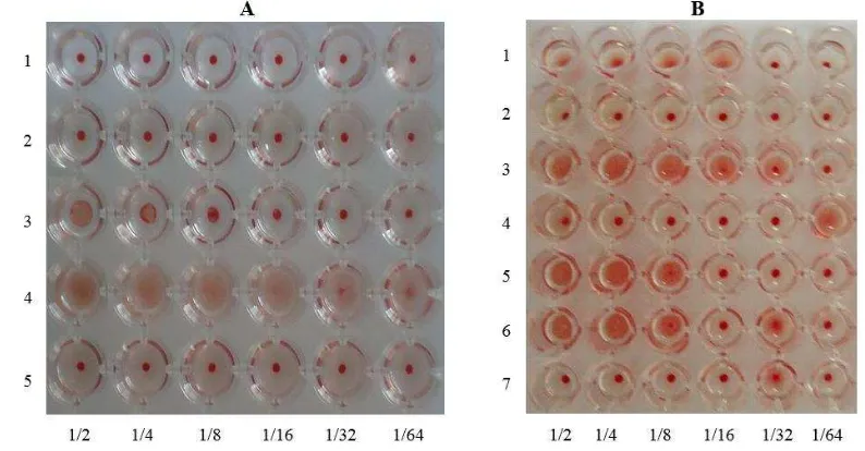
Hemaglutination assay was performed according to Hanne and Finkelstein (1982). Sample to be tested is whole cell lysate, protein pili, and Outer membrane protein. Serial dilution of samples was prepared as double serial dilutions from at 1: 2, to 1: 64 from original concentration on V micro plate. To test agglutinin of protein sub unit pili, 50 uL of each dilution of pili fraction was drooped into well of plate A (pili fraction 1, from row A 2 A 6; pili fraction 2 from row A3 to A6 and pili fraction 3, from row A4 to A6 respectively. As negative control 50 uL of PBS pH 7.2 was drooped into each well, row A1 to A6 and from row A5 to A6 (Fig. 2A). Similar reaction was also done for OMP proteins in Plate B. Subsequently, a drop of 50 uL of mice sensitized red blood cell with concentration of 0,5% was added into each well, shaken with plate rotator for 1 minute and incubated at room temperature for one hour. The titer was determined by observing the agglutination of red blood cells.
Isolation protein haemaglutinin sub unit pili 49,6 kDa H.pylori
Haemagglutinin protein of 49,6 kDa sub-unit pili was isolated from gel by cutting a long side close to the protein positions, around 49,6 kDa. The isolated gel was sliced and inserted into the dialisa membrane soaked with PBS buffer. Subsequently, protein of interes was electro eluted by placing the membrane in the negative electrode with current 20 mA for 15 minutes. Total protein was measured using a method derived DC Protein Assay (Biorad), suspended to a concentration of about 10 ng per ml and kept at -20oC until used.
The production of rabbit polyclonal antibodies to prepare IgG against the sub unit pili 49,6 kDa protein Polyclonal antibodies was prepared in a New Zealand healthy rabbit by immunizing it with immunogen, three times one week interval. At the first, the animal was immunized intramuscularly with 1 ml of emulsified immunogen contained 20 ng in Freunds complete adjuvant. The second and third immunization were given with the same dose of antigen but with Freunds incomplete adjuvant. One week after the last vaccination, blood was taken for serum. To purify IgG, serum was precipitated using ammonium sulfate, aliquoted 100 µL each and stored at -20 oC until used.
Western blotting analysis
Western blotting analysis was performed according to Towbin (1979). Protein of interes was subjected into SDS-PAGE gel and transfered to PVDF membran using mini Transblot Cell (Biorad, USA). The cell was soaked with Tris Borate buffer pH 8.3, run at 100 V contants voltage for one hour. Subsequently, membran-contained tranfered proteins was blocked with 5% nonfat skim milk for 30 minutes at room temperature, to block non-spesific binding site. The first antibody (IgG) with 1:100 dilution with PBS was then added into the membrane, incubated for 1 h at room temperature. After washing with TBE Tween20 twice, the membran was incubated with goat-anti rabbit HRP-conjugated antibody solution, as recomended by the manufacture. Finally, the membrane was again washed before colour development beeing done, washed with H2O, dried and photograph.
Mice gastric epithelial cell preparation
Confirmation of adherence Protein Hemagglutinin sub-
unit Pili with MW 49.6 kDa…
www.iosrjournals.org 61 | Page was then soaked with a solution (pH 7.3) containing 1,5 mM KCl, 9,6 mM NaCl, 2,7 mM Na-citrate, 8 mM KH2PO4 and 5,6 mM cold Na2HPO4, before being incubated at 37oC for 30 minutes with gentle shaking. The
supernatant was removed, and the tissue was transferred into PBS containing 1.5 mM EDTA and 0,5 dithiothreithol and incubated for a further 15 min at 37oC with vigorous shaking. Again, the supernatant was removed, and the cells were washed with PBS by three centrifugation at 900xg for 5 min at 4oC. Isolated gastric cells were harvested by a further centrifugation at 900xg for 5 min at 4oC and suspended in PBS containing 1% Bovine serum albumin (BSA), counted with a hemcytometer to a concentration of about 106 cells per ml. The suspended cells were kept at 4oC until required for Immunohistochemistry and adherence assays.
Immunocytochemistry method
Immunocytochemistry was performed using reagents LSAB2 System HRP (Dako Cytomation). A total of 100 uL cell suspension was spread over the glass slide, air-dried and fixed with absolute methanol. The slides were then treated with 3% H2O2 in distilled water for 5 min to block endogenous peroxidase, then washed an
additional 3 times with distilled water. Subsequently, 100 µl of the 49,6 kDa subunit pili protein was dropped onto the slide and incubated for 30 minutes at room temperature. After incubation with the antigen, the slides were then washed 3 times with PBS and 100 µl of the rabbit IgG as prepared above was added and incubated for 30 minutes at room temperature. The slides were again washed 3 times with PBS, and two drops of biotinylated link secondary antibody were then added to the slide and incubated for 15 min. The slides were again washed 3 times with PBS, and then 2 drops of peroxidise-labelled streptavidin was added to the slides and incubated for 10 min. After a further 3 washes with PBS the colour reaction was developed by adding 3-3’diaminobenzidine (DAB) for 3-5 min. The slides were then washed with distilled water, dried before being examined by light microscope. Negative controls were provided by omitting either the antigen or IgG solution.
Adherence inhibition assay of H. pylori to mice gastric epithelial cells
A serial dilution of 100 µL IgG suspension from 1:1 to 1:8 was made, each of it was mixed with 100 uL gastric epithelial cells (106 cells/mL). The mixture was then allowed to incubate at 37o C for 30 minutes with gentle shaking. The cells were collected by centrifugation at 900xg for 5 minutes. The cell pellet from each different mixture was suspended with similar amount of 100 uL of H. pylori cells (105 cells/mL), and the non-adherent cells were removed by repeated washing with PBS containing 1% BSA. Finally, 100 uL cell suspension was stained with 0,5% Tryphan Blue, and examine by phase-contrast microscopy at a magnification of 1000x. An adherence index was calculated as the mean number of gastric epithelial cells per bacteria determined in three separate experiments.
III.
Results
SDS-PADE analysis
The SDS-PAGE analysis showed that obviously 5 major protein bands were detected with molecular weight of about 80kDa, 60 kDa, 54 kDa, 49,6 kDa and 47 kDa respectively (Fig 1). A protein band of interest with the molecular weight of 49,6 kDa was consistently observed. This protein band was sliced and electro-eluted for further analysis.
Figure 1. SDS-PAGE analysis of H. pylori pili protein. Lanes 1: A low range molecular weight marker, 2-7: protein profile of H.pylori pili that consisted of five major protein bands namely with molecular
Confirmation of adherence Protein Hemagglutinin sub-
unit Pili with MW 49.6 kDa…
Haemagglutination assay
Both the pili and outer membrane originated proteins of H. pylori were examined for haemagglutination of mouse erythrocytes. Of the 3 pili protein fractions isolated, fraction 3 showed the highest haemagglutination titer of 1/16 (Fig.2 A). Fraction 2 had a low titer of 1/4 but fraction 1 had no haemagglutination reactivity, similar to negative, PBS control. Meanwhile, among the OMP fraction proteins, only fraction 2 showed strong haemagglutination titer of 1/8 (Fig. 2B), but all of negative control (PBS) had no haemagglutination properties.
Figure 2A. Haemagglutination test of H. pylori protein sub unit pili in serial shearing process Figure 2B. Haemagglutination test of H. pylori protein sub unit pili and OMP H. pylori using mice
erythrocytes
Legend : A.1: blanko/PBS B.7: blanko/PBS B.2: pili fraction 2 A.2: pili fraction 1 B.6: OMP fraction 3 B.1: pili fraction 1 A.3: pili fraction 2 B.5: OMP fraction 2
A.4: pili fraction 3 B.4: OMP fraction 1 A.5: blanko/PBS B.3: pili fraction 3
Western blotting analysis
Hyper immunized rabbit against sub unit pili 49,6 kDa proteins was tested by Western blotting to confirm the presence of the expected antibodies. A protein band with molecular weight of about 49.6 kDa showed the strongest reactivity suggesting that this protein was immunogenic and immunodominant, although other minor protein bands were also observed (Fig. 3).
Figure 3. Western blot analysis of protein sub unit pili from sequential cutting H. pylori against rabbit polyclonal anti-H. pylori showed that the polyclonal antibodies reacted strongly with protein sub unit pili with MW of about 49,6 kDa, compared to other protein components . Lanes: 1. Molecular
Confirmation of adherence Protein Hemagglutinin sub-
unit Pili with MW 49.6 kDa…
www.iosrjournals.org 63 | Page Adherence inhibition assay of H. pylori to mice gastric epithelial cells
To confirm directly that H. pylori has the ability to adhere to mice gastric epithelial cells, the inhibitory effects of the isolated sub unit pili 49,6 kDa proteins and the IgG prepared from a polyclonal antiserum produced against the purified sub unit pili 49,6 kDa proteins were examined by incorporating these preparations into the assay model at different concentrations. The pre-treatment of the mice gastric epithelial cells with the purified sub unit pili 49,6 kDa proteins to protect the receptor involved in the adherence repealed in an inhibition of further of the H. pylori cells to mice gastric epithelial cells in dose-dependent manner with respect to the amount of IgG dilution added (Fig. 4 A, B, C). This result indicated that the higher levels of antibodies coated to gastric epithelial cells, the less bacteria attached to the gastric epithelial cells. However, non-immune IgG fraction had no effect on the adherence
A B C
Figure 4. Gastric epithelial cells that had been coated with antibodies and then attached by H. pylori, showed a little adherence of the bacteria on the cells, compared to the non-coated antibody-cells.
Table 1. Calculation results of gastric epithelial cells of mice with antibody and H. pylori (positive) for every 100 field of views.
Treatments Repeated Showed Positive
Results Mean (
x
)Adhesion Index
T1 R1 20
19,33 0,19 (Gastric epithelial cells + ½ dilution antibody
+ 104H. pylori)
R2 10
R3 28
T2 R1 27
25,00 0,25 (Gastric epithelial cells + ¼ dilution antibody
+ 104H. pylori)
R2 20
R3 28
T3 R1 49
44,67 0,45 (Gastric epithelial cells + 1/8 dilution antibody
+ 104H. pylori)
R2 55
R3 30
From Table 1 it shows that the higher levels of antibodies that coated to gastric epithelial cells, the less bacteria attached to the gastric epithelial cells. This indicated that the IgG prepared against sub unit pili 49,6 kDa proteins was a homolog antibody against bacterium Helicobacter pylori.
Linear regression statistical test is then performed on the results of this study. Obtained results R2 0,529 with p 0,026 as shown in Chart 1 below.
Confirmation of adherence Protein Hemagglutinin sub-
unit Pili with MW 49.6 kDa…
The statistical test results indicate that there was a positive correlation between concentration antibodies and the number of bacteria attached to the gastric epithelial cells.
Immunocytochemistry assay
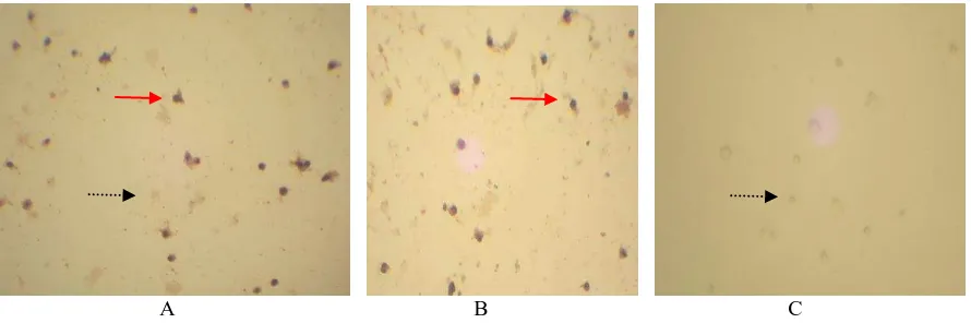
The haemagglutinin role of sub unit pili 49,6 kDa proteins as adhesion protein, was demonstrated using it Immunocytochemistry assay. The sub unit pili 49,6 kDa proteins reacted specifically with mice gastric epithelial cells depending on the amount of protein added, the higher the concentration of protein the stronger the reaction (Fig. 6). This result confirmed that the mice gastric epithelial cells had a specific receptor to the protein or the sub unit pili 49,6 kDa proteins had a specific adhesion molecules to bind the target gastric cells of mice.
A B C
Figure 6. The results of immunocytochemical staining of gastric epithelial cell suspensions of mice were mixed with pili proteins. The positive reactions are indicated by dark brown dots. (A). Gastric epithelial cell with 200µg protein pili, (B). Gastric epithelial cell with 50µg protein pili, and (C).
Gastric epithelial cell with 12,5µg protein pili.
Adhesion index of protein pili of H. pylori in gastric epithelial cells of mice are shown in Table 2.
Table 2. Calculation results of gastric epithelial cells of mice and protein pili of H. pylori (positive) for every 100 field of views. attached by pili proteins showed a darker color.
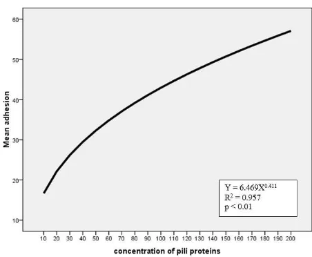
Linear regression statistical test is then performed on the results of this study. Obtained results R2 0,957 with p < 0,01 showed in Figure 7.
Confirmation of adherence Protein Hemagglutinin sub-
unit Pili with MW 49.6 kDa…
www.iosrjournals.org 65 | Page Figure 5. Model Y = aXb. Adhesion index (Y) coefficient (a) Concentration of pili protein (X) attachment rate
(b)
This results shows the significance that the number of pili proteins that attach to gastric epithelial cells is influenced by the number of pili concentration given.
IV.
Discussion
Kaiser (2011) stated that one of the roles of pili of certain bacteria is to allow bacteria to colonize the surface of target cells and are resistant to environmental conditions outside of the attachment. It is known that the initial step of colonization process by H. pylori is their ability to adhere the mucosal surface of gastric epithelial cells. However, information regarding the pili components particularly haemagglutinin and adhesion molecules of the bacteria is limited. In other bacteria, it was reported that a sub unit pili protein of Shigella dysentriae and Salmonella typhi with molecular weight of 49,8 kDA and 48 kDa respectively were hemaglutinin with adhesion properties (Wiwik et al.,, 2012, Sumarno et al., 2012).
In this study, the pathogenic properties associated with a sub unit pili 49,6 kDa protein of H. pylori were investigated. In Western blot analysis, IgG purified from polyclonal antibodies prepared against the protein specifically recognized the similar antigen, indicating that the protein was immunogenic in nature. Confirmation of this protein using haemagglutination assay, showed that the protein consistently agglutinated mouse erythrocytes, suggesting that this protein possessed haemagglutinin properties. Interestingly, a direct reaction between the purified sub unit pili 49,6 kDa protein and mice gastric epithelial cells in immunocytochemical staining was positive, coincided with those of haemagglutination assay. Furthermore, application of the isolated IgG on the pre-treated mice gastric epithelial cells with the purified sub unit pili 49,6 kDa proteins, repealed an inhibition of H. pylori cells to adhere the cells. This further suggested that the sub unit pili 49,6 kDa proteins had a specific adhesion molecules to bind the target gastric cells of mice. The inability of the bacteria to attach the gastric epithelial cells particularly when a high titer of IgG was added, confirming the sub unit pili 49,6 kDa protein was a functional protein that may associated with the pathogenesis of the bacteria.
Sumarno (2011) have found that combinations of protein adhesin sub-unit pili 37.8 kDa V. cholerae with cholera toxin sub-unit B V. cholerae may be can as candidate vaccine of cholera. This results is opened to use as a consideration in developing H. pylori vaccine containing bacterial adhesion molecular component.
V.
Conclusion
In vitro studies using western blotting analysis, hemagglutination test, adherence inhibition assays and immunocytochemical staining repealed that the 49,6 kDa sub unit pili protein of H. pylori was immunogenic. The ability of this protein in agglutinating the mice erythrocytes, indicating it was haemagglutinin in nature. It was also confirmed that the protein possessed adhesion molecules that play a crucial role in the early phase of the pathogenesis H. pylori onto the epithelial cells of gastric. Further study is required to investigate the biological functions of this protein for protecting the infection by this microorganism in causing gastric ulcers.
Acknownledgment
Confirmation of adherence Protein Hemagglutinin sub-
unit Pili with MW 49.6 kDa…
Reference
[1]. Clyne, M., B. Dolan, dan E. P. Reeves. 2007. Bacterial factors that mediate colonization of the stomach and virulence of
Helicobacter pylori. FEMS Microbiol. Lett. 268: 135-143.
[2]. Cover, T. L., dan S. R. Blanke.2005. Helicobacter pylori VacA, a paradigm for toxin multifunctionality. Nat. Rev. Microbiol. 3:320–332 (Abs).
[3]. Groves, F. D., Perez-Perez, G., Zhang, L., Wei-cheng You, Lipsitz, S. R., Gail, M. H., Fraumeni Jr, J. F., danBlaser, M. J.2001. Serum Antibodies to Helicobacter pylori and the CagA Antigen Do Not Explain Differences in the Prevalence of Precancerous Gastric Lesions in Two Chinese Populations with Contrasting Gastric Cancer Rates. Cancer Epidemiol, Biomarkers &prev, 11: 1091-1094.
[4]. Kaiser, G.E. 2011. The Prokaryotic Cell: Bacteria. The Community College of Baltimore Country, Catonsville Campus.
[5]. Laemmli U.K. 1970. Cleavage of Structural proteins during the assembly of the head of bacteriophage T4. Nature227(5259):680-5. [6]. Leite, K. R. M., E. Darini, F. C. Canavez, C. M. de Carvalho, C. A. T. da Silveira Mitteldorf, and L. H. Camara-Lopez. 2005.
Helicobacter pylori and cag A gene detected by polymerase chain reaction in gastric biopsies : correlations with histological findings, proliferation and apoptosis. Sao Paulo Med. J. 123 (3) : 113-118.
[7]. Nagayama, K Oguchi, T Arita, M Honda, T 1995,’Purification and Characterizations of a Cell-Associated Hemagglutinin of Vibrio
parahaemoliticus, Infection and Immunity vol 63, pp 1987-1992.
[8]. Nunes, J. A. 1998. The play of environmental and genetic susceptibility and the differential ecologies of stomach cancer. Center for Social Studies and School of Economics University of Coimbra, Portugal.
[9]. Salyer ,AA & Whitt, DD 2002,’ Bacterial Pathogenesis A Molecular Approach’, ASM Press Washington DC, pp. 115-127.
[10]. Sumarno R.P, Uun Y, Winarsih S, Islam S and Sanarto S, 2012. Detection of molecule adhesion sub-unit pili 48 kDa Salmonella Typhi by immunochemistry method using sera patients suffering from typhoid fever. J. Basic. Appl. Sci. Res., 2(9)8527-8532. [11]. Towbin, H., Stahelin, T. and Gordon, J. 1979. Electrophoretic transfer of protein from polyacrylamide gels to nitrocellulose sheets.
Proc. Nat. Acad. Sci. USA 76:4350-4354.
[12]. Wiwik A, Fitri L.E, Raras T.Y.M, Siswanto B, and Sumarno R.P. 2012. Antibody Protein Hemagglutinin Subunit Pili with MW 49,8 kDa Shigella dysenteriae can inhibit Shigella dysenteriae adhesion on Mice Enterocyte. IOSR Journal of Pharmacy., 2 (5) .13-20.
[13]. Sumarno, R.P., Susanto, A., Ismanoe, G. and Winarsih S. (2011). Combinations of Protein Sub-Unit PILI 37.8 kDa V. Cholerae