http: // www.ijesmr.com
(C)International Journal of Engineering Sciences & Management Research
[16]A NOVEL APPROACH FOR RED BLOOD CELL COUNTING USING FUZZY C-MEANS
SEGMENTATION
Silpa Murukan*, Neenu Suresh
Dept of Computer Science and Engineering, Sree Narayana Gurukulam College of Engineering, M.G University, Kerala
Keywords: Red blood cells, Image segmentation, HSV, Fuzzy C-Means, morphological operations
ABSTRACT
Red blood Cell count is a preliminary process in major applications related to disease diagnosing and medical research. This paper is the study of automated methods of cell counting processed through computational algorithms. HSV and Fuzzy C-Means (at 2nd stage) were efficient for processing of image. The input image of cell is cleaned and morphological operations are optimized to generate the number of red blood cells. Results show the efficiency of algorithm over manual calculations in terms of minimized time frame and equivalent accuracy. The model was developed on virtual platform (MATLAB)..
I.
INTRODUCTION
Blood is the vital element of living species that transports necessary fluids across the nerves and organs of an individual. Blood cells are composed of erythrocytes (red blood cells, RBCs), leukocytes (white blood cells, WBCs) and thrombocytes (platelets). The most abundant small reddish cells are erythrocytes and called red blood cell [1]. The elementary operation of red blood cells is to propagate oxygen across the tissues. The red blood cells than transport the carbon dioxide from the cells because of breaking down the nutrients. An erythrocyte is a disc-shaped cell with a thick rim and a thin sunken center where the nucleus used to be [3]. The normal red blood cell count of a healthy person is divided on basis of age groups by American Cancer Society (2009) [2].
Red blood cells usually exist in ‘hundred-thousand’ range per cubic millimeter. This measurement is made with a microscope and a specially ruled chamber (hemacytometer). Counting RBC in this way is costly and time consuming. The method and mathematical formula for manual computation is given in works of Alaa Hamouda [4]. The automated approaches for cell counting are either image processing or hemocytometer. Hemocytometer is a costly device that counts the blood cells by infringes creation due to cells obstructed in path of light. The cost of this device makes it impossible to get installed at number of source and finance constrained resources hence image segmentation method is broadly researched as an alternate option.
The image segmentation method of red blood cell count is an array of various steps initiating from image preprocessing to final cell count. The input image is captured from a digital device and contains noise due to electrical impulses and device error. Further the cell image has platelets and white blood cells. Finally the image is converted to grey and binary form for counting process. The steps involved for these actions are described by a number of researchers with different methods. In [5], the authors adapted neural network along with Genetic Algorithms (GA) for red blood cell classification for Thalassemia diagnostic tool. Similarly, the authors of [6] studied the usage of different neural network technique for extracting the red blood cell components from microscopic images and classify them using neural networks. Roy A. Dimayuga et. al [8] used the histogram thresholding to distinguish the nucleus of the leukocyte or white blood cells from the rest of the cells in the image. Ramin Soltanzadeh [9] has
purposed feature extraction technique based on morphology in his three blood cell’s experiments. Based on morphology of the
cells, the mass center of each cell in the images and then find the distance of each pixel on an edge from the center. Heidi Berge
[10] has purposed the segmentation red blood cells in a thin blood smear image which is based on the Zack’s Method [11]. This
method is one of the approached to determine the red blood cells thresholding where a line is drawn between the two peaks and between these two peaks, they used the point which is furthest from the drawn line as a threshold for red blood cell. In the conclusion for this technique, the segmentation result is better to the blood smear which in case red blood cells is sparse and in the image. However, in images with high Red blood cell concentrations, large clumps may result and this method is less accurate. Guitao et al. [12] purposed the Hough transform in detecting and extracting the red blood cells in the urine micrograph. Based on Hough transform [7], Guitao has used the geometrical feature to detect the circle center in the image.
In this paper, the input image is pre-processed for noise reduction and saturation is performed for color enhancement. The blood cells are separated from background and based on area, the overlapping and cells at edge of image were taken into consideration. The results show that total number of cells is 81 in the image. The result when compared with manual reading demonstrated more than 95% accuracy and is higher than the counting done by Hough Transform.
http: // www.ijesmr.com
(C)International Journal of Engineering Sciences & Management Research
[17]II.
I
MAGEP
ROCESSINGT
OOLSImage Acquisition
The first stage of any vision system is the image acquisition stage. After the image has been obtained, various methods of processing can be applied to the image to perform the many different vision tasks required today. However, if the image has not been acquired satisfactorily then the intended tasks may not be achievable, even with the aid of some form of image enhancement.
Median Filtering
Median filtering is a noise reduction tool and is efficient for impulsive and pepper & salt noise cases. It also preserves edges in an image while reducing random noise. Impulsive or salt-and pepper noise can occur due to a random bit error in a communication channel. In a median filter, a window slides along the image, and the median intensity value of the pixels within the window becomes the output intensity of the pixel being processed.
Let { �} is the m-directional sequence with index � ∈ . A sliding window ∈ has �= { �+�: � ∈ } at position �.
The erosion of the colour image A by the structuring element B is defined by: ⊝ = {���| �⊆ } (3)
Where, � is the translation of B by the vector z, i.e. �= {� + �|� ∈ }, ∀�∈ � (4)
When the structuring element B has a centre (e.g., B is a disk or a square), and this centre is located on the origin of E, t hen the erosion of A by B can be understood as the locus of points reached by the centre of B when B moves inside A. For example, the erosion of a square of side 10, centred at the origin, by a disc of radius 2, also centred at the origin, is a square of side 6 centred at the origin.
The erosion of A by B is also given by the expression:
⊝ = ∩� ∈ −� (5)
HSV Transformation
In all of the colour spaces, the HSV colour space was been chosen in this paper due to the three advantages as follows: first ly, it is closer to human perception, secondly it is user-oriented and thirdly, it gives better colour conversion accuracy [12].
The HSV model consists of three components: hue, saturation and value. The hue component H, of which the range is from 0 to 360 degree, denotes the spectral composition of colour, such as the green primary at 120 degree. The saturation component S defines the relative purity of the colour, which ranges from 0 to 1.0. It indicates how much white light dilutes a pure colour. The larger the S value is, the purer the colour is. The value component V refers the luminance value of the colour, which also varies from 0 to 1, namely, from black to white. Larger V value means brighter colour.
The precise transformation between RGB and HSV colour space is reference in [12].
Fuzzy C-Means clustering
In fuzzy clustering approach, the pixels belong to degree of freedom contradicting with property of fuzzy logic. Any point x has a set of coefficients giving the degree of being in the ��ℎ cluster � . With fuzzy c-means, the centroid of a cluster is the mean of all points, weighted by their degree of belonging to the cluster [14].
Fuzzy clustering is a healthy approach as it considers the cell particles at the edge of image. Proposed by Bezdek in 1981 [14] it is an indispensible tool in this century for clustering of objects.
http: // www.ijesmr.com
(C)International Journal of Engineering Sciences & Management Research
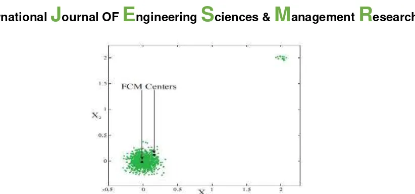
[18]Figure.1: Illustrative representation of Cluster centers obtained with the FCM algorithm for data with two groups. The larger and the smaller groups have 1000 and 15 points, respectively.
The fuzzy clustering separates the red blood cells from the background pixels. The importance of this approach lies in the fact that at digital scale the colour values of pixels may match for cells and background.
A 2nd level fuzzy clustering is applied at image to unrelated the red blood cells with white blood cells (blue cells in figure 3).
Morphological Operations
Morphology is a technique of image processing based on shape and form of objects. Morphological methods apply a structuring element to an input image, creating an output image at the same size. The value of each pixel in the input image is based on a comparison of the corresponding pixel in the input image with its neighbours. By choosing the size and shape of the neighbour, you can construct a morphological operation that is sensitive to specific shapes in the input image. The morphological operat ions can first be defined on colour images where the source image is planar.
The morphological operations are divided in four segments:
Erosion: It is the process of colour enhancement and highlights the red blood cells of given image.
Dilation: Change a background pixel to foreground if it has a foreground pixel as a 4-neighbor.
Opening: Eliminates all pixels in regions that are too small to contain the structuring element.
Closing: Consists of a dilation followed by erosion and can be used to fill in holes and small gaps.
III.
P
ROPOSEDM
ETHODOLOGYThe input image for this analysis is a raw image imported from an open source. Figure 3 is the original image of a blood sample. The blue portion in the center is white blood cells. The pre-processing of image is processed through median filter for noise reduction. The enrichment of color is gained through erosion method. The image is saturated by HSV transformation to highlight the red color in this image. Fuzzy segmentation separates the red and blue color from background. 2nd level fuzzy c-means
separates the red color from blue color. The image is converted to grey and binary form for computational ease in counting process. As the dilation process of morphological operations differentiates the background and foreground on basis of image pixels, grey and binary conversion clips the possibilities of confusion in this step. Morphological operations classify the blood cells from each other and differentiate from each other and also consider the cells at edge based on proposed method. The counting is offered via simple formula at MATLAB platform.
http: // www.ijesmr.com
(C)International Journal of Engineering Sciences & Management Research
[19]Figure 2: Flowchart of RBC Counting Process
IV.
R
ESULTSThe steps of proposed algorithm yields results followed in this section. The original image is a digital image captured with microscopic device attached. The following results were obtained via simulation of proposed architecture.
Figure 3: Original Image of Red blood cells obtained, representing various elements of blood
The image taken for the research is of a healthy individual and shows the red blood cells (red circles), white blood cells (blue part of image) and noise.
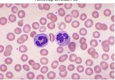
Figure 4: The image is filtered with a median filter as the stage of pre-processing
The noise reduction of image however cannot be visualized in this figure. It may be possible that error is too small to be filtered as we did not present any special mark for such.
Original Image
Filtered Image with Median Filter
Image Pre-Processing
Fuzzy clustering for segmentation
Cell differentiation by
Morphological Operations
http: // www.ijesmr.com
(C)International Journal of Engineering Sciences & Management Research
[20]Figure 5: The conversion of original image as the Eroded image and HSV for color enhancement
The color enhancement of denoised image leaves the cells with small holes (vacant point of cells) and enhanced cell circles. The visible white circles in previous image are shrunk and cell gained their density. The color of cells in the image was highlighted using HSV color transform.
Figure 6: Calculation of Saturation component using fuzzy clustering algorithm
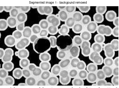
Figure 7: Image with removed background of a blood cell after saturation
The saturation of image clipped the background from the image. Only white and red blood cells are visible. Eroded Image
Saturation Component
http: // www.ijesmr.com
(C)International Journal of Engineering Sciences & Management Research
[21]Figure 8: Removal of white blood cells and platelets as second stage of saturation Second level of clustering discards the white blood cells and platelets in image.
Figure 9: Conversion of a saturated image into binary image
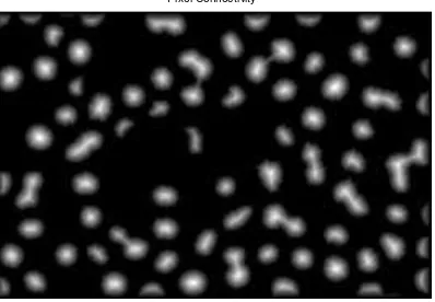
Figure 10: Connectivity of Pixels through centroid calculation of cells
Segmented image 2 - RBC only
Binary Image After Morphological Operations
http: // www.ijesmr.com
(C)International Journal of Engineering Sciences & Management Research
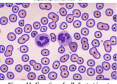
[22]Figure 11: Counting of cells for test input
Morphological operations (four step process) is applied for two purposes. Based on the area of cells, the cell regions were marked. This was essential as many cells were overlapped and needed to be classified. Another, at the edge of images, the operations determines whether the cells should be considered in counting or not. Figure 11 shows that the cells that have holes are considered for counting and rest are discarded. Also the cells that were overlapped not completely are taken into account for counting. In comparison with the Hough Transformation for cell counting [2], the manual observations of both the results demonstrate that Hough Transform did not efficiently consider the cells at edge of image. Many cells were dropped in Hough transform that should have been considered. This comparison is done at manual basis as the input image for both the researches were different and also the environments of experimentations were distinct. We do not compare our technique with K-means presented by Alaa Hamouda [4] as the image considered in that research did not had any cells at boundary of image. Compared with manual counting, the proposed algorithm showed more than 95% accuracy and least amount of time.
V.
C
ONCLUSIONThe method present for red blood cell counting is an efficient approach in its segment. The red blood cells were differentiated from the white blood cells, noise and background of image that enhanced the accuracy of architecture. The pre-processing of input image clears the image for segmentation and discards the white blood cells and platelets based on number accountability and size of cells respectively. Also in comparison with Hough Transform, it is found that Morphological operations are superior as they consider the cells at boundary of image and overlapping of cells.
Since diseases and health of bio-degradable objects are variable in nature and with elapse of time new terminologies are surfaced, the research in this field is a non-ending process and will continue for a long time. Also with new discoveries, new methods will be innovated with different properties and parameters of evaluation. The blood cell count in this research is only constrained to red blood cells, but white blood cells and platelets are seemingly having same importance. And also the foreign objects in blood that adverse the nature of normal blood also comes under the scope of this subject.
VI.
REFERENCES
[1] K.S. Saladin, Anatomy and Physiology: The Unity of Form and Function, McGraw-Hill, NY, 4th, chap. 18, pp.680-696, 2007 [2] Mahmood, N. H., & Mansor, M. A. (2012). Red Blood Cells Estimation Using Hough Transform Technique. Signal & Image Processing: An International Journal (SIPIJ), 3(2), 53-64
[3] Walsh, D., & Raftery, A. E. (2002). Accurate and Efficient Curve Detection in Images: The Importance Sampling Hough Transform. Pattern Recognition, 35(7), 1421-1431.
[4] Hamouda, A., Khedr, A. Y., & Ramadan, R. A. (2012), Automated Red Blood Cell Counting. Int. J. Comput. Sci, 1
[5] W. Wongseree and N. Chaiyaratana, “Thalassemic Patient Classification Using a Neural Network and Genetic Programming,” IEEE, pp. 2926-2931, 2003
[6] J. Poomcokrak1 and C. Neatpisarnvanit, Red Blood Cells Extraction and Counting, The 3rd International Symposium on Biomedical Engineering (ISBME 2008)
[7] Tahir Rabbani and Frank van den Heuvel, Efficient Hough Transform for Automatic Detection of Cylinders in Point Clouds. In Proceedings of the 11th Annual Conference of the Advanced School for Computing and Imaging (ASCI '05), The Netherlands, June 2005
[8] Roy A. Dimayuga, Gerwin T. Ong, Rainier Carlo S. Perez, Gefferson O. Siy, Saman C. Sohrabi Langroudi and Miguel O.Gutierrez, Leukemia Detection Using Digital Image Processing in Matlab. ECE Student Forum, De La Salle University, Manila. March 26, 2010
http: // www.ijesmr.com
(C)International Journal of Engineering Sciences & Management Research
[23] [9] Ramin Soltanzadeh, Classification of Three Types of Red Blood Cells in Peripheral Blood Smear Based on Morpho logy. Proceedings of ICSP, 2010[10] Heidi Berge, Dale Taylor, Sriram Krishnan, and Tania S. Douglas, Improved Red Blood Cell Counting in Thin Blood Smears. Proceedings of ISBI, 2011. pp.204-207
[11] Zack G.W., Rogers W.E. and Latt S.A, Automatic-Measurement of Sister Chromatid Exchange Frequency. Journal of Histochemistry & Cytochemistry 25, 1977, 741-753
[12] Guitao Cao, Cai Zhong,Ling Li and Jun Dong, Detection of Red Blood Cell in Urine Micrograph. The 3rd International Conference on Bioinformatics and Biomedical Engineering (ICBBE). 2009
[13] Melkamu Hunegnaw Asmare, Vijanth Sagayan Asirvadam, and Lila Iznita Izhar, Color Space Selection for Color Image Enhancement Applications. ICSAP, 2009, pp. 208-212
[14] G. Pai, Empirical analysis of Software Fault Content and Fault Proneness Using Bayesian Methods. IEEE Transactions on software Engineering, 33(10), pp. 675-686, 2007
[15]Blood cell histology-http://www.unomaha.edu/hpa/blood.html