Hydrolytic enzyme activity of Paenibacillus sp. strain
B2 and effects of the antagonistic bacterium on cell
integrity of two soil-borne pathogenic fungi
S.W. Budi
1, D. van Tuinen, C. Arnould, E. Dumas-Gaudot,
V. Gianinazzi-Pearson, S. Gianinazzi
∗UMR INRA/Universite de Bourgogne BBCE-IPM, Centre de Microbiologie du Sol et de l’Environnement, INRA, BV 1540, 21034 Dijon Cedex, France
Received 31 May 1999; received in revised form 1 December 1999; accepted 23 March 2000
Abstract
Paenibacillus sp. strain B2, isolated from the mycorrhizosphere of Sorghum bicolor and having an antagonistic activity
towards soil-borne fungal pathogens, possessed extracellular cellulolytic, proteolytic, chitinolytic and pectinolytic enzyme activities. The eventual role of these lytic enzymes in cellular interactions between Paenibacillus sp. strain B2 and Phytophthora
parasitica and Fusarium oxysporum was investigated by electron microscopy and molecular cytology. Electron microscopic
observations showed that the presence of Paenibacillus sp. strain B2 resulted in disorganisation of cell walls and/or cell contents of P. parasitica and F. oxysporum. However, when P. parasitica was treated with commercial purified cellulase, protease, chitinase and pectinase, only protease had an inhibitory effect on mycelial growth. It is proposed that the inhibitory effect of Paenibacillus sp. strain B2 on the growth of soil-borne fungal pathogens is probably derived from more than one mechanism. © 2000 Elsevier Science B.V. All rights reserved.
Keywords: Hydrolytic enzymes; Paenibacillus sp. strain B2; Cell integrity; Wall metabolism; Phytophthora parasitica; Fusarium oxysporum
1. Introduction
Pesticides applied to circumvent damage caused by pathogens and insects in order to increase plant pro-duction are a major chemical input in modern agricul-ture. However, their excessive use has led to problems
∗Corresponding author. Tel.:
+33-3-80-69-31-46, fax:+33-3-80-69-32-46.
E-mail addresses: vivienne.gianinazzi-pearson@epoisses.
inra.fr (V. Gianinazzi-Pearson),
[email protected] (S. Gianinazzi)
1Present address: Sylviculture Laboratory, Department of Forest Management, Faculty of Forestry, Bogor Agricultural University, Campus Jaranaga, PO Box 168, Bogor 6600, Indonesia.
of environmental degradation and pollution. Such chemicals can, for example, be lethal to useful soil insects and to key micro-organisms in the rhizosphere (e.g. mycorrhizal fungi), and they may also enter the food chain. Moreover, there are increasing examples where their efficiency is decreased due to the devel-opment of resistant pathogens (Rosenberger, 1991). The potential of biological control of soil-borne plant pathogens by antagonistic micro-organisms (Dunn et al., 1997) offers a non-polluting complement, or alter-native, to existing disease management strategies that depend heavily on chemical pesticides. However, the use of antagonistic micro-organisms in agricultural practices should be compatible with the persistence
and function of symbiotic mycorrhizal fungi which play a central role in plant health and survival (Paulitz and Linderman, 1989; Barea et al., 1998).
We have isolated previously a gram positive bac-terium Paenibacillus sp. strain B2 from the myc-orrhizosphere of Sorghum bicolor inoculated with surface sterilised sporocarps of Glomus mosseae, and demonstrated that although it possesses a broad spec-trum of antagonistic activity against many important pathogenic fungi, it has no detrimental effect on AM fungi in vitro and in vivo. In particular, it inhibits in vitro sporangia production, germination and hyphal growth of zoosporangia of Phytophthora parasitica and reduces root necrosis caused by P. parasitica in tomato seedlings (Budi et al., 1999).
A variety of mechanisms have been reported to contribute to the biocontrol activity of microbes and it is, for example, known that cell wall degrading enzymes, such as b-1,3-glucanases, cellulases, pro-teases and chitinases are involved in the antagonistic activity of some biological control agents against phy-topathogenic fungi (Ordentlich et al., 1988; Shapira et al., 1989; Harman et al., 1993; Chernin et al., 1995; Dunn et al., 1997). Understanding the mode of action of biocontrol agents is a prerequisite for: (i) developing rational procedures in order to select more effective antagonistic microbial strains, (ii) develop-ing appropriate production and formulation methods that enhance biocontrol activity, and (iii) fulfilling some requirements of the toxicological and registra-tion packages needed for commercial development (Jijakli and Lepoivre, 1998).
In this paper, we describe the ability of the isolated Paenibacillus sp. strain B2 to produce hydrolytic en-zymes that may contribute to its antagonistic activity, and the effect of the bacterium on cell integrity of two pathogenic fungi P. parasitica and Fusarium oxysporum.
2. Material and methods
2.1. Micro-organisms and culture conditions
Eight isolates of bacteria were isolated originally from an in vitro pot culture of Sorghum bicolor L. inoculated with surface disinfected sporocarps of Glo-mus mosseae BEG12 (Budi et al., 1999). The isolates
were kept on Luria Broth (LB) media supplemented with 10% glycerol at −80◦C. When needed, they were transferred to LB agar media plates and grown overnight at 37◦C.
2.2. Plate assays for hydrolytic (cellulolytic, pectinolytic, proteolytic and chitinolytic) activities
Bacterial isolates were grown in Erlenmeyer flasks for 24 h on a culture shaker at 30◦C in LB buffered medium. Ten microlitres of each bacterial suspension 10−3–18−8CFU/m/l was spotted onto plates contain-ing the substrate of the enzyme to be tested and grown at 25◦C for 48 h.
Cellulolytic activity was assessed as described by Teather and Wood (1982) using a solid medium, con-taining MgSO4·7H2O (0.1 g/l), CaCl2·2H2O (0.2 g/l), FeSO4·7H2O (0.04 g/l), NaCl (0.2 g/l), KH2PO4 (0.3 g/l), K2HPO4 (0.5 g/l), CMC (carboxymethyl-cellulose) (Sigma) (5 g/l), yeast extract (0.1 g/l) and Bacto agar (15 g/l). For visualisation of b-d-glucan
hydrolysis, the agar medium containing CMC was flooded with an aqueous solution of congo red (1 mg/ml) for 15 min.
Pectinolytic activity was studied as described by Hankin et al. (1971). The reaction medium contained (NH4)2SO4 (2.0 g/l), KH2PO4 (4 g/l), Na2HPO4 (6 g/l), FeSO4·7H2O (1 mg/l), MgSO4(0.2 g/l), CaCl2 (1 mg/l), H3BO3(10mg/l), MnSO4(10mg/l), ZnSO4 (70mg/l), CuSO4 (50mg/l), MoO3 (10mg/l), apple pectin (Sigma) (5 g/l), yeast extract (1 g/l) and Bacto agar (15 g/l). Pectinolytic activity was revealed by flooding plates for 10 min with a 1% solution of hex-adecyl trimethyl ammonium bromide in water. En-zyme activities were identified by the development of a zone of clearing (halo) around the bacterial colonies. Proteolytic activity was performed in an agar medium containing skimmed milk (Difco) (100 g/l), yeast extract (1.5 g/l) and Bacto agar (15 g/l) accord-ing to Dunn et al. (1997) and visualised directly on the plates after 48 h. Chitinolytic activity was revealed according to the method of O’Brien and Colwell (1987). Bacteria were grown on LB agar medium for 24–48 h. One to five colonies from culture plates were blotted vigorously onto Whatman No. 1 filter paper and 20ml of the 4-methylumbelliferyl-N-acetyl-b-d-glucosaminide-buffered substrate solution was
bacteria plus the solvent. After incubation at 37◦C for 10 min, each test spot was covered with a drop of a saturated sodium bicarbonate solution and ex-posed to UV light. The reactions were graded as positive, weakly positive or negative. In positive reac-tions, substrate–bacteria mixtures produced a strong light-blue fluorescence.
2.3. Susceptibility of P. parasitica to commercial cellulase, protease, chitinase and pectinase
The effect of commercial hydrolytic enzymes on P. parasitica growth was studied in vitro. Agar plugs (5 mm in diameter) of P. parasitica were taken from the periphery of growing mycelia maintained on malt agar medium and incubated in sterile water (con-trol), cellulase from Aspergillus niger (Sigma EC 3.2.1.4, 1.0 U/ml), proteinase K from Tritirachium al-bum (Boehringer, 1.0 U/ml), chitinase from Serratia marcescens (Sigma EC 3.2.1.14, 1.0 U/ml) and pecti-nase from A. niger (Merck EC 3.2.1.15, 1.0 U/ml) for 24 or 48 h at 24◦C. The plugs were then rinsed with sterile water and transferred to malt agar medium. Fungal growth was measured as colony diameter after incubating plates for 5 days at 24◦C.
2.4. Ultrastructural observations
Phytophthora parasitica (syn. P. parasitica B. de Haan var. parasitica (Dastur) Waterh) isolate 201 (kindly provided by P. Bonnet, INRA, Antibes, France) and F. oxysporum isolate Foeu 1 (kindly provided by G. Lori, La Plata University, Argentina) were used. Agar plugs of P. parasitica and F. oxys-porum cultures were placed on nutrient agar either containing or not containing CMC (5 g/l) or ap-ple pectin (5 g/l), and in the presence or not in the presence of drops of a suspension of Paenibacillus sp. strain B2. The cultures were incubated in the dark at 27◦C for 3 days. Mycelia were then sampled at the fungal colony margin adjacent to the bacterium, fixed overnight at 4◦C in 2% glutaraldehyde buffered in 0.1 M Pipes (pH 7.2), and washed four times in Pipes buffer. Half of the samples of each treatment was post-fixed 1 h in 1% osmium tetroxide. All sam-ples were subsequently dehydrated through a graded ethanol series, and embedded in Epon or LR White medium grade resin as described by Gianinazzi and
Gianinazzi-Pearson (1992). Ultrathin sections of 85–95 nm were cut onto carbon-coated gold grids using a Reicher Ultracut microtome. For ultrastruc-tural observations, sections of osmium tetroxide-fixed, epon-embedded samples were subjected to the periodic acid-thiocarbohydrazide-silver proteinate (PATAg) reaction to detectb(1-4) andb(1-6) glucans (Thiéry, 1967). An indirect immunogold labelling technique was carried out to localize b(1-3) glu-cans. For this, LR White embedded sections were incubated overnight at 4◦C with a mouse mono-clonal antibody raised againstb(1-3) glucopyranose polymers (Biosupplies Australia, Australia) and di-luted 1:50 in saline Tris-buffer containing 1% bovine serum albumin. After washing, sections were in-cubated 1 h in a gold-labelled (15 nm) polyclonal goat antimouse antibody. Immunological controls were performed by omitting the primary antibodies. N-acetyl glucosamine residues (chitin) were localised in F. oxysporum mycelium by incubating LR White embedded sections 1 h at room temperature in 10 nm gold-labelled wheat germ agglutinin lectin (WGA, Sigma) diluted 1:50. All treated sections were coun-terstained for 10 min with 2% aqueous uranyl acetate before being observed using a Hitachi 600 electron transmission microscope at 75 kV.
2.5. Statistical analysis
In vitro experiments were performed with four replicates and repeated twice. Data were analysed by one-way ANOVA and Newman–Keuls test at a probability of p<0.05.
3. Results
3.1. Plate assays for hydrolytic activities
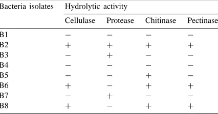
Table 1
Presence (+) or absence (−) of cellulolytic, proteolytic, chitinolytic and pectinolytic activity in different strains of bacteria isolated from the mycorrhizosphere of Sorghum bicolor
Bacteria isolates Hydrolytic activity
incubation at 25◦C). The other isolates B6 and B8 which did not display any antagonistic activity to-wards soil-borne fungal pathogens (Budi et al., 1999) also produced cellulolytic and pectinolytic activities revealed by the formation of clear zones of 17 and 20 mm diameter for cellulolytic activity and 12 and 22 mm diameter for pectinolytic activity, respectively. No proteolytic activity was observed for these two isolates. The two other isolates B3 and B7, which have a very weak antagonistic activity (Budi et al., 1999), also produced proteolytic activity with the formation of clear zones of 14 and 15 mm diameter, respectively. No cellulolytic and pectinolytic activities were ob-served for these two isolates. The chitinolytic activity was detected under UV light as a strong light-blue fluorescence for Paenibacillus sp. strain B2 and a weak light-blue fluorescence for isolates B6 and B8. No fluorescence was observed for isolates B3 and B7.
3.2. Effect of commercial hydrolytic enzymes on P. parasitica
The effect of commercial hydrolytic enzymes on P. parasitica mycelial growth are shown in Table 2. No effects were observed on the growth of P. parasit-ica up to 48 h of incubation in cellulase, chitinase or pectinase, but protease inhibited mycelial extension by approximately 25%.
3.3. Ultrastructural observations
In the absence of Paenibacillus sp. strain B2, hyphae of P. parasitica taken at colony margins had active cell
Table 2
Effect of commercial hydrolytic enzymes on P. parasitica mycelial growth assessed 5 days after immersion for 24 or 48 h in the solutions
Treatment Mycelial growth (cm)a
24 h 48 h
aValues followed by the same letter in each column do not differ significantly from each other (p<0.05).
contents characterised by large individual vacuoles, a polysome-rich cytoplasm and numerous organelles (Figs. 1 and 3). Hyphal cells were delimited by a thin, electron-translucent wall (Fig. 1), the cellulosic com-ponent of which was strongly stained by the PATAg reaction (Fig. 3). Immunolabelling ofb(1-3) glucans
Fig. 1. Transmission electron micrographs of hyphae of P.
para-sitica grown in the absence of Paenibacillus sp. strain B2.
Fig. 2. Transmission electron micrographs of hyphae of P.
parasit-ica grown in the presence of Paenibacillus sp. strain B2. Sections
were contrasted with uranyl acetate. Fungal cell contents are ex-tremely electron dense (arrow) in the presence of the bacterium and paramural deformations (arrowheads) develop into the cyto-plasm. Bar=2mm.
Fig. 3. Transmission electron micrographs of hyphae of P.
para-sitica grown in the absence of Paenibacillus sp. strain B2.
Sec-tions were stained by the PATAg reaction. The fungal wall reacts strongly to the PATAg reaction (arrowhead) and the cytoplasm is rich in mitochondria (m) and vacuoles (v). Bar=2mm.
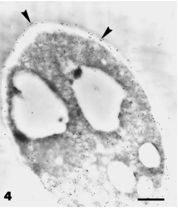
Fig. 4. Transmission electron micrographs of hyphae of P.
parasit-ica grown in the absence of Paenibacillus sp. strain B2. Sections
were stained by the immunogold labelled withb(1-3) antibod-ies: Gold particles (arrowheads) indicate the presence of ab(1-3) glucan wall component. Bar=2mm.
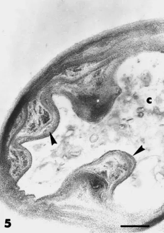
was associated with the cell wall (Fig. 4). Hyphal extension was inhibited about 70% by the presence of Paenibacillus sp. strain B2 (see also Budi et al., 1999). When mycelium of P. parasitica was sampled in apical regions, where contact between the micro-organisms had not occurred, hyphal contents showed different degrees of alteration. In some hyphae, the cytoplasm was extremely electron-dense, vacuoles were hardly visible and large lipid droplets accumulated (Fig. 2), whilst in others, cell contents were completely de-graded (Fig. 5). The wall of most hyphae was distorted and paramural deformations developed intensely into the fungal cell (Figs. 2 and 5). Like the normal cell wall, these wall proliferations were PATAg-positive and containedb(1-3) glucans (Figs. 5 and 6).
Fig. 5. Transmission electron micrographs of hyphae of P.
parasit-ica grown in the presence of Paenibacillus sp. strain B2. Sections
were stained by the PATAg reaction. PATAg reactive wall prolif-erations (arrowheads) protrude into the degraded cell lumen (c). Bar=0.5mm.
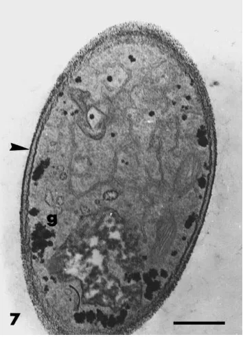
cases, cytoplasmic contents were dense, and rich in organelles and membrane systems, with vacuolisation occurring only in older parts (not shown). The outer part of the fungal wall stained with the PATAg reac-tion (Fig. 7) and WGA labelling indicated the presence of chitin (Fig. 8). Glycogen granules were present in most cells of F. oxysporum growing in bacteria-free conditions (Fig. 7) but they were less abundant or absent in hyphae growing in the presence of Paeni-bacillus sp. strain B2 (Fig. 9). A consistent modifica-tion induced by the bacterium was the deposimodifica-tion of polymorphic vesicle inclusions in an amorphous ma-trix between the cell wall and the fungal protoplast (Figs. 9 and 10). In these cells, the wall was distorted and the plasma membrane was retracted and convo-luted around the inclusions. This material, easily dis-tinguished from the cell wall by its lack of reaction to PATAg (Fig. 9), was labelled intensely with WGA in-dicating its cell wall origin (Fig. 10). These different alterations in the cell integrity and wall organisation
Fig. 6. Transmission electron micrographs of hyphae of P.
para-sitica grown in the presence of Paenibacillus sp. strain B2.
Sec-tions were immunogold labelled with b(1-3) antibodies. b(1-3) glucans (arrowheads) are located in the paramural wall deposits. Bar=0.5mm.
of P. parasitica and F. oxysporum were induced sim-ilarly by Paenibacillus sp. strain B2 in the presence and in the absence of cellulose or pectin in the nutrient medium.
4. Discussion
Fig. 7. Transmission electron micrographs of hyphae of F.
oxyspo-rum grown in the absence of Paenibacillus sp. strain B2. Sections
were stained by the PATAg reaction. The outer fungal wall (arrow-head) reacts strongly to the PATAg reaction. The cytoplasm is rich in glycogen (g), organelles and membrane systems. Bar=0.5mm.
The electron microscope investigations from this study revealed that growth inhibition of P. parasitica and of F. oxysporum induced in the presence of the bacterium is accompanied by marked cellular changes in the two fungi. Effects on P. parasitica were much more pronounced with rapid disorganisation of hyphal contents and cell death. However, bacteriinduced al-terations in both fungi were associated with the depo-sition of material on the inner side of the hyphal wall. The occurrence of cell wall components in these ab-normal deposits raises the question of why and how the fungi react in reorganising wall material in such an un-usual way. Disturbances in the regulation of enzymes involved in the biosynthesis of wall compounds may explain deposition of wall polymers at sites where this does not normally occur. The cellulolytic and chiti-nolytic activity of Paenibacillus sp. strain B2 could affect the structural integrity of the walls of P. par-asitica and of F. oxysporum, respectively, leading to the release of molecules which may be responsible
Fig. 8. Transmission electron micrographs of hyphae of F.
oxyspo-rum grown in the absence of Paenibacillus sp. strain B2. Sections
were lectin-gold labelled with wheat germ agglutinin. Scattered gold particles (arrowheads) indicate the presence of chitin in the fungal wall. Bar=0.2mm.
Fig. 9. Transmission electron micrographs of hyphae of F.
oxys-porum grown in the presence of Paenibacillus sp. strain B2.
Sec-tions were stained by the PATAg reaction. A PATAg non-reactive paramural matrix with vesicle inclusions (arrowhead) is bordered by a retracted plasma membrane (arrow). The cytoplasm is rich in mitochondria (m) and membrane systems. Bar=0.5mm.
Fig. 10. Transmission electron micrographs of hyphae of F.
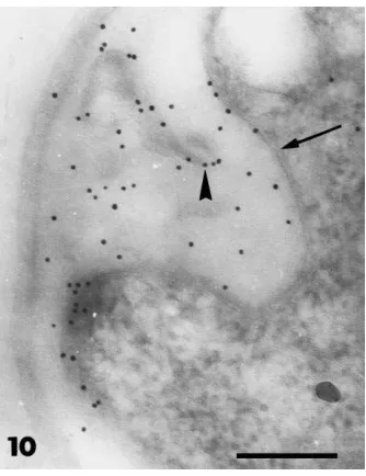
oxys-porum grown in the presence of Paenibacillus sp. strain B2.
Sec-tions were lectin-gold labelled with wheat germ agglutinin. The matrix between the fungal wall and the retracted plasma mem-brane (arrow) is labelled for chitin (arrowhead). Bar=0.2mm.
Acknowledgements
We are grateful to the Center Grand Program, Di-rectorate General of Education, Republic of Indone-sia and the INCO-DC EU Project No. ERBIC18 CT 970180 for supporting this work. The authors thank Denise Dubois for technical assistance.
References
Barea, J.M., Andrade, G., Bianciotto, V., Dowling, D., Lohrke, S., Bonfante, P., O’Gara, F., Azcon-Aguilar, C., 1998. Impact on arbuscular mycorrhiza formation of Pseudomonas strains used as inoculants for biocontrol of soil-borne fungal plant pathogens. Appl. Environ. Microbiol. 64, 2304–2307. Benhamou, N., 1992. Ultrastructural and cytochemical aspects of
chitosan on Fusarium oxysporum f. sp. radicis-lycopersici, agent of tomato crown and root rot. Phytopathology 82, 1185– 1193.
Budi, S.W., van Tuinen, D., Martinotti, D., Gianinazzi, S., 1999. Isolation from the mycorrhizosphere of Sorghum bicolor of a bacterium compatible with arbuscular mycorrhizal development and antagonist against soil-borne fungal pathogens. Appl. Environ. Microbiol. 65, 5148–5150.
Chernin, L., Ismailov, Z., Haran, S., Chet, I., 1995. Chitinolytic
Enterobacter agglomerans antagonistic to fungal plant pathogens. Appl. Environ. Microbiol. 61, 1720–1726. Dunn, C., Crowley, J.J., Moënne-Loccoz, Y., Dowling, D.N., de
Bruijn, F.J., O’Gara, F., 1997. Biological control of Pythium
ultinum by Stenotrophomonas maltophilia W18 is mediated by
an extracellular proteolytic activity. Microbiology 143, 3921– 3931.
El Gaouth, A., Arul, J., Grenier, J., Benhamou, N., Asselin, A., Bélanger, R., 1994. Effect of chitosan on cucumber plants: suppression of Pythium aphanidermatum and induction of defense reactions. Phytopathology 84, 313–320.
Fuller, M.S., Roberson, R.W., Gisi, V., 1990. Effects of the sterol demethylase inhibitor, cyproconazole, on hyphal tip cells of Sclerotium rolfsii. Pest. Biochem. Physiol. 36, 115– 126.
Gianinazzi, S., Gianinazzi-Pearson, V., 1992. Cytology, histo-chemistry and immunohisto-chemistry as tools for studying structure and function in endomycorrhiza. In: Norris, J.R., Read, D.J., Varma, A.K. (Eds.), Methods in Microbiology, Vol. 24. Academic Press, New York, pp. 109–139.
Hankin, L., Zucker, M., Sand, D.C., 1971. Improved solid medium for the detection and enumeration of pectolytic bacteria. Appl. Microbiol. 22, 205–209.
Harman, G.E., Hayes, C.K., Lorito, M., Broadway, R.M., Di Pietro, A., Peterbaues, C., Tronsmo, A., 1993. Chitinolytic enzymes of Trichoderma harzianum: purification of chitobiosidase and endochitinase. Phytopathology 83, 313–318.
Jijakli, H.M., Lepoivre, P., 1998. Characterization of an
antago-nist of Botrytis cinerea on apples. Phytopathology 88, 335– 343.
O’Brien, M., Colwell, R.T., 1987. A rapid test for chitinase activity that uses 4-methylumbelliferil-N-acetyl-b-d-glucosaminide. Appl. Environ. Microbiol. 53, 1718–1720.
Ordentlich, A., Elad, Y., Chet, I., 1988. The role of chitinase of Serratia marcescens in biocontrol of Sclerotium rolfsii. Phytopathology 78, 48–88.
Paulitz, T.C., Linderman, R.G., 1989. Interactions between fluorescent pseudomonads and VA mycorrhizal fungi. New Phytol. 113, 37–45.
Rosenberger, R.G., 1991. Postharvest diseases (blue mold–grey mold). In: Jones, A.L., Aldwinckle, H.S. (Eds.), Compendium
of Apple and Pear Diseases. The American Phytopathological Society, St. Paul, MN, pp. 55–58.
Shapira, R., Ordentlich, A., Chet, I., Openheim, A.B., 1989. Cont-rol of plant diseases by chitinase expressed from cloned DNA in Escherichia coli. Phytopathology 79, 1246– 1249.
Teather, R.N., Wood, P.J., 1982. Use of congo red-polysaccharide interactions in enumeration and characterization of cellulolytic bacteria from the bovine rumen. Appl. Environ. Microbiol. 43, 777–780.