Brain Research 884 (2000) 192–195
www.elsevier.com / locate / bres
Short communication
Effect of 6-fluoro-m-tyrosine on dopamine release and metabolism in
rat striatum using in vivo microdialysis
*
Thor D. Stein, Onofre T. DeJesus
Department of Medical Physics, University of Wisconsin Medical School, 1530 Medical Sciences Center, 1300 University Avenue, Madison,
WI53706, USA
Accepted 29 August 2000
Abstract
18
6-[ F]Fluoro-m-tyrosine (FMT) is a positron emission tomography (PET) imaging agent for the aromaticL-amino acid decarboxylase
enzyme. Its parent compound,L-m-tyrosine (LMT) induces behavioral effects in rodents via dopamine release. To assess the potential
pharmacologic effect of FMT, its role in dopamine release and metabolism in rat striatum was compared with LMT andL-DOPA using in
vivo microdialysis. Results indicate that FMT will not have the same dopamine-induced behavioral effects as LMT. 2000 Elsevier Science B.V. All rights reserved.
Theme: Neurotransmitters, modulators, transporters and receptors
Topic: Catecholamines
Keywords: 6-Fluoro-m-tyrosine; In vivo microdialysis; Dopamine release
L-m-Tyrosine (LMT) is a monohydroxy analog of 3, an indirect agonist effect whereby MTA is thought to enter
4-dihydroxyphenylalanine (L-DOPA) which initially drew storage vesicles inducing the displacement and release of
interest as a therapeutic agent for Parkinson’s disease (PD) stored dopamine into the synapse.
because of its anti-reserpine activity. The pharmacologic Recently, a fluorinated m-tyrosine analog,
6-fluoro-m-actions of LMT included (1) an awakening effect on tyrosine (FMT), was suggested and developed as an
reserpinized mice [2], (2) protective effects on reserpine- imaging agent to assess monoamine systems using positron
induced depletion of catecholamine stores [5], (3) reversal emission tomography (PET) [7,10]. FMT is attractive as a
of reserpine-induced suppression of the conditioned avoid- PET radiotracer because of its shared biochemistry with
ance response [14] and (4) other behavioral effects similar LMT including brain uptake via the large neutral amino
to that of L-DOPA action in animal models of PD [1,24]. acid (LNAA) transporter [9], high affinity for AAAD as
Unfortunately, clinical trials in PD patients proved m- substrate [8], and poor affinity for the
dopamine-deactivat-tyrosine ineffective in ameliorating the symptoms associ- ing enzyme catechol-O-methyl-transferase (COMT) due to
ated with parkinsonism [6,20]. its non-catechol structure. More recent studies support the
Nevertheless, LMT is an excellent substrate of the utility of FMT as a selective PET tracer to assess AAAD
aromaticL-amino acid decarboxylase (AAAD) enzyme [8] activity [4,11].
and its behavioral and pharmacologic effects in rodents In order to further characterize the pharmacology of
have been shown to result from the action of its AAAD FMT, we performed in vivo microdialysis studies to
product, m-tyramine (MTA) [12,21]. A possible mecha- monitor the effect of systemically administered FMT on
nism proposed for the biological activity of MTA involves the concentration of dopamine and its metabolites,
3,4-dihydroxyphenylacetic acid (DOPAC) and homovanillic acid (HVA), in the extracellular space in the striatum of anesthetized rats. By comparing the effect of FMT on
*Corresponding author. Tel.: 11-608-263-8929; fax: 1
1-608-262-dopamine release and metabolism in rodents with those of
2413.
E-mail address: [email protected] (O.T. DeJesus). better characterized analog drugs, LMT and L-DOPA, the
T.D. Stein, O.T. DeJesus / Brain Research 884 (2000) 192 –195 193
potential stimulant effect of FMT in human subjects can be tubes containing 20 ml 1 N perchloric acid. The
micro-evaluated. dialysis probes were calibrated in vitro using a mixture of
L-DOPA, DL-m-tyrosine, and a-chymotrypsin were ob- 1 mM each of DOPAC, DA, and HVA to determine
tained from Sigma Chemicals (St. Louis, MO) while efficiency. Only probes with efficiencies.20% were used
DOPAC, DA, and HVA were obtained from Aldrich Chem. in these studies. Samples were directly analyzed using an
Co. (Milwaukee, WI). LMT was resolved and separated HPLC system consisting of a reversed phase C18 column
from the racemic commercial DL-m-tyrosine using the (3m, 10034.6 mm) (Alltech Associates, Deerfield, IL), ion
method of Tong et al. [23]. DL-FMT was prepared by a pairing mobile phase and BAS LC-4C electrochemical
synthetic scheme adapted from the method of Snyder et al. detector (Bioanalytical Systems, West Lafayette, IN) set at
[22]. The purified product was analyzed by high resolution 10.8 V. Peak areas were calculated and used as the
1 13 19
mass spectrometry and multinuclear ( H, C, F) NMR measure of DOPAC, DA, and HVA concentration. Basal
spectroscopy and shown to be the desired compound. The levels of DOPAC, DA, and HVA in each study were
L-FMT stereoisomer was similarly isolated from the determined as the means of at least three microdialysate
racemic product using the method of Tong et al. [23]. The samples taken prior to the injection of the drugs-L-DOPA
experiments in this study were done using the active levo (60 mg / kg), L-m-tyrosine (LMT) (60 mg / kg) and FMT
stereoisomers of FMT, LMT, andL-DOPA. (60 and 90 mg / kg).
Following an experimental protocol approved by the The time-courses of extracellular concentrations of
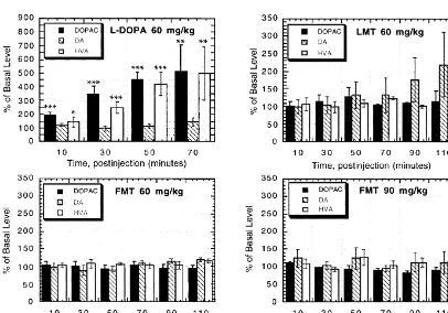
University of Wisconsin Animal Care and Use Committee, DOPAC, dopamine, and HVA as expressed as percentage
microdialysis probes (CMA Microdialysis, Solna, Sweden) of their respective basal concentrations following the
were placed, using a Kopf stereotaxic frame, into the left administration of L-DOPA, LMT, and FMT are shown in
striatum of 250–400 g male HSD rats (Harlan Sprague Fig. 1. The error bars are S.E.Ms obtained from two to
Dawley, Madison, WI) anesthetized with urethane (1.5 four animals per drug treatment. When given without a
g / kg, i.p.). The coordinates used for probe placement were peripheral AAAD inhibitor, the increase in extracellullar
AP: bregma 10.5 mm, L: 2.5 mm, DV: dura 27 mm DA after acute L-DOPA (50 mg / kg, i.p.) has been reported
based on the rat atlas of Paxinos and Watson [19]. to be transient (occurring within 20 min postinjection) [3].
Artificial CSF (flow-rate52ml / min) was perfused through Although we did not observe this short-lived increase in
the probe and 20-min samples (40 ml) were collected in DA in our study, we did find continually increasing
194 T.D. Stein, O.T. DeJesus / Brain Research 884 (2000) 192 –195
extracellular concentrations of DA metabolites, DOPAC Acknowledgements
and HVA, similar to that observed by Brannan et al. [3].
Since both metabolites are derived from dopamine, it is We gratefully acknowledge the technical assistance of
reasonable to assume that the observed increase in P. Lefeber and funding support from NIH Grant 2 RO1
DOPAC1HVA levels correspond to the increased forma- NS26621.
tion of DA originating from the exogenousL-DOPA and its
rapid metabolism. Thus, in theL-DOPA panel in Fig. 1, in
vivo microdialysis results show that 70 min after 60 References
mg / kgL-DOPA i.p. injection, there is a 10-fold increase in
extracellular dopamine. In comparison, an equal dose of [1] N.-E. Anden, S. Butcher, J. Engel, Central dopamine and
norad-renaline receptor activity of the amines formed from m-tyrosine,
FMT caused no change in DA, DOPAC, and HVA levels at
a-methyl-m-tyrosine and a-methyldopa, J. Pharm. Pharmacol. 22
70 min, 90 min and 110 min (Fig. 1). Similarly, no change
(1970) 548–550.
was observed at a higher FMT dose of 90 mg / kg (Fig. 1). [2] H. Blaschko, T.L. Chrusciel, The decarboxylation of amino acids
In contrast to bothL-DOPA and FMT, LMT (60 mg / kg) related to tyrosine and their awakening action in reserpine-treated
was found to increase DA in the extracellular space greater mice, J. Physiol. 151 (1960) 272–284.
[3] T. Brannan, P. Knott, H. Kaufmann, L. Leung, M. Yahr,
Intracereb-than two-fold 110 min postinjection, while DOPAC and
ral dialysis monitoring of striatal dopamine release and metabolism
HVA levels were unchanged at all times. However, this
in response toL-DOPA, J. Neural Transm. 75 (1989) 149–157.
increase in dopamine levels did not reach significance [4] W.D. Brown, O.T. DeJesus, R.W. Pyzalski, A.D. Roberts, S.E.
(P.0.05). Shelton, H. Uno, D. Houser, R.J. Nickles, J.E. Holden, Localization
The increases in DOPAC and HVA found afterL-DOPA of trapping of 6-[18F]fluoro-L-m-tyrosine. A presynaptic AAAD
tracer for PET, Synapse 34 (1999) 111–123.
administration are likely due to the inability of vesicles to
[5] A. Carlsson, M. Lindquist, Metatyrosine as a tool for selective
store the relatively excessive amounts of DA produced
protection of catecholamine stores against reserpine, Eur. J.
Phar-from exogenous L-DOPA. Although LMT-induced DA
macol. 2 (1967) 187–192.
increase in extracellular space observed in this study was [6] G.C. Cotzias, P.S. Papavasiliou, I. Mena,L-m-tyrosine and
Parkin-not statistically significant, the effect on DA tissue levels sonism, J. Am. Med. Assoc. 223 (1973) 83.
[7] O.T. DeJesus, J. Mukherjee, Radiobrominated m-tyrosine analog as
was not determined. A study by Smyth et al. [21] using a
a potential CNS L-DOPA PET tracer, Biochem. Biophys. Res.
higher LMT dose (150 mg / kg) observed tissue DA levels
Commun. 150 (1988) 1027–1031.
60 min after i.p. injection to be reduced to about half that [8] O.T. DeJesus, D. Murali, R.J. Nickles, Synthesis of brominated and
of basal level DA. This LMT-induced DA reduction can be fluorinated ortho-tyrosine analogs as potential DOPA decarboxylase
blocked by AAAD inhibition in support of the role of tracers, J. Label. Comp. Radiopharm. 37 (1995) 147–149.
[9] O.T. DeJesus, J.E. Holden, C.J. Endres, D. Murali, T.R. Oakes, S.E.
MTA as the causative agent [21]. Furthermore, the
sug-Shelton, H. Uno, D. Houser, L. Freund, S.B. Perlman, R.J. Nickles,
gestion by Smyth et al. [21] that MTA may displace DA is
Visualization of dopamine nerve terminals by positron emission
supported by previous findings that MTA is taken up by 18
tomography using [ F]fluoro-b-fluoromethylene-m-tyrosine, Brain
chromaffin granules [13,17]. The displaced DA is likely Res. 597 (1992) 151–154.
quickly metabolized since the systemic injection of m- [10] O.T. DeJesus, C.J. Endres, S.E. Shelton, R.J. Nickles, J.E. Holden, Evaluation of fluorinated m-tyrosine analogs as PET imaging agents
tyrosine produced intense behavioral stimulation only
of dopamine nerve terminals: comparison with 6-fluoroDOPA, J.
when DA metabolism by MAO was blocked [12].
Nucl. Med. 38 (1997) 630–636.
On the other hand, the results of this study show that [11] O.T. DeJesus, C.J. Endres, S.E. Shelton, R.J. Nickles, J.E. Holden,
DA and its metabolites in the extracellular space are not Noninvasive assessment of aromatic L-amino acid decarboxylase
affected by the systemic injection of FMT up to an i.p. activity in aging rhesus monkey striatum, Synapse (in press).
[12] L.E. Dyck, C.W. Kazakoff, C.T. Dourish, The role of
catechol-dose of 90 mg / kg. This finding is supported by our
amines, 5-hydrotryptamine and m-tyramine in the behavioral effects
previous observation that decarboxylated FMT, FMTA, is
of m-tyrosine in the rat, Eur. J. Pharmacol. 84 (1982) 139–149.
poorly taken up by chromaffin granules compared to [13] C.J. Endres, S. Swaminathan, O.T. DeJesus, M. Sievert, A.E.
fluorodopamine and MTA [13]. In vivo studies previously Ruoho, D. Murali, S.G. Rommelfanger, J.E. Holden, Affinities of
demonstrated that after its administration, FMT is rapidly dopamine analogs for monoamine granular and plasma membrane
transporters: implications for PET dopamine studies, Life Sci. 60
decarboxylated to form FMTA which, in turn, is rapidly
(1997) 2399–2406.
oxidized by MAO as shown by the rapid formation, the
[14] J. Engel, Metatyrosine-induced reversal of the suppression of the
dominance and the persistence of the MAO product of conditioned avoidance response in reserpine-treated rats, Acta
FMTA, fluoro-hydroxyphenylacetic acid, in both extracel- Pharmacol. Toxicol. 30 (1971) 278–288.
18
lular space [16] and whole striatal tissues in rodents [18] [15] G. Firnau, R. Chirakal, C. Nahmias, E.S. Garnett, [ F]Fluoro-meta-18
tyrosine is a better PET tracer than [ F]fluoro-L-dopa for the
and in non-human primates [15]. The rapid MAO
oxida-delineation of dopaminergic structures in the human brain, J. Label.
tion of FMTA is consistent with its lack of vesicular
Comp. Radiopharm. 30 (1991) 266–268.
uptake and protection [13]. Thus, unlike LMT, even with [16] S. Jordan, K.S. Bankiewicz, J.L. Eberling, H.F. Van Brocklin, J.P.
MAO inhibition, FMT would not be expected to have O’Neil, W.J. Jagust, An in vivo microdialysis study of striatal
18
T.D. Stein, O.T. DeJesus / Brain Research 884 (2000) 192 –195 195
[17] J. Knoth, J.O. Peabody, P. Huettl, D. Njus, Kinetics of tyramine [21] R.G. Smyth, J.H. Tong, A. D’Iorio, Studies on the depletion of brain transport and permeation across chromaffin-vesicle membranes, amines by m-tyrosine, Eur. J. Pharmacol. 42 (1977) 267–273. Biochemistry 23 (1984) 2011–2016. [22] H.R. Snyder, J.F. Shekleton, C.D. Lewis, Synthetic amino acids. [18] W. Melega, M.M. Perlmutter, A. Luxen, C.H. Nissenson, S.T. Syntheses from acetamidomalonic acids, J. Am. Chem. Soc. 67
18
Grafton, S.C. Huang, M.E. Phelps, J.R. Barrio, 4-[ F]fluoro-L-m- (1945) 310–312.
tyrosine: an L-3,4-dihydroxyphenyl-alanine analog for probing pre- [23] J.H. Tong, C. Petitclerc, A. D’Iorio, N.I. Benoiton, Resolution of synaptic dopaminergic function with positron emission tomography, ring-substituted phenylalanines by the action alpha-chymotrypsin on J. Neurochem. 53 (1989) 311–314. their ethyl esters, Can. J. Biochem. 49 (1971) 877–881.
[19] C. Paxinos, G. Watson (Eds.), The Rat Brain Atlas, 2nd Edition, [24] U. Ungerstedt, K. Fuxe, M. Goldstein, A. Battista, M. Ogawa, B. Academic Press, New York, 1986. Anagnoste, Action of m-tyrosine in experimental models: evidence [20] M. Sandler, S.J. Corne, R. Stephens, K.M. Shaw, K.R. Hunet, G.M. for possible antiparkinsonian activity, Eur. J. Pharmacol. 21 (1973)