JAIDS Journal of Acquired Immune Deficiency Syndromes Publish Ahead of Print DOI: 10.1097/QAI.0b013e3182926f95
ACCEPTED
Prevalence and Outcomes of Cryptococcal Antigenemia in HIV-seropositive PatientsHospitalized for Suspected Tuberculosis in Uganda
A.O. Andama1,2,3, S. den Boon1, D. Meya2,3, A. Cattamanchi4, W. Worodria1,3, J.L. Davis4, N.D.
Walter5, S.D. Yoo6, N. Kalema1, B. Haller7, L. Huang4,8 on behalf of the International
HIV-associated Opportunistic Pneumonias (IHOP) Study
1. Makerere University – University of California, San Francisco (MU-UCSF) Research
Collaboration, Kampala, Uganda
2. College of Health Sciences, Makerere University, Kampala, Uganda
3. Department of Medicine, Mulago National Referral Hospital, Kampala, Uganda
4. Division of Pulmonary and Critical Care Medicine, University of California, San
Francisco, San Francisco, CA, USA
5. Division of Pulmonary Sciences and Critical Care Medicine, University of Colorado,
Denver, CO, USA
6. Health Tutors’ College Mulago, Kampala, Uganda
7. Department of Laboratory Medicine, University of California, San Francisco, San
Francisco, CA, USA
8. HIV/AIDS Division, University of California, San Francisco, San Francisco, CA, USA
Key words: Cryptococcus neoformans, antigenemia, tuberculosis, HIV, AIDS
Body: 2,860 words.
ACCEPTED
Financial Support: This work was supported by grant numbers K24 HL087713 (LH), R01 HL090335 (LH), U01 AI089244-01 (COAT study), K23 AI080147 (JLD) from the National
Institutes of Health.
ACCEPTED
ABSTRACTBackground
Cryptococcal infection occurs in HIV-seropositive patients and is associated with high mortality.
However, limited information is available on the prevalence and outcomes of cryptococcal
antigenemia among hospitalized HIV-seropositive patients in sub-Saharan Africa.
Objectives
To determine the prevalence of and risk factors for cryptococcal antigenemia among
HIV-seropositive patients presenting to Mulago Hospital (Kampala, Uganda) with unexplained cough
≥2 weeks and suspected TB, and also to determine if antigenemia is associated with an increased
mortality.
Methods
Between September 2009 and September 2010, we enrolled consecutive HIV-seropositive adults
hospitalized at Mulago Hospital with cough ≥2 weeks and suspected TB. Banked serum was
tested for cryptococcal antigen. We compared demographic, clinical characteristics and 2-month
mortality in patients with and without cryptococcal antigenemia.
Results
Of 563 HIV-seropositive patients, 32 (5.7%) were CrAg-positive. None had Cryptococcus
neoformans detected on fungal culture of BAL fluid (n=116). CrAg-positive patients had a lower
median CD4-count compared to CrAg-negative patients (25 vs. 55 cells/uL, p=0.02) and a
ACCEPTED
test was not associated with increased mortality during the 2-month follow-up period (HR: 0.99,
95% CI: 0.63–1.54, p=0.95) after adjusting for CD4 count and ART use at enrollment and/or
follow-up.
Conclusions
Occult cryptococcal antigenemia occurs commonly among hospitalized HIV-seropositive
patients with suspected TB. CrAg testing should be considered in hospitalized, HIV-seropositive
patients with CD4 count <50 cells/uL, coupled with longer follow-up to evaluate the diagnostic
value of CrAg and therapeutic interventions in patients with asymptomatic cryptococcal
antigenemia.
INTRODUCTION
Cryptococcosis is a serious fungal infection that is a common cause of meningitis and
death among patients with advanced human immunodeficiency virus (HIV) infection.
Pneumonia with or without accompanying meningitis and isolated cryptococcal antigenemia are
also common presentations. Recent estimates suggest that there are about 1 million new cases
and at least 500,000 deaths annually due to HIV-associated cryptococcosis worldwide [1]. In
more affluent countries, the incidence of HIV-associated cryptococcosis has decreased
dramatically since the introduction of antiretroviral therapy (ART) [2], and mortality associated
with this disease is generally <10%. Although the incidence has also decreased in sub-Saharan
Africa with the increased use of ART [3], cryptococcosis remains a significant cause of
ACCEPTED
The burden of cryptococcosis has previously been reported to be particularly high in
patients with or suspected of having pulmonary TB. In South Africa, cryptococcal disease was
associated with 75% of deaths from opportunistic infections in gold miners with pulmonary
tuberculosis [5]. However, few previous studies have evaluated the burden and impact of
cryptococcosis among hospitalized HIV-seropositive TB suspects in sub-Saharan Africa.
In sub-Saharan African settings, hospitalized HIV-seropositive patients without
neurological symptoms are rarely tested or treated for cryptococcosis. Thus, the diagnosis may
be missed in patients with isolated cryptococcal antigenemia. We hypothesized that
unrecognized cryptococcosis may be prevalent in this population and may contribute to early
mortality.
Therefore, we performed cryptococcal antigen testing on banked serum specimens from
consecutive HIV-seropositive adult patients (≥18 years) with suspected tuberculosis (defined as a
cough ≥2 weeks) at Mulago Hospital in Kampala, Uganda, who had been enrolled between
September 2009 and September 2010. These patients were not suspected of cryptococcosis by
their treating clinicians. Our objectives were to determine the prevalence and risk factors
associated with cryptococcal antigenemia among HIV-seropositive patients presenting with
cough. We also determined if cryptococcal antigenemia is associated with an increased mortality.
METHODS
Participants and procedures
The current study is a sub-study of the International HIV-associated Opportunistic
Pneumonias (IHOP) Study, a prospective observational cohort. The general study protocol has
ACCEPTED
and were interviewed to obtain demographic and clinical data. Chest radiographs and blood were
obtained at enrollment. Blood was used for HIV testing and, if HIV-positive, for CD4+
T-lymphocyte count measurement. Patients submitted two sputum specimens (1 spot and 1 early
morning) for acid-fast bacilli (AFB) smear and culture on Lowenstein-Jensen media, and those
with negative sputum AFB results (using Ziehl Neelsen method) were referred for bronchoscopy
with bronchoalveolar lavage (BAL). Bronchoscopy included inspection for endobronchial
Kaposi sarcoma (KS). BAL fluid was examined for mycobacteria, Pneumocystis jirovecii (using
modified Giemsa, Diff Quik method), Cryptococcus neoformans and other fungi (using
Sabouraud medium). The follow-up period was two months after enrollment to assess vital
status. After this 2-month follow-up period, final diagnoses were assigned by 2 study doctors
according to pre-specified criteria (see Appendix, Supplemental Digital Content,
http://links.lww.com/QAI/A416), which included TB culture results, response to treatment, and
other previously collected data [6, 7].
At time of enrollment, 3-4 mL of blood was collected from each patient into a vacutainer tube
with clot activator and processed for serum as follows: blood was allowed to clot at room
temperature for 30 minutes and centrifuged at 3000 RPM for 5 minutes. The resulting serum was
used to confirm HIV status (200 µ L) and the remainder (0.5-1.0 mL) was promptly transferred
into cryovials and stored at -20oC. For this study, banked serum was thawed and tested for
cryptococcal antigen.
Serum CrAg testing
ACCEPTED
and microbiologic data. CrAg testing was performed using a commercially-available test kit
(Immuno-Mycologics, Inc. OK, USA) according to the manufacturer’s instructions. Testing
included positive and negative controls with each batch, and testing was repeated if the results
were indeterminate (i.e., failed positive or negative control). The qualitative test result was
interpreted as non-reactive (negative) if it showed a smooth, milky suspension with absence of
agglutination, and reactive (positive) if it showed distinct large clumps against a clear or lightly
milky background or small but definite clumps against a milky background. CrAg titers were
subsequently obtained in reactive specimens using standard serial dilutions. The titer of a
particular sample was taken to be the highest dilution that showed agglutination.
Sample size determination and data analysis
The first objective of this study (to determine the prevalence of cryptococcal co-infection
in HIV seropositive patients with suspected TB in Mulago Hospital) was used for the sample size
and precision estimates. An 8% prevalence was reported in a cohort of adult ART-naive AIDS
patients who were subsequently started on ART in Kampala, Uganda [8]. Using the Kish and
Leslie formula [9], 452 serum samples would provide a prevalence estimate with 90% precision
and a 95% confidence interval. However, to further increase our precision, we elected to analyze
all 563 available banked serum samples from our HIV-seropositive patients.
Data analysis was performed using STATA release 10 (Stata Corporation, College
Station, Texas, USA). We compared categorical variables using the chi-squared or Fisher’s exact
tests, and continuous variables using Student’s t-test or the Mann–Whitney rank sum test. We
performed backwards multivariate logistic regression analysis to determine factors associated
ACCEPTED
defined as a p-value less than 0.05, were removed from the model. We conducted survival
analysis to determine whether cryptococcal antigenemia is independently associated with
mortality. We controlled for both CD-4 count and ART use, variables that are known to impact
survival. We defined significance in reference to the probability of a 2-tailed, type I error
(p-value) less than 0.05.
Ethical considerations
Written informed consent was obtained from study participants and included permission
to use banked specimens collected at enrollment for future studies. The Makerere University
Faculty of Medicine Research and Ethics Committee, the Mulago Hospital Institutional Review
Board, the Uganda National Council for Science and Technology, and the University of
California San Francisco Committee on Human Research approved the protocol.
RESULTS
Patient characteristics
Of 753 adult patients with cough >2 weeks and suspected TB who were enrolled, 563
tested positive for HIV antibodies and had banked serum available for this study (Figure 1).
Two-month follow-up data was available for 430 patients. There were no significant differences
between patients with and patients without 2-month follow-up data available for the following
variables: gender (p=0.71), median age (p=0.35), median CD4-count (p=0.24), ART use on
admission (p=0.88), cotrimoxazole prophylaxis use on admission (p=0.20), fever (p=0.87),
ACCEPTED
with patients who completed the 2-month follow-up period, patients who were lost to follow-up
less often knew their HIV status at enrollment (59% vs. 69%, p=0.04) and more often had an
unknown diagnosis for their cough (44% vs. 19%, p<0.001).
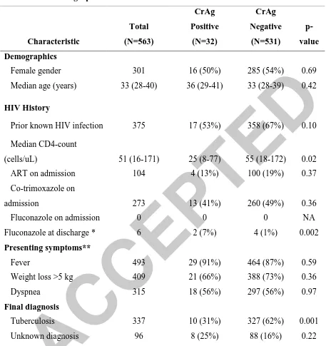
Of the 563 HIV-seropositive patients who were included in this study, the majority were
female (n=301, 53%) and young (median age=33 years, interquartile range, IQR=28-40 years)
(Table 1). Approximately two-thirds (66%) were known to have HIV infection at the time of
their presentation and the remaining one-third had HIV infection diagnosed during this
hospitalization. Most patients had advanced HIV disease (median CD4-count=51 cells/uL,
IQR=16-171 cells/uL). Despite this, only 28% (n=104) of the 375 patients known to have HIV
infection were receiving antiretroviral therapy at the time of presentation; 73% (n=273) were
receiving co-trimoxazole for PCP prophylaxis and none were receiving fluconazole. The
majority of patients presented with fever (88%), weight loss >5 kg (73%), and dyspnea (56%).
Cough, which was an inclusion criterion for the prospective study, was present in 100%. In most
cases, these non-specific complaints were determined to be due to tuberculosis (60%). Other
common diagnoses included bacterial pneumonia (11%), PCP (1%) and pulmonary Kaposi’s
sarcoma (2%). A substantial proportion of patients had an unknown final diagnosis (17%).
Prevalence of and risk factors for cryptococcal antigenemia
Overall, 32 (5.7%) of the 563 patients had a positive CrAg test. Among the 116 patients
who underwent bronchoscopy, none had Cryptococcus neoformans detected on fungal culture of
BAL fluid. Thus, if CrAg testing had been done prospectively, the positive serum CrAg result
would have represented the sole diagnosis for cryptococcal disease.
CrAg-ACCEPTED
positive patients had a lower median CD4-count (25 cells/uL, interquartile range, (IQR): 8-77
cells/uL) compared to those who had a negative CrAg test (55 cells/uL, IQR: 18-172 cells/uL,
p=0.02) (Table 1). A substantial proportion of CrAg-positive patients had concurrent TB (31%)
and thus, TB is a frequent co-pathogen in patients with cryptococcemia. The proportion of
CrAg-positive patients who had TB was lower than the proportion in CrAg-negative patients (31% vs.
62%, p=0.001). Other clinical factors including gender, age, prior diagnosis of HIV infection,
and taking ART or co-trimoxazole prophylaxis at admission were all not associated with a
positive CrAg test. Presenting symptoms were also similar in CrAg-positive and CrAg-negative
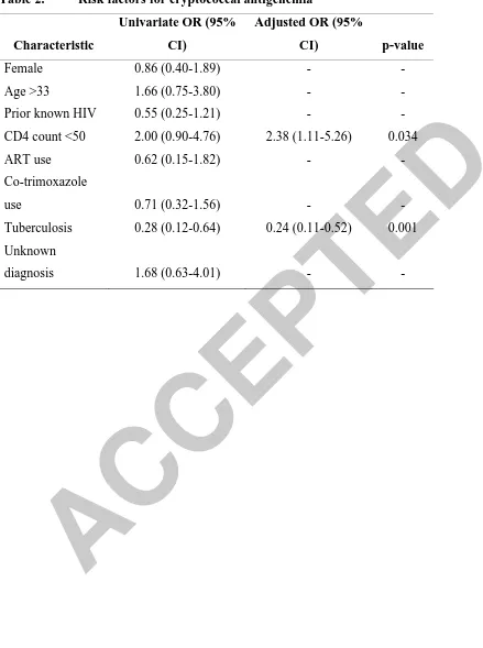
patients. In the final multivariate model, a CD4-count of <50 cells/ L was significantly
associated with increased odds of cryptococcal antigenemia (adjusted Odds Ratio=2.38,
p=0.034) while a diagnosis of tuberculosis was associated with a lower odds of antigenemia
(adjusted Odds Ratio=0.24, p=0.001) (Table 2).
Overall, 10 CrAg-positive patients were diagnosed with TB and eight were classified as
having an unknown final diagnosis but potentially having disseminated cryptococcal infection as
the etiology of their presentation. In the absence of a specific diagnosis of cryptococcosis, 6
patients (2 CrAg-positive and 4 CrAg-negative) were treated with fluconazole during
hospitalization because they had oral thrush or possible esophageal candidiasis, and were
discharged on this medication. CrAg-positive patients were more often initiated on empiric
fluconazole treatment during their hospitalization compared to CrAg-negative patients (7% vs.
ACCEPTED
Cryptococcal antigenemia and factors associated with 2-month mortalityAmong the 430 patients with 2-month follow-up data available, 310 (72.1%) were found
to be alive and 120 (27.9%) were determined to have died (Figure 1). Of the CrAg-positive
patients, 37% had died at 2-month follow-up compared to 27% of the CrAg-negative patients,
but this difference was not statistically significant (p=0.28). In survival analysis, there was no
significant difference between CrAg-positive and CrAg-negative patients in time to death
(Figure 2, p=0.47). In bivariate analysis, we did not find a significant association between CrAg
positivity and 2-month mortality (HR: 1.0, 95% CI: 0.64–1.55, p=1.0), although we could not
exclude the possibility of a clinically important increase or decrease in mortality because of the
imprecision of our estimate. After adjusting for CD4-count, ART use at admission and/or
follow-up, and TB diagnosis and/or unknown diagnosis, the association between CrAg result and
mortality remained non-significant (HR: 0.99, 95% CI: 0.63–1.54, p=0.95).
Among patients with a positive CrAg test, we compared mortality in 21 patients with low
titers (titer ≤1:512) with mortality in 11 patients with high titers (titer >1:512). CrAg titer was
not significantly associated with mortality in bivariate analysis (HR: 0.58, 95% CI: 0.17–1.97,
p=0.38) nor after adjusting for CD4-count, ART use at admission and/or follow-up, and TB
diagnosis and/or unknown diagnosis (HR: 1.02, 95% CI: 0.22–4.79, p=0.98).
DISCUSSION
In this study, we found an overall cryptococcal antigenemia prevalence of 5.7% in
HIV-infected adults hospitalized at Mulago Hospital (Kampala, Uganda) with cough >2 weeks and
suspected tuberculosis. None of the 116 patients who underwent bronchoscopy as part of their
ACCEPTED
did not identify a difference in mortality or time to death between positive and
CrAg-negative patients at 2 months of follow-up.
The moderate prevalence of cryptococcosis observed in this cohort is lower than that
reported among hospitalized patients in South Africa and Cambodia [10, 11] where cryptococcal
antigenemia prevalence was reported to be 13% and 20%, respectively. The lower rates that we
observed may be due to the fact that the majority of patients enrolled in these previous studies
were terminally ill and had symptoms of cryptococcosis. In contrast, our study selected a
different population: although hospitalized, patients in our cohort did not have
Cryptococcus-specific symptoms and were not clinically suspected to have cryptococcal infection.
Furthermore, patients with an altered mental status or confusion were excluded from our study
because they were unable to provide informed consent for study participation. Therefore, patients
with symptomatic cryptococcal neurologic disease would not have been enrolled. Surprisingly,
the prevalence that we noted in our hospitalized cohort was comparable to cryptococcal
antigenemia prevalences previously described among asymptomatic HIV-seropositive
outpatients in Uganda. In the Infectious Diseases Institute in Kampala and in a rural community
clinic setting in Tororo, cryptococcal antigenemia prevalences were 8% and 5.8%, respectively
[8, 12].
None of the CrAg-positive patients who underwent bronchoscopy in our prospective
study were diagnosed with concurrent cryptococcal pneumonia. This finding was unexpected as
we found Cryptococcus neoformans in 11.4% of HIV-infected patients undergoing
bronchoscopy in a previous cohort (September 2007 through July 2008) of 132 patients with
cough >2 weeks and suspected tuberculosis [6]. Nevertheless, our findings demonstrate that
ACCEPTED
culture results.
Even in the absence of diagnosis and specific treatment of cryptococcosis, CrAg-positive
patients in our cohort were not more likely to die before 2-month follow-up than CrAg-negative
patients. These findings differ from other studies that reported on HIV-positive patients with and
without cryptococcal antigenemia. Jarvis and colleagues reported on patients accessing an ART
program in South Africa and found that CrAg-positive patients were at far higher risk of
mortality than antigen-negative patients during a one-year follow-up period (HR=4.75, 95% CI
2.6-8.8, p<0.001) [10]. Similarly, French et al reported a poor survival of patients after diagnosis
of cryptococcal antigenemia (median survival 26 days, range 0-138 days), though this study was
done in the period before ART was available in Africa [3]. The fact that we failed to show an
association with mortality may be because we had a relatively small sample size and a
comparatively short follow-up period.
Overall, CrAg testing identified 32 patients with cryptococcal antigenemia, including 10
with concurrent TB and eight classified as having an unknown final diagnosis but potentially
having disseminated cryptococcal infection as the etiology of their presentation. In these
individuals, CrAg testing at the time of presentation may have been beneficial. However,
evaluation of the possible benefits of such an intervention requires an understanding of the
clinical course of patients with cryptococcal antigenemia. Cryptococcal antigenemia in the
context of advanced HIV infection has been assumed to indicate disseminated disease and the
progression to symptomatic cryptococcosis has been thought to be inevitable unless appropriate
anti-fungal treatment is given [13, 14]. During our relatively short 2-month follow-up period, we
were unable to confirm symptomatic cryptococcosis in any of these individuals, and it is unclear
ACCEPTED
individuals.
Despite the lack of association between cryptococcal antigenemia and mortality in our
study, the preponderance of evidence suggests that prospective testing for cryptococcal
antigenemia in severely immunosuppressed HIV-seropositive patients with suspected TB in the
absence of prior cryptococcal disease should have a role in the diagnostic evaluation. With the
new point-of-care lateral flow assay (LFA) for cryptococcal antigen, this testing could even be
performed at peripheral health facilities that the majority of patients visit prior to hospitalization
[15]. In addition, such testing with subsequent intervention may prevent unmasking of
subclinical cryptococcal infection that could present as meningitis or pneumonitis after ART
initiation. Screening for serum cryptococcal antigen is highly sensitive and specific [16], and
Meya et al., described the cost benefit of this strategy in preventing death in HIV-infected
patients with CD4 counts ≤100 cells/µ L [8]. This screening strategy may also be beneficial in
hospital-based cohorts such as ours that are composed of HIV-infected patients with advanced
immunosuppression and high 2-month mortality [7].
Our study had limitations. First, the screening test used (CrAg) does not differentiate
between acute and old infections. Cryptococcal antigen can persist in the blood up to 48 months
after successful treatment for cryptococcosis [17]. It is therefore possible that some of the
patients who tested positive might have had prior cryptococcal infection, since we mainly relied
on patient recall to rule out any history of cryptococcal infection and fluconazole use. That said,
none of the patients reported current fluconazole use at the time of hospital admission. Second,
the short period (two months) of follow-up, and the fact that the CrAg analyses were conducted
six months after the last patient was seen made it impossible to determine if the cases we
ACCEPTED
pneumonitis but with negative BAL cultures.
In summary, we report a moderately high prevalence (5.7%) of cryptococcal antigenemia
among HIV-infected patients who were hospitalized with suspected TB. Cryptococcal disease
was not suspected in these patients prior to CrAg testing and none of the CrAg-positive patients
who underwent bronchoscopy had concurrent cryptococcal pneumonia diagnosed. As a result,
the positive CrAg test may have represented the sole diagnosis of disseminated cryptococcal
infection and our results suggest a potential role for serum CrAg screening in this hospitalized
population. No significant association between CrAg positivity and 2-month mortality among
ACCEPTED
Acknowledgments:We would like to thank Sylvia Kaswabuli, Patrick Byanyima, John Kiidha, Rachel Kyeyune and
Leatitia Kampiire for enrolling and caring for the patients included in this study. We would also
like to thank the Mulago Hospital administration and staff for their approval and support of the
ACCEPTED
References1. Park, B.J., Wannemuehler, K.A., Marston, B.J., Govender, N., Pappas, P.G. and Chiller, T.M.
2009. Estimation of the current global burden of cryptococcal meningitis among persons living
with HIV/AIDS. AIDS; 23: p. 525-30.
2. CDC, 2000. Cryptococcosis.
3. French, N., Gray, K., Watera, C., Nakiyingi, J., Lugada, E. and Moore, M, 2002. Cryptococcal
infection in a cohort of HIV-1-infected Ugandan adults. AIDS; 16:1031–1038.
4. Kambugu, A., Meya, D.B., Rhein, J., O'Brien, M., Janoff, E.N., Ronald, A.R. and Kamya,
M.R. 2008. Outcomes of cryptococcal meningitis in Uganda before and after the availability of
highly active antiretroviral therapy. Clin Infect Dis; 46 (11): p. 1694-701.
5. Churchyard, G.J., Kleinschmidt, I., Corbett, E.L., Murray, J., Smit, J. and de Cock, K.M.
2000. Factors associated with an increased case-fatality rate in HIV-infected and non-infected
South African gold miners with pulmonary tuberculosis. Int J Tuberc Lung Dis; 4:705–712.
6. Yoo, S.D., Worodria, W., Davis, J. L., Cattamanchi, A., den Boon, S., Kyeyune, R., Kisembo,
H. and Huang L. 2010. The Prevalence and Clinical Course of HIV-Associated Pulmonary
Cryptococcosis in Uganda. J Acquir Immune Defic Syndr; 54:269–274
7. Kyeyune, R., den Boon, S., Cattamanchi, A., Davis, J.L., Worodria, W., Yoo, S.D. and Huang,
L. 2010. Causes of early mortality in HIV-infected TB suspects in an East African referral
hospital. J Acquir Immune Defic Syndr; 55(4):446-50.
8. Meya, D.B., Munabe, Y.C., Castelnuovo, B., Bethany, A.C., Ali, M.E. and Kambugu, A.
2010. Cost-Effectiveness of Serum Cryptococcal Antigen Screening to Prevent Deaths among
HIV-Infected Persons with a CD4+ Cell Count _100 Cells/mL Who Start HIV Therapy in
ACCEPTED
9. Kish and Leslie. 1965. Survey Sampling. New York: John Wiley and Sons, Inc.
10. Jarvis, J.N., Lawn, S.D., Vogt, M., Bangani, N., Wood, R. and Harrison, T.S. 2009.
Screening for cryptococcal antigenemia in patients accessing an antiretroviral treatment program
in South Africa. Clin Infect Dis; 48(7): p. 856-62.
11. Micol, R., Lortholary, O., Sar, B., Laureillard, D., Ngeth, C. and Dousset, J.P. 2007.
Prevalence, determinants of positivity, and clinical utility of cryptococcal antigenemia in
Cambodian HIV-infected patients. J Acquir Immune Defic Syndr; 45(5): p. 555-9.
12. Liechty, C.A., Solberg, P., Were, W., Ekwaru, J.P., Ransom, R.L. and Weidle, P.J. 2007.
Asymptomatic serum cryptococcal antigenemia and early mortality during antiretroviral therapy
in rural Uganda. Trop Med Int Health; 12(8): p. 929-35.
13. Feldmesser, M., Harris, C., Reichberg, S., Khan, S. and Casadevall, A. 1996. Serum
cryptococcal antigen in patients with AIDS. Clin Infect Dis; 23(4): 827–30. [PubMed: 8909854]
14. Yuen, C., Graziani, A., Pietroski, N., Macgregor, R. and Schuster, M. 1994. Cryptococcal
antigenemia in HIV-infected patients [abstract no. 93]. Clin Infect Dis ; 19:579.
15. Lindsley, M.D., Mekha, N., Baggett, H.C., Surinthong, Y., Autthateinchai, R., Sawatwong,
P., Harris, J.R., Park, B.J., Chiller, T., Arunmozhi, S.B. and Poonwan, N. 2011. Evaluation of a
Newly Developed Lateral Flow Immunoassay for the Diagnosis of Cryptococcosis. Clin Infect
Dis. 15; 53 (4): 321–325.
16. Uwe K.F., Nishimura S.L., Li N.C., Sugai K., Majko D.M., Hadly K. and NG V.L. 1993.
Evaluation of an Enzyme Immunoassay for Detection of Cryptococcal Capsular Polysaccharide
ACCEPTED
17. Lortholary, O., Poizat, G., Zeller, V., Neuville, S., Boibieux, A., Alvarez, M., Dellamonica,
P., Botterel, F., Dromer, F. and Chêne, G. 2006. Long-term outcome of AIDS-associated
ACCEPTED
Table 1: Demographic and Clinical CharacteristicsCharacteristic
Data presented as number (%) or median (interquartile range)
* Fluconazole use at discharge only known for 520 patients
** Cough present in 100% (parent study inclusion criteria)
ACCEPTED
Table 2. Risk factors for cryptococcal antigenemiaCharacteristic
Univariate OR (95%
CI)
Adjusted OR (95%
CI) p-value
Female 0.86 (0.40-1.89) - -
Age >33 1.66 (0.75-3.80) - -
Prior known HIV 0.55 (0.25-1.21) - -
CD4 count <50 2.00 (0.90-4.76) 2.38 (1.11-5.26) 0.034
ART use 0.62 (0.15-1.82) - -
Co-trimoxazole
use 0.71 (0.32-1.56) - -
Tuberculosis 0.28 (0.12-0.64) 0.24 (0.11-0.52) 0.001
Unknown