O
ne-carbon (C1) metabolism is essential to all organisms. In plants, it supplies the C1units needed to synthesize proteins, nucleic acids, pantothenate and many methylated mol-ecules1. Fluxes through C1pathways are particularly high in plants that are rich in methylated compounds such as lignin, alkaloids and betaines because methyl moieties make up several percent of their dry weight2
. Transfers of C1units are also central to the massive photorespiratory fluxes that occur in all C3plants
3
. In spite of the fundamental significance of these roles, and the interest in the meta-bolic engineering of lignin2
, betaines4
and photorespiration3 , there is much that is not understood about the enzymes, pathways and regu-latory mechanisms of plant C1metabolism. In part this is because of the obstacles that C1metabolism presents for classical biochemistry and genetics: its enzymes can be of low abundance and/or exist as
several isoforms, mutants are lacking, and its key intermediates – C1 substituted folates – are labile and hard to quantify. Fortunately, classical approaches to C1metabolism can now be complemented by genomics-driven approaches that exploit the fast-growing DNA sequence databases. Accordingly, this review has three aims: • To illustrate how genomics-based approaches are advancing
our knowledge of plant C1biochemistry.
• To bring together biochemical and genomics-derived data to show which C1pathways might operate in plants, and where they operate in the cell.
• To examine progress towards engineering C1metabolism. Nucleotide sequence information – from genomes, cDNAs and ESTs – can be used to complement biochemical approaches in sev-eral ways. Because most enzymes of C1 metabolism are highly 20 Fukuda, Y. (1997) Interaction of tobacco nuclear proteins with an
elicitor-responsive element in the promoter of a basic class I chitinase gene. Plant Mol. Biol.34, 81–87
21 Triezenberg, S.J. (1995) Structure and function of transcriptional activation domains. Curr. Opin. Genet. Dev.5, 190–196
22 Hanna-Rose, W. and Hansen, U. (1996) Active repression mechanisms of eukaryotic transcription repressors. Trends Genet.12, 229–234 23 Hoecker, U.et al.(1995) Integrated control of seed maturation and
germination programs by activator and repressor functions of Viviparous-1 of maize. Genes Dev.9, 2459–2469
24 Garcia-Bustos, J.et al.(1991) Nuclear protein localization. Biochim. Biophys. Acta1071, 83–101
25 Landschulz, W.H.et al.(1988) The leucine zipper: a hypothetical structure common to a new class of DNA-binding proteins. Science240, 1759–1764 26 Somssich, I.E. and Hahlbrock, K. (1998) Pathogen defense in plants – a
paradigm of biological complexity. Trends Plant Sci.3, 86–90
27 Gus-Mayer, S.et al.(1998) Local mechanical stimulation induces components of the pathogen defense response in parsley. Proc. Natl. Acad. Sci. U. S. A.95, 8398–8403
28 Quirino, B.F.et al.(1999) Diverse range of gene activity during Arabidopsis thalianaleaf senescence includes pathogen-independent induction of defense-related genes. Plant Mol. Biol.40, 267–278
29 Rouster, J.et al.(1997) Identification of a methyl-jasmonate-responsive region in the promoter of a lipoxygenase-1 gene expressed in barley grain.
Plant J.11, 513–523
30 Cao, H.et al.(1997) The Arabidopsis NPR1gene that controls systemic acquired resistance encodes a novel protein containing ankyrin repeats. Cell
88, 57–63
31 Li, X.et al.(1999) Identification and cloning of a negative regulator of systemic acquired resistance, SNI1, through a screen for suppressors of npr1-1. Cell98, 329–339
32Walker, A.R.et al.(1999) TheTRANSPARENT TESTA GLABRA1locus, which regulates trichome differentiation and anthocyanin biosynthesis in
Arabidopsis, encodes a WD40 repeat protein. Plant Cell11, 1337–1349 33Gatz, C. and Lenk, I. (1998) Promoters that respond to chemical inducers.
Trends Plant Sci.3, 352–358
34Sablowski, R.W.M. and Meyerowitz, E.M. (1998) A homolog of NO APICAL MERISTEMis an immediate target of the floral homeotic genes
APETALA3/PISTILLATA. Cell92, 93–103
35Martin, C. and Paz-Ares, J. (1997) MYB transcription factors in plants. Trends Genet.13, 67–73
36Gan, S. and Amasino, R.M. (1997) Making sense of senescence. Plant Physiol.113, 313–319
37Altschul, S.F.et al.(1997) Gapped BLAST and PSI-BLAST: a new generation of protein database search programs. Nucleic Acids Res.25, 3389–3402 38Bailey, T.L. and Elkan, C. (1994) Fitting a mixture model by expectation
maximization to discover motifs in biopolymers. In Proceedings of the Second International Conference on Intelligent Systems for Molecular Biology
(Altmann, R., ed.), pp. 28–36, AAAI Press
39Lupas, A. (1996) Coiled coils: new structures and new functions. Trends Biochem. Sci.21, 375–382
Thomas Eulgem, Paul Rushton, Silke Robatzek and Imre Somssich*are at the Max-Planck-Institut für Züchtungsforschung, Abteilung Biochemie, Carl-von-Linné-Weg 10, D-50829 Köln, Germany; Thomas Eulgem is currently at the Dept of Biology, 108 Coker Hall CB#3280, University of North Carolina, Chapel Hill, NC 27599-3280, USA.
*Author for correspondence (tel 149 221 5062310;
fax 149 221 5062313; e-mail [email protected]).
Plant one-carbon metabolism
and its engineering
Andrew D. Hanson, Douglas A. Gage and Yair Shachar-Hill
conserved, homology with bacterial, yeast or animal sequences can identify DNA sequences for known plant C1enzymes. The proteins that these sequences encode can then be characterized by expression in heter-ologous systems. Homology searches can suggest the presence (or absence) of enzymes for which there is no biochemical information in plants. The differential expression of genes can be inferred from variation in the count of their cognate ESTs, and this can provide valuable clues about the levels at which pathways might be operating5
. ESTs, cDNAs and genomic sequences can also give information about the organellar targeting of enzymes via their characteristic signal sequences, and about the sizes of gene families. Lastly, the avail-ability of sequences from a wide range of plants can speed up the preparation of the cDNAs that are needed to make constructs for metabolic engineering.
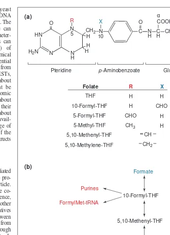
Role of folates in one-carbon metabolism
Here we briefly introduce folate-mediated C1metabolism (reviewed in Ref. 1) to pro-vide background for the rest of the article. Many C1 transfers are mediated by the co-enzyme tetrahydrofolate (THF). In essence, catab-olism of serine, glycine and other molecules generates specific C1 derivatives of THF that are then interconverted between different oxidation states, ranging from 10-formyl-THF (most oxidized) through 5,10-methenyl- and 5,10-methylene- to 5-methyl-THF (most reduced; Fig. 1). These interconversions of C1-substituted folates form the core of C1metabolism, from which C1 units are withdrawn by anabolic reac-tions. The largest anabolic flux is the use of 5-methyl-THF to convert homocysteine to methionine, which is incorporated into pro-teins or converted to S-adenosylmethionine (AdoMet), the donor for methylations. In other anabolic reactions, 10-formyl-THF is used to synthesize purines and formylmeth-ionyl-tRNA (for translation initiation in plas-tids and mitochondria), and 5,10-methyl-ene-THF is used to produce thymidylate and pantothenate (Fig. 1).
Compartmentation of plant one-carbon metabolism
Folate-mediated and other C1reactions in plants are highly compart-mented (Fig. 2). The overall picture is like that for other eukaryotes6 inasmuch as many C1 enzymes occur in both the cytosol and the mitochondria. However, most of them are also found in plastids, and two enzymes are specific to peroxisomes. The data point to some surprising deductions about gaps in pathways and the inter-organel-lar traffic in C1-related metabolites that these gaps imply. Although the deductions presented here are robust inasmuch as they are based on concordance between DNA sequence data and biochemical find-ings, both types of information are subject to caveats. Thus, the algo-rithms available for predicting proteins from genomic DNA and
organellar targeting from the protein sequence are fallible, plant EST collections are far from a complete inventory of the transcriptome, and the Arabidopsis genome is only ~80% complete. ‘Missing’ enzymes, especially organellar ones, might therefore yet be found. Also, clearcut results on enzyme compartmentation can be hard to achieve because of the difficulties in obtaining highly purified sub-cellular fractions. The following deductions should therefore be viewed as provisional and as stimuli for further investigation. • The cytosol, mitochondria and plastids all contain the activities
required to convert formate to 10-formyl-THF, to interconvert 10-formyl-, 5,10-methenyl- and 5,10-methylene-THF, and to generate 5,10-methylene-THF from serine. However,
Fig. 1.Tetrahydrofolate (THF) and its role in C1interconversions and transfers. (a) Chemical
structures of THF and its C1-substituted derivatives. Plant folates, like those of other
organisms, are predominantly present as g-glutamyl conjugates in which a short g-linked polyglutamyl chain of up to about eight residues is added to the glutamate moiety1
. Most folate-dependent enzymes have greater affinity for the polyglutamate forms than for the corresponding monoglutamyl folates1
. (b) Major C1unit interconversions and transfers
involv-ing THF derivatives. Sources of C1units are shown in blue and their metabolic fates in red. Trends in Plant Science
N H N HN
N O
CH2
H
H H
R
N C
O
N
H CH CH2 COOH
H2N
X
CH2 COOHγ
5 10
Pteridine p-Aminobenzoate Glutamate
α
Folate R X
THF
10-Formyl-THF
5-Formyl-THF
5-Methyl-THF
5,10-Methenyl-THF
5,10-Methylene-THF
CH
CH2
H
CHO
H
H H
H
CHO
CH3
Formate
10-Formyl-THF
5,10-Methenyl-THF
5,10-Methylene-THF
AdoMet 5-Methyl-THF
Glycine
Serine
Purines FormylMet-tRNA
Thymidylate Pantothenate Methionine
(a)
5,10-methylene-THF reductase, which is required to convert 5,10-methylene-THF to 5-methyl-THF, is confined to the cytosol. • The cytosol and mitochondria contain cobalamin-independent methionine synthase and can therefore form methionine from homocysteine and 5-methyl-THF. This suggests that mitochon-dria must import 5-methyl-THF and homocysteine, which is synthesized de novo only in plastids7
and is produced from S-adenosylhomocysteine (AdoHcy) only in the cytosol. • Although plastids carry out all steps in methionine synthesis
up to homocysteine, and have a pool of 5-methyl-THF (in its monoglutamyl form, presumably imported)7
, they do not contain cobalamin-independent methionine synthase. Plastids
might therefore import methionine from the cytosol. Alterna-tively, plastids could have a methionine synthase of the cobalamin-dependent-type found in bacteria and animals7
. This possibility is attractive in light of evidence for the monoglu-tamyl form of 5-methyl-THF in plastids because the cobalamin-dependent enzyme can use this substrate. However, there are no published plant DNA sequences that encode this enzyme, which is highly conserved between bacteria and animals.
• Neither chloroplasts nor mitochondria have AdoMet syn-thetase, implying that they import AdoMet from the cytosol. A related deduction, albeit based only on sequence data (the absence of transit peptides), is that these organelles also lack
Fig. 2.The reactions of plant one-carbon (C1) metabolism and their probable compartmentation as deduced from biochemical and DNA
sequence evidence. Compartmentation was inferred from sequence data by examining the predicted polypeptides encoded by C1-related genes,
cDNAs and ESTs for well-defined organellar targeting signals. The enzymes corresponding to the numbered reactions are listed in Table 1, together with corresponding literature citations and/or GenBank Accession numbers. The abundance of ESTs for these enzymes in dBEST is given in Figure 4. Different types of evidence were used to assign compartmentation, as indicated by the different colors: bold, black type indi-cates biochemical and DNA sequence data; red indiindi-cates DNA sequence data only; blue indiindi-cates biochemical data only. Boxed metabolites are those most likely to be transported (or to diffuse) across organellar membranes. The single asterisk marks the glyoxylate decarboxylation reaction, which is probably mainly non-enzymatic in leaves, but can also be mediated by the peroxidatic action of catalase (enzyme 25) (Refs 18,20). The double asterisk marks the reaction mediated by 5-formimino-THF cyclodeaminase (EC 4.3.1.4), evidence for which has not yet been found in plants. Question marks denote enzymes for which no inferences about compartmentation are yet possible. Abbreviations: THF, tetrahydrofolate; 10-HCO-THF, 10-formyl-THF; 5-HCO-THF, 5-formyl-THF; 5,105CH-THF, 5,10-methenyl-THF; 5,10-CH2-THF, 5,10-methylene-THF;
5-CH3-THF, 5-methyl-THF; DHF, dihydrofolate; Hcy, homocysteine; GSH, glutathione; HM-GSH, S-hydroxymethylglutathione; FGAR,
formylglycinamide ribonucleotide; FAICAR, formamidoimidazolecarboxamide ribonucleotide; SMM, S-methylmethionine; X, methyl acceptor; methyl-X, methylated acceptor.
Trends in Plant Science HCOOH
AdoHcy hydrolase, and therefore must export AdoHcy to the cytosol for hydrolysis to homocysteine and adenosine. AdoHcy hydrolysis is crucial to the regulation of methylation reactions, because AdoHcy is a potent competitive inhibitor of AdoMet-dependent methyltransferases.
Other deductions from a combination of DNA data and biochemi-cal results are (i) that the two 10-formyl-THF-dependent steps in purine synthesis occur in both mitochondria and plastids, and (ii)
that plants have a bifunctional thymidylate synthase–dihydrofolate reductase, not two separate enzymes, and that this enzyme is prob-ably present in both mitochondria and plastids.
Plants have two unique one-carbon reactions and two unusual ones
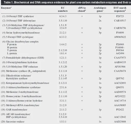
In terms of plant-specific enzymes, there are only a few major evolutionary novelties in plant C1 metabolism. The deduced Table 1. Biochemical and DNA sequence evidence for plant one-carbon metabolism enzymes and their compartmentation
Enzymesa
EC cDNAs Arabidopsis EST search Refse
numbers clonedb
genesc
sequencesd
(1) 10-Formyl-THF synthetase 6.3.4.3 1 1p P28723 32,33
(2) 10-Formyl-THF deformylase 3.5.1.10 2x CAB10517
(3) 5,10-Methylene-THF dehydrogenase/ 1.5.1.5/
5,10-methenyl-THF cyclohydrolase 3.5.4.9 1 3p CAB56756 32–34
(4) Serine hydroxymethyltransferase 2.1.2.1 1 5p P34899 33,35,36
(5) 5-Formyl-THF cycloligase 6.3.3.2 AW065622 27
(6) Glycine decarboxylase complex
P-protein 1.4.4.2 1 2p P26969 37
H-protein 1 2p P16048– 37
T-protein 2.1.2.10 1 1p P49364 37
L-protein 1.8.1.4 1 2p A42494 37
(7) Formaldehyde dehydrogenase (GSH) 1.2.1.1 1 1p CAA57973 38–41
(8)S-Formylglutathione hydrolase 3.1.2.12 1x AAB84335 41
(9) 5,10-Methylene-THF reductase 1.5.1.20 1 1p AF181966 13
(10) Methionine synthase (B12-independent) 2.1.1.14 1 2p CAA58474 42–45
(11) Dihydrofolate reductase/ 1.5.1.3/
thymidylate synthase 2.1.1.45 1 2p Q05762 34,46
(12) Ketopantanoate hydroxymethyltransferase 2.1.2.11 1x AAC62893
(13)S-Adenosylmethionine synthetase 2.5.1.6 1 3p Q96551 42,47
(14) Methionine S-methyltransferase 2.1.1.12 1 1p AAD49574 8,9,48
(15) Homocysteine S-methyltransferase 2.1.1.10 1 2p AF219222 8,f
(16)S-Adenosylhomocysteine hydrolase 3.3.1.1 1 2p AAC14714 7
(17) Methionyl-tRNA transformylase 2.1.2.9 AAA58085 49
(18) GAR transformylase 2.1.1.2 1 1p P52422 50,51
(19) AICAR transformylase/ 2.1.2.3/
IMP cyclohydrolase 3.5.4.10 1x AAC12842 50
(20) Sarcosine oxidase 1.5.3.1 1x AAD23888 16
(21) Glyoxylate synthetase – 10,11
(22) Polypeptide deformylase 3.5.1.31 1x AAD39667
(23) Formate dehydrogenase 1.2.1.2 1 Q07511 15–17
(24) Glutamate formiminotransferase 2.1.2.5 1x AAD20912
(25) Catalase (peroxidatic activity) 1.11.1.16 1 3p – 20
aEnzyme numbers 1–25 are as in Figure 2. Abbreviations: GSH, glutathione; B
12, cobalamin; GAR, glycinamide ribonucleotide; AICAR, aminoimidazolecarboxamide
ribonucleotide.
bPlus indicates that at least one plant cDNA has been demonstrated by a functional assay to encode the corresponding enzyme activity.
cValues are the number of sequenced genes (as of October 1999) in the Arabidopsisgenome that are highly homologous to Arabidopsisor other plant cDNAs (p), or to
cDNAs or genes from other organisms (x), which have been demonstrated to encode the corresponding enzyme.
dDenotes the sequences used to search dBEST for the EST abundance data of Figure 4. GenBank protein sequences were used in all cases except enzyme 5, for which the
open reading frame of an EST was used.
amino acid sequences of most plant C1 enzymes are strikingly similar to those of other organisms, even though the polypeptides specifying individual activities are sometimes fused together differently. Thus, although other eukaryotes have a trifunctional C1-THF synthase and plants have a 10-formyl-THF synthetase plus a bifunctional 5,10-methylene-THF dehydrogenase–5,10-methenyl-THF cyclohydrolase1
, the two plant enzymes are clearly homologous to the domains of the trifunctional protein. Likewise, the domains of the bifunctional dihydrofolate reductase–thymidy-late synthase are similar to their monofunctional counterparts in other organisms1
. However, one enzyme is unique to plants, and there is some biochemical evidence for a second unique enzyme. A third enzyme is not known in other eukaryotes; a fourth occurs also in fungi, but its compartmentation is different in plants and it is regulated in an unusual way. These four enzymes are described below.
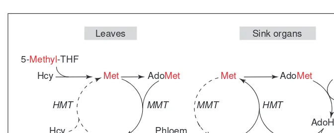
Methionine S-methyltransferase (MMT)
Methionine S-methyltransferase (MMT; Fig. 2, step 14) catalyzes the synthesis of S-methylmethionine (SMM), and is unique to plants8
. SMM can serve as a methyl donor for another methyltransferase, homocysteine S-methyltransferase (HMT; Fig. 2, step 15), which converts homocysteine to methionine8
. (HMT also occurs in other organisms, where it confers the ability to use SMM of plant origin.) MMT and HMT acting in tandem, together with AdoHcy hydrolase, constitute what was thought to be a futile cycle, known as the SMM cycle8
. However, SMM is now known to be a major amino acid in the phloem, thus the halves of this cycle could be at least partially separated in space and time, with SMM synthesis dominant in leaves and SMM reconversion to methionine dominant in developing seeds or other sinks9
(Fig. 3). Phloem transport of SMM enables methyl groups to be produced in one place and consumed in another, which emphasizes that some facets of C1metabolism must be viewed in a whole-plant as well as in a cellular context.
MMT (Ref. 9) and HMT (F. Bourgis, et al., http://www. rycomusa.com/aspp1999/public/cgi-bin/showabs.cgi?0363) were recently cloned; in both cases extensive use was made of ESTs in the cloning process, and the recombinant enzymes were expressed in E. colifor characterization. Interestingly, MMT ESTs are far less abundant than those for AdoMet synthetase and AdoHcy hydrolase
(Fig. 4), implying that the major trans-methylation flux from AdoMet involves methyltransferases other than MMT. This has been shown in Lemna8
, substantiating the idea that useful inferences about meta-bolic fluxes can be drawn from EST abun-dance data5
.
Because MMT is an evolutionary inno-vation, it is noteworthy that it exists as a homotetramer (uncommon among methyl-transferases) of an exceptionally large poly-peptide with a modular structure9
. The N-terminal domain shares homology with methyltransferases and has motifs associ-ated with AdoMet-binding. The C-terminal region shares homology with aminotrans-ferases that act on amino acids of the aspar-tate family, but lacks the conserved lysine residue that binds the catalytic pyridoxal 59 -phosphate cofactor in aminotransferases. Therefore it is likely that the evolution of MMT involved a gene fusion event9
. The functions of the two domains are unknown.
Glyoxylate synthetase
Glyoxylate synthetase (Fig. 2, step 21) has only been reported in potato tuber chloroplasts10,11
and has not been cloned. It appears to mediate a THF-dependent condensation of two formate mol-ecules to give glyoxylate, a reaction with no precedent in other organisms. By creating a route from C1to C2compounds, such an enzyme, in conjunction with aminotransferase and serine hydroxymethyltransferase activities, would enable the elab-oration of C3(and thence larger) structures solely from C1units, as occurs in methylotrophs. Therefore, glyoxylate synthetase is potentially important. The direct reduction of CO2to formate is also reported to occur in potato tuber chloroplasts12; this reaction is known to occur in anaerobic bacteria but has not been reported in eukaryotes.
NADH-dependent, AdoMet-insensitive 5,10-methylene-THF reductase
The 5,10-methylene-THF reductases (MTHFRs; Fig. 2, step 9) from Arabidopsisand maize were recently cloned by genomics-based methods, and characterized by expression in yeast. Both enzymes were found to be NADH-dependent and AdoMet-insen-sitive13
, unlike the mammalian and yeast enzymes, which use NADPH and are allosterically inhibited by AdoMet – the AdoMet-binding site being in a C-terminal, regulatory domain14
. The pyridine nucleotide specificity is crucial because the NADPH-dependent MTHFR reaction is physiologically irre-versible, because of the large free energy change involved in the reduction of 5,10-methylene-THF and the high cytosolic NADPH:NADP ratio13,14
. An NADPH-dependent reaction thus commits C1units to methyl group synthesis, and potentially can deplete the 5,10-methylene-THF pool; feedback inhibition by AdoMet checks such depletion. By contrast, because the cytosolic NADH:NAD ratio in plants is low, the NADH-dependent MTHFR reaction might well be reversible; this would obviate the need for feedback-inhibition by AdoMet. The NADH-dependence and AdoMet-insensitivity of plant MTHFRs have important implications for the control of C1fluxes in plants and their engi-neering. MTHFRs provide cautionary examples of how proteins sharing high homology can have critically different properties that cannot be predicted from their primary sequences.
Fig. 3. Long-distance transport of methyl groups via movement of S-methylmethionine (SMM) in the phloem. SMM can be formed in leaves by methionine S-methyltransferase (MMT), transported in the phloem to seeds or other sink organs, and there reconverted to methionine (Met) by the action of homocysteine S-methyltransferase (HMT)9. The
methion-ine so formed can give rise to S-adenosylmethionine (AdoMet) for use in methylation reac-tions (X → methyl-X). The hydrolysis of the S-adenosylhomocysteine (AdoHcy) produced by these methylation reactions releases homocysteine (Hcy), which can then sustain further conversion of SMM to methionine. Unbroken arrows indicate major fluxes, broken arrows minor ones. The path taken by the methyl groups is indicated in red.
Trends in Plant Science
5-Methyl-THF
Hcy Met AdoMet Met AdoMet X
Hcy
Hcy
SMM Phloem SMM
transport
AdoHcy
Adenosine Methyl-X
MMT MMT
HMT HMT
Mitochondrial formate dehydrogenase
Formate dehydrogenase (FDH; Fig. 2, step 23) occurs in fungi as a cytosolic enzyme, but both biochemical and DNA sequence data show it to be a mitochondrial matrix enzyme in plants15
. Moreover, FDH is one of the most abundant soluble proteins in mitochondria from non-green tissues (e.g. up to 9% of the total protein content in potato tuber mitochondria15
) although it is a minor protein in mitochondria from illu-minated, unstressed leaves16
. FDH is remarkable for being strongly induced in leaves by darkness, by C1-related com-pounds, by environmental stresses16
, and in roots by hypoxia and by Fe deficiency17
. These patterns of developmental and envi-ronmental regulation strongly suggest that formate is a major metabolite in certain tis-sues and conditions.
Genomic evidence points to unsuspected one-carbon reactions There is evidence that formate can be formed in the leaves of illuminated C3plants as a result of a chemical reaction between glyoxylate and H2O2 (Refs 18,19) (perhaps facilitated by the peroxidatic activity of catalase20
). However, it is not at all clear how formate is produced in the dark, in non-photosynthetic organs, or by C4 species. Until now, the most plausible route was con-sidered to be from serine or glycine via 5,10-methylene-THF and 5,10-methenyl-THF to 10-formyl-5,10-methenyl-THF, followed by rever-sal of the 10-formyl-THF synthetase reaction (Fig. 1). Therefore, it is significant that DNA sequence data point to three addi-tional possibilities for the origin of formate that have received little or no attention. (1) Hydrolysis of 10-formyl-THF: the
Arabidopsis genome contains two homologs of the E. coli purUgene, encoding 10-formyl-THF deformy-lase. Both the putative PurU proteins appear to be mitochondrial (Fig. 2, step 2). Cognate ESTs indicate that these genes are expressed in Arabid-opsis and other plants at a modest level (Fig. 4). The 10-formyl-THF deformylase, which in E. coliis acti-vated by methionine and inhibited by glycine21
, releases formate from 10-formyl-THF in an essen-tially irreversible reaction. Therefore, if 10-formyl-THF deformylase is active in plants, it could drive flux out of C1 -substituted folate pools into formate. It is noteworthy that if the C1 folates were derived via serine and the 3-phospho-glycerate from glycolysis, a flux from C1folates to formate (and ultimately CO2) could operate as an energy-yielding dissimilatory route, bypassing the Krebs cycle.
(2) Oxidation of formaldehyde as its glutathione adduct. The relatively high abundance of ESTs encoding glutathione-dependent formaldehyde dehydrogenase (FADH, also known as class III alcohol dehydrogenase), coupled with the
presence of an Arabidopsisgene and cognate ESTs that puta-tively specify S-formylglutathione hydrolase (SFGH, also known as esterase D) (Fig. 2, steps 7 and 8) suggest another possible origin of formate. FADH converts the glutathione adduct of formaldehyde (which forms spontaneously) to S-formylglutathione, from which formate is released by SFGH. FADH and SFGH are not highly specific enzymes and so might in principle be playing roles unrelated to each other or to formaldehyde oxidation. However, at least for bacteria, this appears not to be the case because FADH and SFGH are members of a preserved operon in many species. There are various possible sources of formaldehyde; these
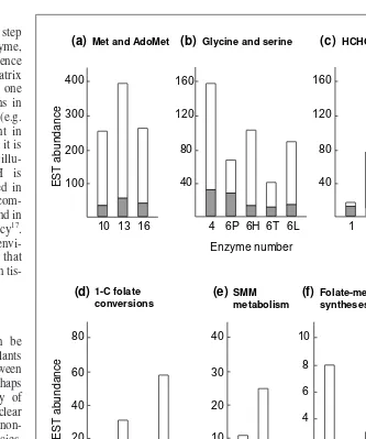
Fig. 4.EST abundance in dBEST for C1metabolism enzymes in Arabidopsis(gray) and in
all other higher plants (white) (as of October 1999). The enzymes, which are numbered as in Figure 2 and Table 1, are divided into functional groups related to (a) methionine and
S-adenosylmethionine (AdoMet) metabolism, (b) glycine and serine metabolism, (c) formaldehyde and formate metabolism, (d) C1 substituted folate interconversions, (e)
S-methylmethionine (SMM) metabolism and (f) folate-mediated syntheses. Note that the EST abundance scales vary by a factor of up to 40 between the six functional groups, but that the abundances of ESTs within each group are generally fairly similar. Note also that the ESTs for methionine and AdoMet metabolism are by far the most abundant, making up 51% of the total. Enzyme 7 (glutathione-dependent formaldehyde dehydrogenase, EC 1.2.1.1) shares <50% amino acid homology with alcohol dehydrogenase (EC 1.1.1.1). ESTs were therefore taken to represent EC 1.2.1.1 only if they showed greater similarity to this enzyme (CAA59773) than to an EC 1.1.1.1 sequence (P06525).
Trends in Plant Science Met and AdoMet
(a)
100 200 300 400
10 13 16
HCHO and HCOOH (c)
40 80 120 160
1 7 8 20 23
Glycine and serine (b)
40 80 120 160
4 6P 6H 6T 6L
1-C folate conversions (d)
20 40 60 80
2 3 5 9 24
SMM metabolism
10 20 30 40 (e)
14 15
Folate-mediated syntheses (f)
2 4 6 8 10
Enzyme number Enzyme number
11 12 17 18 19 22
EST
abundance
EST
include spontaneous dissociation of 5,10-methylene-THF formed from glycine or serine3
, and catalase-mediated oxida-tion of methanol derived from pectin hydrolysis20,22
. (3) Oxidation of sarcosine or other N-methylamino acids. The
Arabidopsisgenome includes a homolog of bacterial and mammalian monomeric sarcosine oxidases, and there are cognate ESTs from Arabidopsisand other plants (Fig. 4). Sarcosine oxidase (Fig. 2, step 20) converts sarcosine to glycine and formaldehyde, which can be oxidized to for-mate. Caution is necessary with this genomic evidence because (a) mammalian sarcosine oxidases also attack pipecolic acid and proline (without release of formalde-hyde), (b) a sarcosine oxidase-like enzyme in E. coliacts on N-methyltryptophan, and (c) it is not clear if there are major sources of sarcosine or other N-methylated amino acids in plants. It is therefore significant that sarcosine is a strong inducer of FDH in potato leaves16
, because this suggests that sarcosine is indeed metabolized to formate.
Towards engineering of one-carbon metabolic fluxes
Apart from the photorespiratory flux through glycine and serine3 , little is known about C1fluxes in plants. Therefore a major reason to engineer plant C1metabolism is to modify the fluxes through the main pathways (Fig. 2), to understand how they are controlled at the enzyme and gene levels23
. A key part of such work is the use of isotope tracer techniques to measure fluxes24
, and research in yeast25
that combined genetic modification with 13
C NMR analy-ses of C1 fluxes provides an excellent paradigm for plants. Although such studies have yet to be made using engineered plants, the utility of 13
C NMR techniques in plant C1metabolism has been demonstrated with wild-type Arabidopsisplants26,27
and sycamore (Acer pseudoplatanus) cell cultures28
. This work estab-lished, inter alia, that:
• The glycine decarboxylase complex and the mitochondrial ser-ine hydroxymethyltransferase are tightly coupled via a pool of 5,10-methylene-THF that does not equilibrate with the overall pool26,28
.
• Serine catabolism is probably limited by the THF supply26,28 . • Plants convert supplied 5-formyl-THF to other C1substituted
folates27 .
Engineered modification of C1fluxes in plants has begun in the area of AdoMet metabolism. When cosuppression was used to reduce AdoMet formation in tobacco, leaf tissue accumulated high levels of free methionine (400-fold that of the wild type) and a catabolic route producing methanethiol was induced29
. This shows that methionine itself exerts little control over its own syn-thesis, and supports an earlier finding that plants have an inducible methionine-g-lyase that can liberate excess methylthio groups29
. In another approach to lowering AdoMet levels, the bacteriophage T3 enzyme AdoMet hydrolase was expressed in tomato30
(AdoMet hydrolase converts AdoMet to homoserine and methylthioadenosine). The transformants produced less ethylene (a metabolite of AdoMet), which suggests that AdoMet pools were depleted. Lastly, when AdoHcy hydrolase activity in tobacco was lowered by antisense RNA expression, the plants were morphologically abnormal and showed hypomethylation of DNA (Ref. 31). The most remarkable aspect of these C1 engi-neering experiments is that the engineered plants were viable, implying that plants can somehow maintain essential C1fluxes in spite of major perturbations.
Future engineering challenges will be to attack folate-mediated C1metabolism, for instance by preventing C1units derived from formate and serine entering the C1 folate pool and by stopping methyl groups from leaving it. In addition, the activity of the
SMM cycle could be reduced, and the demand for methyl groups increased or decreased by adding or subtracting secondary path-ways. Such engineering research, in conjunction with studies of how C1gene expression patterns change in the engineered plants, will provide insight into how C1fluxes are controlled in plants. To date, we know in a general way that C1 metabolism is highly regulated at both metabolic and gene levels, and that its capacity can vary greatly as a function of development and environment. However, little is known about either the mechanisms or the extent of this plasticity.
Acknowledgements
We thank Michael J. Ziemak for preparing the figures. This work was supported by National Science Foundation grants IBN-9813999 to A.D.H. and BES-9902876 to D.A.G., by a grant from the National Institute of Standards and Technology to Y.S-H., and by the Florida Agricultural Experiment Station (Journal Series no. R-07307).
References
1Cossins, E.A. and Chen, L. (1997) Folates and one-carbon metabolism in plants and fungi. Phytochemistry45, 437–452
2Whetten, R. and Sederoff, R. (1995) Lignin biosynthesis. Plant Cell7, 1001–1013
3Douce, R. and Neuburger, M. (1999) Biochemical dissection of photorespiration. Curr. Opin. Plant Biol.2, 214–222
4McNeil, S.D. et al.(1999) Betaines and related osmoprotectants. Targets for metabolic engineering of stress resistance. Plant Physiol.120, 945–949 5Mekhedov, S. et al.(2000) Towards a functional catalog of the plant genome:
a survey of genes for lipid synthesis. Plant Physiol.122, 389–402 6Appling, D.A. (1991) Compartmentation of folate-mediated one-carbon
metabolism in eukaryotes. FASEB J.5, 2645–2651
7Ravanel, S. et al.(1998) The specific features of methionine synthesis and metabolism in plants. Proc. Natl. Acad. Sci. U. S. A.95, 7805–7812 8Mudd, S.H. and Datko, A.H. (1990) The S-methylmethionine cycle in
Lemna paucicostata. Plant Physiol.93, 623–630
9Bourgis, F. et al.(1999) S-methylmethionine plays a major role in phloem sulfur transport and is synthesized by a novel type of methyltransferase.
Plant Cell11, 1485–1497
10Janave, M.T. et al.(1993) Purification and characterization of glyoxylate synthetase from greening potato-tuber chloroplasts. Eur. J. Biochem.214, 889–896
11Janave, M.T. et al.(1999) Studies on determination of active site amino acid residues in glyoxylate synthetase from potato tuber chloroplasts. Plant Physiol. Biochem.37, 121–129
12Arora, S. et al.(1985) Partial purification and some properties of a latent CO2 reductase from green potato tuber chloroplasts. Eur. J. Biochem.153, 509–514 13Roje, S. et al.(1999) Isolation, characterization and functional expression of
cDNAs encoding NADH-dependent methylene tetrahydrofolate reductase from higher plants. J. Biol. Chem.274, 36089–36096
14Matthews, R.G. et al.(1998) Methylenetetrahydrofolate reductase and methionine synthase: biochemistry and molecular biology. Eur. J. Pediatr.
157, S54–S59
15Colas des Francs-Small, C. et al.(1993) Identification of a major soluble protein in mitochondria from nonphotosynthetic tissues as NAD-dependent formate dehydrogenase. Plant Physiol.102, 1171–1177
16Hourton-Cabassa, C. et al.(1998) Stress induction of mitochondrial formate dehydrogenase in potato leaves. Plant Physiol.116, 627–635
17Suzuki, K. et al.(1998) Formate dehydrogenase, an enzyme of anaerobic metabolism, is induced by iron deficiency in barley roots. Plant Physiol.116, 725–732
18Wingler, A. et al.(1999) Photorespiratory metabolism of glyoxylate and formate in glycine-accumulating mutants of barley and Amaranthus edulis.
19 Brisson, L.F. et al.(1998) Manipulation of catalase levels produces altered photosynthesis in transgenic tobacco plants. Plant Physiol. 116, 259–269 20 Havir, E.A. and McHale, N.A. (1990) Purification and characterization of an
isoenzyme of catalase with enhanced-peroxidatic activity from leaves of
Nicotiana sylvestris. Arch. Biochem. Biophys.283, 491–495
21 Nagy, P.L. et al.(1995) Formyltetrahydrofolate hydrolase, a regulatory enzyme that functions to balance pools of tetrahydrofolate and one-carbon tetrahydrofolate adducts in Escherichia coli. J. Bacteriol.177, 1292–1298
22 Fall, R. and Benson, A.A. (1996) Leaf methanol – the simplest natural product from plants. Trends Plant Sci.1, 296–301
23 Igamberdiev, A.U. et al.(1999) Origins and metabolism of formate in higher plants. Plant Physiol. Biochem.37, 503–513
24 Stephanopoulos, G. (1999) Metabolic fluxes and metabolic engineering.
Metabolic Engin.1, 1–11
25 West, M.G.et al. (1996) Metabolic role of cytoplasmic isozymes of 5,10-methylenetetrahydofolate dehydrogenase in Saccharomyces cerevisiae.
Biochemistry35, 3122–3132
26 Prabhu, V. et al.(1996) 13C nuclear magnetic resonance detection of
interactions of serine hydroxymethyltransferase with C1-tetrahydrofolate synthase and glycine decarboxylase complex activities in Arabidopsis. Plant Physiol.112, 207–216
27 Prabhu, V. et al.(1998) Effects of sulfanilamide and methotrexate on 13C
fluxes through the glycine decarboxylase/serine hydroxymethyltransferase enzyme system in Arabidopsis. Plant Physiol.116, 137–144
28 Mouillon, J-M. et al.(1999) Glycine and serine catabolism in non-photosynthetic higher plant cells: their role in C1metabolism. Plant J.20,
197–206
29 Boerjan, W. et al.(1994) Distinct phenotypes generated by overexpression and suppression of S-adenosyl-L-methionine synthetase reveal developmental patterns of gene silencing in tobacco. Plant Cell6, 1401–1414
30 Good, X. et al.(1994) Reduced ethylene synthesis by transgenic tomatoes expressing S-adenosylmethionine hydrolase. Plant Mol. Biol.26, 781–790
31 Tanaka, H. et al.(1997) Morphological changes and hypomethylation of DNA in transgenic tobacco expressing antisense RNA of the S -adenosyl-L-homocysteine hydrolase gene. Plant Mol. Biol.35, 981–986
32 Chen, L. et al.(1997) Distribution of folate derivatives and enzymes for synthesis of 10-formyltetrahydrofolate in cytosolic and mitochondrial fractions of pea leaves. Plant Physiol.115, 299–309
33 Shingles, R. et al.(1984) Effects of glycolate pathway intermediates on glycine decarboxylation and serine synthesis in pea (Pisum sativumL.). Plant Physiol.74, 705–710
34 Neuburger, M. et al.(1996) Mitochondria are a major site for folate and thymidylate synthesis in plants. J. Biol. Chem.271, 9466–9472
35 Besson, V. et al.(1995) Evidence for three serine hydroxymethyltransferases in green leaf cells. Purification and characterization of the mitochondrial and chloroplastic isoforms. Plant Physiol. Biochem.33, 665–673
36 Gardeström, P. et al.(1985) The localization of serine
hydroxymethyltransferase in leaves of C3and C4species. Physiol. Plant.
64, 29–33
37 Oliver, D.J. (1994) The glycine decarboxylase complex from plant mitochondria. Annu. Rev. Plant Physiol. Plant Mol. Biol.45, 323–337 38 Martínez, M.C. et al.(1996) Arabidopsisformaldehyde dehydrogenase.
Molecular properties of plant class III alcohol dehydrogenase provide further insights into the origin, structure and function of plant class P and liver class I alcohol dehydrogenases. Eur. J. Biochem.241, 849–857
39 Shafqat, J. et al.(1996) Pea formaldehyde-active class III alcohol dehydrogenase: common derivation of the plant and animal forms but not of the corresponding ethanol-active forms (classes I and P). Proc. Natl. Acad. Sci. U. S. A.93, 5595–5599
40 Wippermann, U. et al.(1999) Maize glutathione-dependent formaldehyde dehydrogenase: protein sequence and catalytic properties. Planta
208, 12–18
41Uotila, L. and Koivusalo, M. (1979) Purification of formaldehyde and formate dehydrogenases from pea seeds by affinity chromatography and S
-formylglutathione as the intermediate of formaldehyde metabolism. Arch. Biochem. Biophys.196, 33–45
42Wallsgrove, R.M. et al.(1983) Intracellular localization of aspartate kinase and the enzymes of threonine and methionine biosynthesis in green leaves.
Plant Physiol.71, 780–784
43Eichel, J. et al.(1995) Vitamin-B12-independent methionine synthase from a higher plant (Catharanthus roseus). Molecular characterization, regulation, heterologous expression, and enzyme properties. Eur. J. Biochem.230, 1053–1058
44Clandinin, M.T. and Cossins, E.A. (1974) Methionine biosynthesis in isolated
Pisum sativummitochondria. Phytochemistry13, 585–591
45Petersen, M. et al.(1995) Full-length cDNA clone from Coleus blumeiwith high similarity to cobalamine-independent methionine synthase. Plant Physiol. 109, 338
46Luo, M. et al.(1997) Multiple transcription start sites of the carrot dihydrofolate reductase-thymidylate synthase gene, and subcellular localization of the bifunctional protein. Plant Mol. Biol.33, 709–722 47Fuesler, T.P. et al.(1982) Separation of protoporphyrin IX and
Mg-protoporphyrin IX monomethyl ester synthesized de novoby developing cucumber etioplasts. Plant Physiol.69, 421–423
48Trossat, C. et al.(1996) Evidence that the pathway of
dimethylsulfoniopropionate biosynthesis begins in the cytosol and ends in the chloroplast. Plant Physiol.111, 965–973
49Cossins, E.A. (1987) Folate biochemistry and the metabolism of one-carbon units. In The Biochemistry of Plants(Vol. 11) (Conn, E.E. and Stumpf, P.K., eds), pp. 317–353, Academic Press
50Atkins, C.A. et al.(1997) Reexamination of the intracellular localization of
de novopurine synthesis in cowpea nodules. Plant Physiol.113, 127–135 51Schnorr, K.M. et al.(1994) Molecular characterization of Arabidopsis
thalianacDNAs encoding three purine biosynthetic enzymes. Plant J.6, 113–121
Andrew D. Hanson*is at the Horticultural Sciences Dept, University of Florida, Gainesville, FL 32611-0690, USA; Douglas A. Gage is at the Biochemistry Dept, Michigan State University, East Lansing, MI 48824-1319, USA; Yair Shachar-Hill is at the Dept of Chemistry and Biochemistry, New Mexico State University, Las Cruces, NM 88003, USA.
*Author for correspondence (tel 11 352 392 1928; fax 11 352 392 6479; e-mail [email protected]).
Want to subscribe or need help
with your subscription?
If you want to subscribe to Trends in Plant Science or if you have any questions about your subscription then contact
our customer services by mail, fax, telephone or e-mail.
Current Trends Subscriptions, PO Box 331, Haywards Heath,