An Approach to Exudates Detection using Color
Reference Segmentation in Retinal Fundus Image
Diana Tri Susetianingtias
Gunadarma University
Margonda Raya Street 100
Depok
–
Indonesia
Sarifuddin Madenda
Gunadarma University
Margonda Raya Street 100
Depok
–
Indonesia
Rodiah
Gunadarma University
Margonda Raya Street 100
Depok
–
Indonesia
ABSTRACT
The result of retinal images from fundus cameras are often provide unclear blood vessel of retinal images. As consequent, the ophthalmologists find it difficult to analyze the retinal images. Fundus images analyses require a longer time to achieve the test result especially for the patient with diabetic retinopathy. The research conducted in this paper introducing a space color reference approach to perform exudates segmentation. Exudates are one of the symptoms that cause diabetic retinopathy. The existence of exudates can be characterized by the appearance of fundus image in yellowish color with varying size and shape. The difficulties in exudates detection are due to the similar color intensity with optic disc (retinal blind spot), but with smaller size compare to optic disc. The study in this paper proposes color space approach where the object of interest area is used as exudates color references for retinal segmentation. The results of the experiments show that exudates detection based on color space reference are successfully segmented where the optic disc is not segmented as a part of exudates.
Keywords
Exudates, Fundus Image, Region of Interest, RGB Color Space, Segmentation
1.
INTRODUCTION
Retina is an important part of the vision that has light stimulus receptor receiver. It carries visual information before transmitted to visual cortex through the optical nerve [1]. In a diabetic retinopathy there is some decay that occurs gradually (i.e., retinal muscular damage, the leak of optic disc layer). Those kinds of conditions, eventually can lead blurred vision and even blindness. If the retinal damage is severe, a patient with diabetic can become permanently visual impaired even if an attempt is made. Diabetic retinopathy is divided into two types namely the Non-Proliferation of diabetic retinopathy (NPDR) and proliferation diabetic retinopathy (PDR). In the more common form of NPDR, a new vein do not grow (evolve) in the wall of the vein in a weakened retina. Moreover a small protrusions (microaneurysm) protruding from the wall of the smaller vein and sometimes fluid as well as blood leaked into the retina. A large retinal blood vessel can swell and its diameter becoming irregular. NPDR can progress from mild to a severe state since many blood vessel are clogged. One way to detect the existence of diabetic retinopathies is by the emergence of the exudates. Exudates appear when there are lipid or fat that leaked on the abnormal blood vessel or aneurysms[2].
Based on visual observation of an ophthalmologist, the exudates appear in a yellowish white color with different size, shape and location. The exudates sometime can be seen as an individual form or in the form of cluster. This research carried out by localized the existence of exudates as one characteristic
26 the value of the image contrast that is too small. On the
contrary, when the contrast of the image values are enormous, the pixels of blood vessel that are detected also massive. When the blood vessel that being segmented is small, the detection of image cut-points becoming minor as well, thus the accuracy is less. On the other side, when the values of the image contrasts are too huge, the errors of image cut-points detection becoming high thus the obtained accuracy values are small as well [6]. The research conducted by [7] employed a feature extraction using Scale Invariant Feature Transform (SIFT) with 13 x 13 and 25 x 25 ROI fundus image windows size. The Gaussian Support Vector Machine (SVM) is used in their study to classify each patch and to obtain the probability of each batch that contain the exudates. The study in their paper are utilized the median filter for the first channel of the HIS image. Then for the next step, they are using Contrast-limited Adaptive Histogram Equalization methods. This method is done before applying the method of Otsu Thresholding. The last step is eliminating the optical disc.
In this paper, the exudates segmentation on the area of image fundus based on color references is proposed. Based on visual observation of an ophthalmologist, exudates appear in a yellowish white color with different size, shape and location. This research is expected to detect the exudates quickly and accurately. The colors in exudates can be used as references for diabetic retinopathy segmentation.
2.
MATERIALS AND METHODS
The experiment in this paper utilized the image fundus from STARE database. The Ophthalmoscope used camera fundus TopCon TRV-50 with the angle 35° FOV. The size of the image is 700 × 605 pixels.
2.1
Determining ROI Exudates
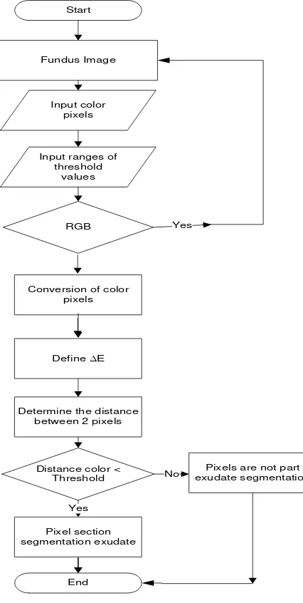
The exudates sometime can be seen as individual fundus or in the form of clusters with yellowish white color. To perform exudates area segmentation on fundus image, semi-automatic segmentation on exudates area as Region of Interest is carried out. The steps of the algorithms can be seen below [8]:
1. Read the fundus image (Automatically the image is stored in 3D RGB matrix).
2. Determine the color pixel of the object of interest area as exudates color reference for segmentation.
3. Determine the range of threshold distance color.
4. When the color that are used for the experiments are other than the one in RGB color space, convert the image and reference pixel color from RGB space color to a color space used for segmentation.
5. For each pixel in fundus image, the color distance calculated using city block distance, which subsequently expressed in equation (1) and (2) where: ( , , )
1 1 G B
R and
(
R
2,
G
2,
B
2)
are stands for Red, Green and Blue color component.b. If the color distance value is less than threshold value, the pixel is the component of the segmented exudates area. Since the pixel is not part of area segmentation, therefore this area is removed.
In general, the semi-automatic segmentation algorithm towards exudates area as Region of Interest (ROI) can be seen in Figure 1 below.
Figure 1. General Chart for Determining ROI Exudates
2.2
Color Space Conversion Process
Exudates segmentation consists of some segment:
1. RGB color space and Euclidian distance formula.
2. HSV space and distance Dcyl with equation (3)[8]
)
The steps to transform RGB to HSV for detection of exudates in color fundus of retinal images can be seen below:
1. Read the RGB fundus image
Pixels are not part exudate segmentation
Yes
No
End
2. Transpose RGB image to HSV image using MATLAB. The pseudocode for transposing the image is:
image = rgb2hsv(rgbImage); h = image(:, :, 1);
s = image(:, :, 2); v = image(:, :, 3);
3. Performing image filter based on the reference values (for determining the value of T) and as a tolerance value with x as HSV color on the existing pixel.
4. If the colors are not include in the range of T, they are colored with black.
5. Re-transpose the RGB image
6. Display the color filter results ranging from T until x + T where x is the color references (exudates).
In general, the RGB color space conversion process using Euclidian distance equation can be seen in Figure 2.
Figure 2. General Chart of RGB Color Space Conversion Process
2.3
Optic Disc Eliminating Process
In this experiment, the color reference of value x is determined with some tolerance value T to specify the similarity level of exudates color to be segmented. There are
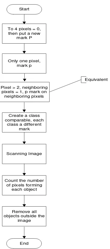
some difficulties in exudates detection since exudates have almost similar color intensity with optical disc as optic disc is a part of retinal image object. The pixel in optic disc is not required and must be removed since this optic disc is considered as noise. The experiments in this paper tag the connected component to remove pixel optic disc that have color intensity similarity with the exudates. The tagging is carried on by examining pixel (P) by pixel (from left to right and top to down) to identify the area of connected pixel (conn). Conn is an area from the adjacent pixel that have similar intensity or the intensity values are within the set of V (In binary image V = {1}). Component connectivity is linked to the input image with connectivity rules (4, 8 or m-connectivity). The standard connectivity for image fundus 2D is pixels. The component marking connection for 8-connected is similar to 4-8-connected. Only the neighboring pixels are examined; beside left and right pixels, the two upper diagonal pixels of P also examined. Therefore, there are 4 neighboring pixels P that are tested. They are upper, left, left diagonal and right diagonal pixel [9] as can be seen in Figure 3.
Figure 3. Optic Disc Eliminating Process Start
Fundus image RGB
Filter with T (variative)
Color is not within the range T
Black Tranpose RGB to
HSV
Yes
NO
The color filter in the range T to x+T
End
Start
To 4 pixels = 0, then put a new
mark P
Only one pixel, mark p
Pixel > 2, neighboring pixels = 1, p mark on
neighboring pixels
Equivalent
Create a class comparable, each
class a different mark
Scanning Image
Count the number of pixels forming
each object
Remove all objects outside the
image
28
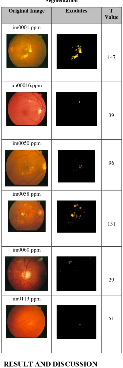
Table 1. The Experiment Results of Exudates Segmentation
Original Image Exudates T Value
im0001.ppm
147
im00016.ppm
39
im0050.ppm
96
im0058.ppm
151
im0060.ppm
29
im0113.ppm
51
3.
RESULT AND DISCUSSION
In general, the experiment of segmentation based on color space approach in this paper can be seen in Figure 5. During the pseudocode execution, the operator should click twice on the same image in the area of the exudates image that going to be segmented. The RGB color of the image pixel fundus is used as references to measure the color distance in RGB color
space. In this paper, the experiment is carried out using image fundus with exudates according to some reference of T values.
Figure 4. Exudates Segmentation with Varied Values of T.
(a). T=29. (b) T= 147
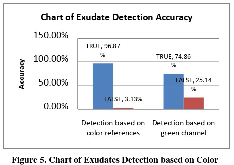
There are many researches working on the area of exudates detection. The previous research use exudates segmentation based on green channel approach. The drawback of this approach is the misinterpretation in segmentation process between exudates and optic disc in retinal fundus image. Both of these objects have similarity of color intensity. Our approach overcomes this drawback by proposing exudates detection based on color references. The exudates are perfectly segmented without the part of optic disc. Based on the test results in Table 2 are used to determine the sensitivity, specificity and accuracy of the system based on the equation presented [10].
Table 2. Sensitivity, Specificity and Accuracy Based on Color Reference Approach
Sensitivity
in %
Specificity
in %
Accuracy
in %
96.87% 78% 95.54%
Table 2 shows the results of sensitivity, specificity, and the percentage of accuracy for the trial image. The test results show that the system can detect exudates to the sensitivity of the system reached 96.87% and specificity of the system reaches 78%. The accuracy of the system is 95.54%. Based on the results in Table 2 clearly shows that the detection of exudates based on reference color space approach provides excellent results with the accuracy of the system (Table 2) compared with the accuracy based on green channel approach. The detection of the reference color space based exudates contributes significantly to increasing the detection accuracy of exudates of 22.01% (Figure 5).
Original Image (im0016)
Original Image (im001)
Exudates using T=39
Exudates using T=147 (a)
Figure 5. Chart of Exudates Detection based on Color Reference Approach and Green Channel Approach
4.
CONCLUSION
Based on the obtained results, our experiments applied varied threshold values which are the limit boundary of the best exudates color similarity that are used for segmentation process according to the color. The value of T can be customized to the preferred level of precession similarity. The higher the threshold value used the harder the exudates, otherwise decrease the value of T for soft exudates (e.g., cotton wool spot). The obtained accuracy value for exudates detection using color references space is 95.54% using ground truth data of image comparison according to the color of the exudates area. This accuracy is achieved by comparing the pixels types of the exudates that are correctly detected and not. For future works, the number of extracted pixels and the establishment of exudates area are calculated. The counted pixels then multiplied by the size of the pixels according to the resolution of image acquisition.
5.
ACKNOWLEDGMENTS
We are indebted to the experts who have contributed towards development of the template.The authors would like to acknowledge to Gunadarma University, dr Rakhma Indria Hapsari.,SpA from Awal Bros Hospital Tangerang (Ophtamologist), Research Center of Medical-Informatics of Gunadarma University
6.
REFERENCES
[1] Vaughan DG, Asbury T, Riordan Eva P. General Ophthalmology. Edisi 17. London: McGraw-Hill. (2007)
[2] Jack J. Kanski, Brad Bowling. Clinical Ophthalmology: A Systematic Approach 7th Edition. Butterworth Heinemann Elsevier. ISBN-13: 978-0702040931. (2011).
[3] B.Ramasubramanian and G.Mahendran. An Efficient Integrated Approach for the Detection of Exudates and Diabetic Maculopathy in Colour fundus Images. Advanced Computing: An International Journal ( ACIJ ), Vol.3, No.5. (2012)
[4] Joussen A.M.2007. Retinal Vascular Diseease. New York: Springer. p. 3-5, 66-70, 129-132, ,228-31, 309, 291-331
[5] Nidhal K. El Abbadi, Enas Hamood Al-Saadi. Automatic Detection of Exudates in Retinal Images. International Journal of Computer Science Issues Vol.10, Issue 2 No.1. ISSN (Print): 0814 | ISSN (Online) : 1694-0784.(2013)
[6] Marwan D. Saleh, C.Eswaran and Ahmed Mueen. (2011). An Automated Blood Vessel Segmentation Algorithm Using Histogram Equalization and Automatic Threshold Selection. Journal of Digital Imaging.DOI: 10.1007/s10278-010=9302-9.
[7] Ratna Bhargavi and V. Rajesh. (2015). Detection And Feature Extraction Using Active Contour Model And Sift In Color Fundus Images. ARPN Journal of Engineering and Applied Sciences. VOL. 10, NO. 6.
[8] Sarifuddin Madenda. Pengolahan Citra & Video Digital. Teori, Aplikasi dan Pemrograman Menggunakan Matlab. Ed. Erlangga.ISBN. 978-602-298-598-3. (2015)
[9] Rodiah, Sarifuddin Madenda, Fitrianingsih. Three-Dimensional (3D) Reconstruction for Detectiong Shape and Volume of Lung Cancer Nodules. IPTEK Journal ITS. No.1. (2014).
[10] Priya, R., Aruna, P., Review of Automated Diagnosis Of Diabetic Retinopathy using The Support Vector Machine, International Journal of Applied Engineering Research, No. 4, Vol. 1, pp: 844-863. 2010
[11] Gonzalez R. C. and Woods E. E., Digital Image Processing. 3rd Edition. Prentice Hal;. (2007)
TRUE, 96.87
% TRUE, 74.86 %
FALSE, 3.13%
FALSE, 25.14 %
0.00%
50.00%
100.00%
150.00%
Detection based on color references
Detection based on green channel
A
cc
u
rac
y