RESEARCH
ELEVATED SERUM S100B PROTEIN LEVEL AS A PARAMETER FOR
BAD OUTCOME IN SEVERE TRAUMATIC BRAIN INJURY PATIENTS
(Peningkatan Kadar Serum Protein S100B Sebagai Tolok Ukur Keluaran Buruk di
Pasien Cedera Kepala Berat)
Ridha Dharmajaya1, Dina Keumala Sari2, Ratna Akbari Ganie3
ABSTRAK
Beratnya suatu cedera kepala akibat trauma akan membuat gangguan saraf pusat. Kerusakan saraf ini dapat dinilai dengan petanda biokimia yang tepat. Pemakaian petanda biokimia terhadap kerusakan otak mendapatkan perhatian yang banyak terutama Protein S100B. Protein S100B adalah suatu ikatan kalsium dan protein yang meningkat cepat sesaat setelah cedera kepala. Kesulitannya adalah untuk memastikan, berapa lama Protein S100B ini harus diukur. Jika berhubungan dengan kerusakan otak, ia tidak selalu terjadi pada 24 jam pertama. Dapat terjadi pada 48–72 jam pasca cedera kepala, bahkan 120 jam pada kecederaan tersebut. Penelitian ini bertujuan untuk mendapatkan kenasaban antara Protein S100B dengan GOS sebagai faktor peramalan yang akurat, mudah, tidak menyakitkan, untuk cedera kepala berat. Pengambilan serum darah untuk pemeriksaan kadar Protein S100B dilakukan pada 24, 48, 72 dan 120 jam pasca trauma. Selanjutnya pengukuran dilakukan dengan menggunakan Enzyme Linked Immunosorbent Assay (ELISA). Keluaran pasien pasca perawatan dinilai menggunakan penggolongan Glasgow Outcome Scale (GOS), tiga bulan pasca kecederaan. Hasil pengukuran kadar Protein S100B pada 120 jam pasca cedera kepala berat menunjukkan hubungan berlawanan yang kuat terhadap keluaran pasien. Pasien cedera kepala berat dengan kadar Protein S100B 120 jam pasca trauma yang tinggi, memiliki hasil keluaran yang buruk.
Kata kunci: Cedera kepala berat, Glasgow Outcome Scale, Protein S100B
ABSTRACT
The severity of traumatic brain injury that makes disorders of Central Nervous System (CNS) can be assessed with biochemical markers. There has been an increased interest in the clinical use of brain markers such as S100B. It is a calcium binding protein that increases rapidly after head injury. The difficulty is to make sure, how long the biomarker should be check. If it is related with brain damage, it will not always come within the first 24 hours. It will come at 48–72 hours post-traumatic brain injury, even within 120 hours after the injury. The objective of this research was to find a correlation between protein S100B serum with Glasgow Outcome Scale (GOS) as a prognostic factor which is simple, minimally invasive for severe brain injury. Protein S100B serum level was measured at 24, 48, 72 and 120 hours after trauma and assessed by using Enzyme-Linked Immunosorbent Assay (ELISA). The outcome was measured by GOS classification three months after the injury. Results found that protein S100B serum level at 120 hours after the injury has a negative strong correlation (r= − 0.6) with GOS (p=0.02). Post-traumatic brain injury patients with high serum levels of protein S100B at 120 hours after trauma may have a bad prognosis.
Key words: Glasgow Outcome Scale, Protein S100B, severe head injury
1 Department of Neurosurgery, Faculty of Medicine, University of Sumatera Utara/Adam Malik General Hospital, Medan, Indonesia. E-mail: [email protected]
2 Department of Nutrition, Faculty of Medicine, University of Sumatera Utara, Medan, Indonesia 3 Department of Clinical Pathology, Faculty of Medicine, University of Sumatera Utara, Medan, Indonesia INTRODUCTION
S100B protein is a calcium and protein bond used as a parameter of glial cell activity and/or death in many disorders of the Central Nervous System (CNS).
S100B protein also plays an important role in the development and repair of CNS damage, including in head injuries.1
The continued effect of a secondary head injury, moreover, can generate pressure within the cranium
cavity. As a result, brain cell death occurs and exacerbates the pathological process of the brain. At a decompensating stage, when the cranium cavity is unable to withstand an increasing volume, there will be a herniation resulting in death.2,3 Therefore, a
secondary head injury process should be prevented to obtain maximum treatment outcomes. However, there are still some difficulties in determining the severity of a secondary head injuries, leading to poor treatment outcomes, or in examining the process whether it is still in a reasonable stage in order to obtain good results. Thus, treatment management is essentially needed to determine prognosis.
Furthermore, impaired delivery and absorption of oxygen also may cause swelling of the brain cells. Increased Intracranial Pressure (ICT) due to the disorder then will disrupt the physiological processes of the brain. Blood flow in the brain becomes disturbed which can further aggravate ischemia process and brain metabolic disorders. This process is considered as a secondary effect of the secondary head injury lasting up to 48 to 72 hours after the injury.2,3
Head injury actually can be divided into four types based on the degree of awareness in patients by using Glasgow Coma Scale (GCS) classification, namely mild, moderate, severe and critical.4,5 Meanwhile, based on
the pathological condition of the head, the head injury can be divided into primary head injury and secondary head injury. Primary head injury is a direct impact of the head injury, while the continued effect of the direct impact is called secondary head injury. Secondary head injury, in other words, is considered as a result of brain perfusion disorders.4-6
Glasgow Outcome Scale is usually used to evaluate the advanced clinical condition of the head in head injury cases. If there is an increase in intracranial pressure within three to ten days, it can suggest a poor prognosis. Nevertheless, although intracranial pressure measurement requires an invasive action, it still cannot evaluate what happens in brain cells. Similarly, radiological parameter also has the same weakness.6,7
Thus, supporting facilities that can evaluate the intracellular conditions of the brain are required to determine the prognosis of head injury. One of the cellular markers that can evaluate the pathological conditions of nerve cells is S100B protein, most developed rapidly in researches. This protein includes the family of the S-100 protein, a calcium-binding protein that is released in the peripheral circulation immediately after brain injury. S100B protein, furthermore, is found in the cytosol of the astroglial and Schwann cells. Serum S100B protein level may
increase rapidly in the first minute after the head injury, up to 5-20 μg/L.8
In addition, serum S100B protein levels in moderate and severe head injuries will elevate as soon after the injuries and then will decrease, but not reach the normal score. In a head injury followed by a secondary head injury, the elevated serum S100B protein level at the onset may continue to increase in subsequent hours. Consequently, the elevated serum S100B protein level in blood may illustrate the severity of brain cell damage and have a high predictor value of patient output, compared with other clinical and radiological parameters.8-11 For these reasons, this research would
evaluate the correlation of serum S100B protein level to the output of the severe traumatic brain injury. This research also aimed to examine the best time of examining this marker.
This research aimed to evaluate the correlation of serum S100B protein level to worsening output (GOS) in three months after a traumatic brain injury.
METHODS
This research was a prospective analytical obser vational study conducted in Emergency Installation, Intensive Care Unit (ICU) and Inpatient Room of Malahayati Islamic Hospital, Medan, North Sumatera. Patients with traumatic brain injury aged 20-60 years old and examined with Glasgow Coma Scale (GCS)5-8 were included as research subjects in
this research. These subjects were represented by their family who was responsible for the patients’ self-willing to approve the installation of intracranial pressure monitor and participate in this research. Meanwhile, patients with mydriasis, bilateral and multiple traumas, operative lesions due to results of CT scans, as well as a history of chronic diseases, cerebral tumors and infections were excluded from this research. Some of the research subjects were also dropped out if their family representing them refused to continue participating in this research.
would be categorized into GOS 5. If the subjects could perform their daily activities independently or had mid disabilities, the results would be categorized into GOS 4. If the subjects were unable to perform routine activities without the help of assistants or severe disabilities, the results would be categorized into GOS 3. If the subjects had a persistent vegetative state, the results would be categorized into GOS 2. And if the subjects were dead, the results would be categorized into GOS 1.11 The serum S100B protein levels then
were associated with GOS to evaluate its influence as a prognostic factor.
In the measurement of serum S100B protein level, the blood of the research subjects was taken as much as 5ml using disposable syringe through vena mediana cubiti after an aseptic action was performed using 70% alcohol.
Next, the blood without anticoagulants was allowed to freeze at room temperature for 30 minutes and centrifuged at 3000 rpm for 15 minutes. The serum then was taken. Afterwards, serum S100B protein level was examined using Elisa Human S100B reagents (Bio-Vendor-Research and Diagnostic Product) in the form of component devices. This measurement using Elisa reader was set at a length wave of 450nm controlled periodically. The level of serum S100B protein at the initial examination was more than 2.5
μg/liter, indicating a high risk of worsening outcome. Meanwhile, the serum S100B protein level of > 2μg/ liter depicted a high risk of poor outcome, or even death.9-14
Data obtained were processed using SPSS version 15. Kolmogorov-Smirnov test was performed to evaluate whether the data were normally distributed or not. If a p-value was more than 0.05, indicating that the distribution of the data was normal, a parametric analytical test was conducted. But, if it was not, non-parametric analytic test would be carried out. Numerical data, moreover, were presented in the mean ± standard and median intersection (minimum-maximum), while categorical data were presented in the number of patients (percentage). The significance limit used in this research was 5%. The data were not significant if a p-value was more than 0.05. On the other hand, the data were significant if a p-value was less than 0.05. Next, Pearson analysis test was performed to examine the numerical correlative hypothesis of the normal data distribution. Meanwhile, to analyze the numerical correlative hypothesis of the abnormal data distribution, Spearman analysis test was conducted. The correlation strength of 0.0- <0.2 was stated to be very weak, 0.2- <0.4 for weak correlation,
0.4- <0.6 for moderate correlation, 0.6- <0.8 for strong correlation and 0.8–1 for very strong correlation.
This research had been approved by the Committee of Health Research Ethics of University of Brawijaya, Malang, East Java, numbered: 066/EC/KEPK-S3-JK/03/2011. Prior to participating in this research, every family member representing the research subjects was asked to fill out the approval sheet after explained about the objectives of the research, examinations required and their advantages and disadvantages. All data and information obtained then were kept confidential. Thus, if the research subjects felt feel aggrieved, then they could state their withdrawal.
RESULTS AND DISCUSSION
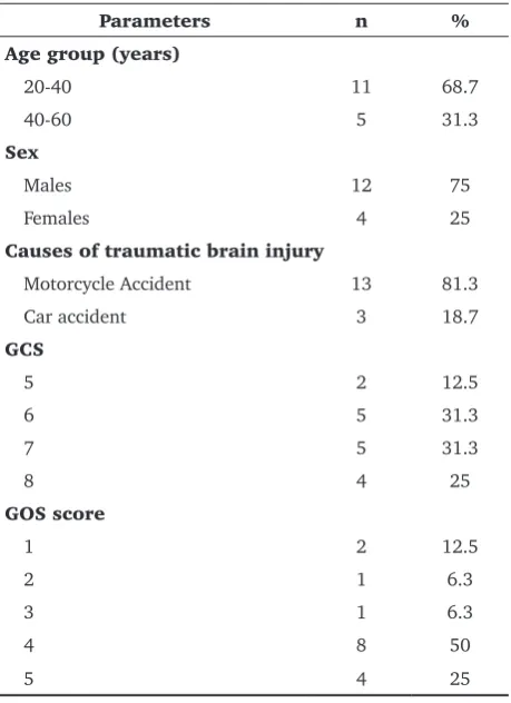
In this research, there were 73 cases of traumatic brain injuries, consisted of 35 of severe head injury cases, 12 of operative lesion cases detected on CT scan, 7 of severe head injury cases with bilateral mydriasis pupils. Thus, the total of the research subjects was 16 (see Table 1) with the most age group of 20–40 years (68.7%). The percentage of male patients with severe traumatic brain injury was higher (75%) with motor
Table 1. Demographic characteristics and research parameters (n=16)
Parameters n %
Age group (years)
20-40 11 68.7
40-60 5 31.3
Sex
Males 12 75
Females 4 25
Causes of traumatic brain injury
Motorcycle Accident 13 81.3
Car accident 3 18.7
GCS
5 2 12.5
6 5 31.3
7 5 31.3
8 4 25
GOS score
1 2 12.5
2 1 6.3
3 1 6.3
4 8 50
αβ, whereas S100B is a homodimer of ββ. S100A is found to be abundant in nerves, muscles, kidneys and other organs, while S100B is localized to neuronal cells and Schwann cells. The protein formerly known as S100α is now known as the S100A protein, whereas the protein formerly known as S100β protein is now called as S100B protein.15,16
Effects of S100B depend on its concentration. At nanomolar concentration, S100B in vitro stimulates neurites to grow large in nerve cells in the cerebral cortex and improves survival of neuronal cells in progression. In contrast to the nanomolar concentration, in micromolar concentration, S100B is found to be destructive since it stimulates the release of proinflammatory cytokines and triggers apoptosis. Recent observations even show that S100B protein at the micromolar concentration can cause apoptotic death by interacting with Receptor for Advanced Glycation End Produces (RAGE), leading to increased reactive oxygen, cytochrome C release and Caspase cascade activation. The high concentrations of S100B protein can also trigger the death of nerve cells through the release of nitric oxide from astrocytes. The biological half-life of S100B is for 30 minutes. This implies an increase in serum S100B protein level for a long time, as an illustration of the continued release of a damaged tissue.15,16
As a marker, S100B protein, moreover, is primarily produced by astrocytic cells in the CNS and indicates
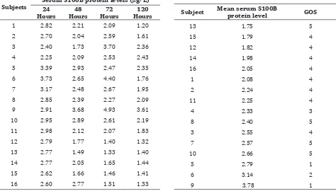
Table 2. Serum S100B protein levels at several hours (n=16)
Subjects
Serum S100B protein levels (μg/L) 24
cycle accident as the most common cause of the injury (81.25%).
Glasgow Coma Scale score was assessed when the patients were admitted after resuscitation. In accordance with the inclusion criteria, all research subjects had severe traumatic brain injury with a GCS score of 6–7 (31.3%). Based on results of the GOS observation after three months, the majority of subjects had moderate disability, i.e GOS 4 (50%).
Table 2 showed that the serum S100B protein levels at 24, 48, 72 and 120 hours after the trauma. Meanwhile, how the mean serum S100B protein level in each patient was associated with GOS can be seen in Table 3.
Calcium is an intracellular second messenger that plays a role in the conduction and transmission of nerve impulses, the contraction of muscles, the motility, growth and development of cells, the expression of the gene, as well as apoptosis and necrosis. As a result of cellular evolution, calcium and protein bonds are formed to regulate the calcium levels in the cytosol and alter the calcium signals. The largest group part of the calcium-protein bond (helix E-loop-helix) is S100 protein. It is called S100 since it can dissolve in 100% ammonium sulfate solution. S100 protein contains a mixture of hetero and homodimer as well as two types or sub units (α, β). Both sub-units have different amino acid compositions. S100A is a heterodimer of
Table 3. The correlation of the mean serum S100B protein level to GOS
Subject Mean serum S100B
the activation of an astrocytic cell. Researches in the field of immunohistochemistry even show that S100B protein astrocytes are mostly produced in gray tissues, while their production in white tissues was performed by oligodendrocyte cells. Outside or inside of cells, S100B protein levels, thus, are used as parameters of astrocytes activation and or death, especially in head injury. In other words, its release tends to be an effect of a state, not as a cause; consequently, it reinforces the role of the S100B protein as a marker of both head injury and central nervous injury.15
After the primary head injury, serum S100B protein level even can increase rapidly to 5-20 μg/L in the first minute due to the release of S100B protein from primary brain cell damage, leading to an increase in severe head injury continued with secondary brain damage.9 In this research, there was an increase in
the serum S100B protein level in the first 24 hours. This is in accordance with some previous researches conducted by Raabe et al14, Townend et al9 and
Mehta11 indicating elevated S100B levels in the first
minute after head injury.9,11,14
However, this research had different results from other researches showing decreased S100B protein levels. For instance, a research conducted by Raabe et al14 found a rapid decrease in S100B protein level,
even reaching the normal level within 4-6 hours.14
Unlike the research conducted by Raabe et al14, in this
research, S100B protein levels decreased at the 48th
hour of the examination, but did not reach the normal one. The decreased S100B protein levels even could be classified into a dangerous level, > 2 μg/L. This decrease was only temporary since it increased again at the 72nd hour of the examination and then decreased
at the 120th hour of the examination, reaching no
harmful level. The differences in the results may be due to the high level of variables of their research subjects.14 Unlike this research, Raabe’s14 research did
not only take subjects with severe head injuries, but all types of the head injury.
Therefore, some previous researches with subjects of severe head injury also show no different results. For instance, a research conducted by Mehta14, classifying
the subjects into mild, moderate and severe head injury groups also found the same results.11 Similarly,
other previous researches on the subjects of severe head injury showed the same results.16-20 S100B levels
increased, high above the normal level up to the 5th
day of examination. The occurrence of the increased secondary injury in the subsequent examination could predict poor outcomes from the subjects with the head injury. On the 6th and 9th day, there was an increase
in S100B protein level at the first 24 hours followed by an elevated S100B protein level at the 72 hours of the examination.
The decreased S100B level to a value below 2 μg/L at the 120 hours of the examination can determine good GOS score for the patients. In this research, the levels of S100B protein decreased at the 48 hours although not reaching the normal GOS score, but still classified as hazardous one (> 2 μg/L). This decrease was only temporary. The levels of S100B protein increased again at the 72 hours of the examination and then decreased at the 120 hours of the examination, but reaching no harmful GOS score. These results indicate the duration of secondary head injury and a good response to treatment. Unlike this research, Raabe et al’s research showed a rapid decrease in S100B levels, even reaching normal GOS score within 4-6 hours.14
Finally, based on the mean serum S100B protein level in this research, it can be said that the lower the mean serum S100B protein level is, the higher the patients’ GOS score would be. However, the higher the mean serum S100B protein level is, the lower the patients’ GOS score would be.
CONCLUSION AND SUGGESTION
In conclusion, in patients with severe traumatic brain injury, serum S100B protein level will elevate at the time of injury and then will fluctuate up to 120 hours after the injury. The elevated serum S100B protein level at the 120th hour after the injury is
significantly associated with a poor outcome. As a result, serum S100B protein level needs to be examined as part of the management of head injury in order to predict GOS in patients with a severe traumatic brain injury after three months.
REFERENCES
Guerrero JJE,
1. Cabezas FM, Carrión JL. The Clinical Utility of the S100Β Protein during Traumatic Brain Injury Management. International Brain Injury Association – IBIA, 2013.
Lindsay KW, Bone I. Neurology and neurosurgery illustrated. 2.
Ed ke-4., United State of America, Edinburgh Churchill Livingstone, 2004; 214–216.
American College of Surgeons Committee on Trauma. 3.
Advanced Trauma Life Support (ATLS) for Doctors: Student Course Manual. Ed ke-7., Chicago, Illinois, United States of America, American College of Surgeons 2004; 154-156. Selladurai BM, Reilly P. Neurosurgical procedures in the 4.
Andrews BT. History, classification, and epidemiology of 5.
cranial trauma. Dalam: Batjer HH, Loftus CM, editor. Textbook of neurological surgery, principles and practice. Philadelphia, Lippincott Williams & Wilkins, 2003; 2795–2798.
Maas AIR, Steyerberg EW, Butcher I, Dammers R, Lu J, 6.
Marmarou A, dkk. Prognostic value of computerized tomography scan characteristics in traumatic brain injury: result from the IMPACT study. J Neurotrauma. 2007; 24(2): 303–314.
Smith M. Monitoring intracranial pressure in traumatic brain 7.
injury. Int Anesthesia Res Soc. 2008; 106(1): 240–248. Raabe A, Kopetsch O, Woszczyk A, Lang J, Gerlach R, 8.
Zimmermann M, dkk. Serum S100B protein as a molecular marker in severe traumatic brain injury. Restor Neurol Neurosc. 2003; 21(3–4): 159–169.
Townend W, Dibble C, Abid K, Vail A, Sherwood R, Lecky F. 9.
Rapid elimination of protein S100B from serum after minor head trauma. J Neurotrauma. 2006; 23(2): 2–7.
ARUP Laboratories. S100B protein, serum for management 10.
of patients with acquired brain injury national reference laboratory. Salt Lake City, Utah. 2009; 1-2.
Mehta SS. Biochemical serum markers in head injury: an 11.
emphasis on clinical utility. Clin Neurosurg. 2010; 57: 134–140.
Townend W, Guy MJ, Pani MA, Martin B, Yates DW. Head 12.
Injury outcome prediction in the emergency department: a role for protein S100B. J Neurol Neurosurg Psychiatry. 2002; 73(5): 542–546.
Bazariana JJ, Beckb C, Blytha B, Ahsenc N, Hasselblatt M. 13.
Impact of creatine kinase correction on the predictive value of S100B after mild traumatic brain injury. Restor Neurol Neurosc. 2006; 24: 163–172.
Raabe A, Gromls C, Seifert V. Serum markers of brain damage 14.
and outcome prediction in patients after severe head injury. British J Neurosurg. 1999; 13(1): 56–59.
Yardan T, Erenler AK, Baydin A, Aydin K, Cokluk C. Usefulness 15.
of S100B Protein in Neurological Disorders. J Pak Med Assoc. 2011; 61: 276–281.
Wilson JTL, Laura EL, Pettigrew LEL. Structured interviews 16.
for the Glasgow outcome scale and the extended Glasgow outcome scale: guidelines for their use. J Neurotrauma. 1998; 15(8): 573–585.
Kleindienst A, Mcginn MJ, Harvey HB, Colello RJ, Hamm 17.
RJ, Bullock MR. Enhanced hippocampal neurogenesis by intraventricular S100B infusion is associated with improved cognitive recovery after traumatic brain injury. J Neurotrauma. 2005; 22(6): 645–655.
Korfias S, Stranjalis G, Boviatsis E, Psachoulia C, Jullien G, 18.
Gregson B, dkk. Serum S100B protein monitoring in patients with severe traumatic brain injury. Intensive Care Med. 2007; 33: 255–260.
Kotlyar S, Larkin GL, Moore CL, D’Onofrio G. S100B 19.
immunoassay: an assessment of diagnostic utility in minor head trauma. J Emerg Med. 2010; 19: 30–37.
Li DR, Zhu BL, Ishikawa T, Zhao D, Michiue T, Maeda H. 20.