R E S E A R C H A R T I C L E
in vitro
Antidiabetic and Antioxidant Activities of Aqueous Extract from
the Leaf and Fruit of
Psidium guajava
L.
Adelina Simamora
1,, Lusia Paramita
1, Nur Azreen Binti Mohamad Hamid
1,
Adit Widodo Santoso
2, Kris Herawan Timotius
11Department of Biochemistry, Krida Wacana Christian University, Jl. Tanjung Duren Raya No. 4, Jakarta, Indonesia 2Department of Herbal Medicine Krida Wacana Christian University, Jl. Tanjung Duren Raya No. 4, Jakarta, Indonesia
Corresponding author. E-mail: [email protected]
Received date: Oct 10, 2017; Revised date: March 28, 2018; Accepted date: Apr 19, 2018
B
ACKGROUND: The leaf and fruit of Psidium guajava L. are potential for neutraceutical beverage especially for antidiabetic drink. The aims of this study were to determine the antidiabetic activity of aqueous extract of leaf (LE) and fruit (FE) from P. guajava.METHODS: Both extracts were investigated for their
inhibitory effect on α-glucosidase activity in vitro. Their antioxidant activities were measured by 2,2-diphenyl-1-picrylhydrazyl (DPPH) free radical scavenging, ferrous ion chelating, reducing power and phosphomolybdate methods.
RESULTS: The IC50 of LE, FE and acarbose as a positive
control were 5.67, 428.00 and 823.99 μg/mL, respectively.
The enzyme kinetic analysis indicated that LE inhibited
α-glucosidase in a competitive inhibition type, similar to that
of acarbose. Both extracts showed antioxidant activities, with LE showed stronger activities than FE in all methods.
In DPPH method, IC50 of LE and FE were 74.77 and 843.84
μg/mL respectively, compared to 53.24 and 21.36 μg/mL for
reference antioxidants butylated hydroxytoluene (BHT) and ascorbic acid (AA), respectively. In ferrous ion chelating activity, the IC50 were 147.07 and 2105.05 μg/mL for LE and FE, whereas ethylenediaminetetraacetic acid (EDTA) as
a control sample was 66.50 μg/mL. In reducing power and
phosphomolybdate methods, at different concentrations, the activities of LE, FE, and standard compounds showed the following order: AA > BHT > LE > FE.
CONCLUSION: LE from P. guajava exhibited excellent
inhibitory activity against α-glucosidase. In addition,
LE had better antioxidant acivities than FE. This study can recommend the aqueous extract from P. guajava as a promising candidate for neutraceutical drink for prediabetic and diabetic patients.
KEywORDS: antioxidant, aqueous extract, α-glucosidase inhibition, guava, Psidium guajava L.
Indones Biomed J. 2018; 10(2): 156-64
Abstract
Introduction
Diabetes mellitus (DM) is a group of metabolic disorder
diseases typified by high level of blood glucose
(hyperglycaemia) over a prolonged period. This may be
due to either deficiency of insulin secretion (as in type 1
DM) or a combination of resistance to insulin action and an inadequate insulin secretion as in type 2 DM (T2DM).(1)
digestive enzymes such as α-glucosidase. Consequently,
inhibitors that target enzymes involved in the digestion of polysaccharides serve as a key strategy in the management
of T2DM. Several synthetic α-glucosidase inhibitors such
as acarbose, voglibose and miglitole are currently used in the treatment of T2DM. However, some unfavourable side effects related to gastrointestinal complications have been reported, such as diarrhoea, abdominal distention
and flatulence.(2) Thus, it is essential to search for safer α-glucosidase inhibitors which are devoid of adverse side
effects of the aforementioned synthetic inhibitors.
One of the major causes of T2DM arises from
the damage of pancreatic β-cells, which are induced by
excess free radicals and the resulting oxidative stress.
The susceptibility of pancreatic β-cells to oxidative
destruction is also due to the minimum antioxidant defence systems.(3) Therefore, diets containing antioxidant compounds may be helpful in protecting cells from the oxidative damages thus preventing the development of DM and its complications.
Plant materials are good source for neutraceutical drink. Many plant materials have been reported to have
inhibitory activities on α-glucosidase (4) and antioxidant activities (5). Moreover, several inhibitors on α-glucosidase
and antioxidant compounds have been successfully isolated from plants to serve as an alternative drug with promising potency and less undesirable side effects than existing drugs.(6)
P. guajava is widely distributed in all tropical and subtropical area. Across the South American and Asian countries including Indonesia, P. guajava has been widely used to treat a number of symptoms, mainly for gastrointestinal and respiratory disturbances and
anti-inflammatory medicine.(7) The leaves are also traditionally
used as antidiabetic remedy.(8) The treatments usually involve decoction or infusion of parts of the plant, such as leaf, bark, fruit and shoot. Recent works have reported compounds from guava leaves elucidated using various solvent systems that have antidiabetic and antioxidant properties. These
include phenolic and flavonoid compounds (gallic acid,
quercetin, kaempferol, guaijaverin, avicularin, myricetin, hyperin and apigenin) and polymerized polyphenol.(9) Despite the numerous traditional use of P. guajava, only a few of these, in particular those involving with water
extraction of the plant materials, are supported by scientific
evidence. Hence, the objectives of the present study were
to determine the α-glucosidase inhibitory activity and
antioxidant activities from leaves and fruit of P. guajava.
Methods
Preparation of P. guajava Extracts
The plant materials P. guajava were collected in February 2017 from Pagarawan village, Merawang, Bangka, Indonesia and the specimen was authenticated by one of the authors. A herbarium of the plant was kept in the Research Laboratory Centre for Herbal Medicine studies, Krida Wacana Christian University, Jakarta, Indonesia, with voucher specimen number KWF015. Leaves and chopped ripe fruit were dried at room temperature and each was ground into a homogenous powder using a mill. The powder was kept at 4oC prior to use.
The extracts were prepared following reported
procedure (5) with some modifications. The leaf extract (LE)
was prepared by decoction by placing 2 grams of the powder in 200 mL of deionised hot water at 90oC. Decoction sample
was gently agitated, and the temperature was kept constant to reduce the solvent until half of the starting volume. After
filtration, extract was lyophilized with a freeze dryer
(MRC-FDN-10N-50-BA) to obtain a light brown powder. The procedure was repeated for fruit extract (FE).
Total Phenolic Content
Total phenolic content of aqueous extracts from P. guajava
was determined by Folin-Ciocalteu method (10) with slight
modification. An aliquot (0.5 mL) of samples (2.02 and 2.75 mg/mL for LE and FE, respectively) was mixed with
2.5 mL of Folin-Ciocalteu (Cat. #F9252) (Sigma-Aldrich,
St Louis, USA) reagent (10%, w/v). The solution was left
to stand for 10 minutes at room temperature. The reaction was then neutralized using saturated sodium carbonate (Cat. #1063860001) (Merck, Darmstadt, Germany) solution
(75 g/L). After incubation for 2 hours in darkness at room
temperature, the absorbance was measured at 765 nm using spectrophotometer Biochrom Libra S-22 (Biochrom, Cambridge, UK). The total phenolic content was estimated from a standard curve of gallic acid 12.5, 25, 50, 100, and
200 μg/mL (Cat. #sc205704) (Santa Cruz Biotechnology,
Dallas, USA). The results were expressed as mg gallic acid
equivalent (mg GAE)/gram dry weight of plant material.
Total Flavonoid Content
Total flavonoid content was determined using aluminium chloride colorimetric method (11) with slight modification.
water (2 mL). The reaction was incubated for 5 mins and was added with 0.15 mL of AlCl3 (Cat. #11019)
(Sigma-Aldrich) solution (10%, w/v). The reaction was incubated
for another 5 minutes and was added with 2 mL of NaOH (1 M). The mixture was left to stand for 15 minutes at room temperature and the absorbance was read at 510
nm with spectrophotometer. The total flavonoid content
was determined from a rutin (Cat. #sc204897B) (Santa Cruz Biotechnology) standard curve and the results were
expressed as mg rutin equivalent (mg RE)/gram of dry
weight of plant material.
α-Glucosidase Inhibitory Activity
The α-glucosidase inhibitory activity of extracts from P. guajava was assayed in vitro according to the literature
procedure with some modification.(12) Appropriate dilution
of samples (50 µL) were mixed with 50 µL phosphate buffer
(50 mM, pH 6.8), 50 µL of α-glucosidase (Cat. #G5003) (Sigma-Aldrich) solution (0.5 unit/mL). The following
concentration ranges were prepared for LE and FE: 3.16,
4.73, 6.31 and 7.89 μg/mL for LE and 137.50, 412.50, 550.00, 687.50 μg/mL for FE. After pre-incubating for 5 minutes at
37oC, 100 µL of 1mM p-nitrophenyl-α-D-glucopyranoside
(Cat. #N1377) (Sigma-Aldrich) as a substrate was added to the reaction mixture and the reaction was further incubated for 20 minutes at 37oC. The reaction was terminated by the
addition of 750 µL of Na2CO3 (100 mM). The α-glucosidase inhibitory activity was determined spectrophotometrically by measuring the amount of p-nitrophenol released from the substrate at 405 nm. The inhibition percentage was calculated using the following equation:
% inhibition of α-Glucosidase = A control-A sample x 100%
A control
Where A control: absorbance of control, A sampel:
absorbance of sample. The α-glucosidase inhibitory activity
was expressed as inhibitory concentration (IC)50 values (µg/ mL) and was determined from the graph plotted against the percentage inhibition. Values were compared with the positive control acarbose United States Pharmacopeia (USP) (Cat. #1000521) (Sigma-Aldrich) the antidiabetic medicine.
Kinetics Inhibition Mode
The mode on inhibition of α-glucosidase by LE was
determined using a Lineweaver-Burk plot.(13) The kinetics assay was performed using increasing concentrations of the
substrate p-nitrophenyl-α-D-glucopyranoside (0.15 - 1 mM).
Substrate was incubated with α-glucosidase in the absence
of inhibitor and presence of LE at different concentrations
(0 to 4.21 µg/mL). A double reciprocal plot (1/[S] and 1/V)
was constructed based on the Lineweaver-Burk. The mode of inhibition was compared with that of the positive control acarbose.
2,2-diphenyl-1-picrylhydrazyl (DPPH) Radical
Scavenging Activity
The free-radical scavenging capacity of extracts from P. guajava was evaluated using DPPH stable radical
following reported method (14) with minor modification.
The assay is based on the ability of a substrate to donate a hydrogen atom in order to scavenge the DPPH radical. DPPH (Cat. #D9132) (Sigma-Aldrich) solution (0.6 mM in ethanol) was prepared and 1 mL of this solution was added to 3 mL of sample in various concentration; 16.83,
25.25, 33.67, 42.08, 50.50, 67.33 and 84.17 μg/mL for LE and 91.67, 183.33, 366.67, 458.33, 641.67 and 916.67 μg/
mL for FE. The mixture was immediately vortexed and incubated for 30 minutes in darkness at room temperature. The decrease in absorbance was measured at 517 nm using spectrophotometer. The percentage of inhibition activity was calculated according to the following equation:
% Inhibition = A control-A sample x 100%
A control
The concentration of the sample and the reference required
to scavenge 50% of the DPPH radical was defined as IC50
and was determined by the graph plotting against percentage
of inhibition. The values were expressed as µg/mL and
values were compared with those of reference solutions i.e.,
butylated hydroxytoluene (BHT) (Cat. #B1378) (Sigma-Aldrich) and ascorbic acid (Cat. #470300-286) (VWR BDH Prolabo Chemicals, Tingalpa, Australia).
Ferrous Ion Chelating Activity
The ability of aqeuous extract from P. guajava and standard to chelate iron(II) was estimated according to the method (15) in the literature. Concentrations of the extracts were
37.88, 75.75, 151.50, 227.25 and 303.00 ug/mL for LE
and 343.75, 687.50, 1031.25, 1375.00, 1718.75 and
2200.00 ug/mL, for FE. In this method, 0.4 mL extract and
ethylenediaminetetraacetic acid (EDTA) (Cat. #E164) (Sigma-Aldrich) at various concentrations (20, 100, 200 and
400 ug/mL) were added with 0.5 mL FeSO4 (Cat. #103965)
was added and the total volume was adjusted to 4 mL with water. The reaction was immediately vortexed and was left to stand in the dark at room temperature for 10 minutes. The absorbance was read at 562 nm using spectrophotometer. Iron chelating ability was calculated using the following equation:
Ferrous ion chelating = A control-A sample x 100%
activity (%) A control
Where A control: absorbance of control, A sample: absorbance of the sample. The concentration of extracts required to chelate 50% of the Fe(II) ion (IC50) was calculated from the graph plotted against the percentage of inhibition. The IC50 value was expressed as μg/mL and values were compared with the standard EDTA.
Reducing Power Activity
Reducing power capacity was determined using method
previously reported (15) with slight modification. Different
concentrations of P. guajava extracts and standards
(ascorbic acid and BHT) in water (50, 100 and 200 µg/
mL) were prepared and 1 mL of each sample solution was mixed with 2.5 mL phosphate buffer (200 mM, pH 6.6) and 2.5 mL K3Fe(CN)6 (Cat. #104971) (Merck) solution
(1% w/v). The mixture was incubated in a water bath for
20 minutes at 50oC. Trichloroacetic acid (Cat. #T6399) (Sigma-Aldrich) solution (2.5 mL, 10% w/v) was added to
the mixture and centrifuged for 10 minutes at 3000 rpm. The upper layer of the solution (2.5 mL) was taken out and mixed with water (2.5 mL) and FeCl3 (Cat. #sc215192)
(Santa Cruz Biotechnology) solution (0.5 mL, 0.1% w/v).
The absorbance of each sample was read at 700 nm by spectrophotometer and was compared with the standards.
Total Antioxidant Activity/Phosphomolydate Method
Total antioxidant capacity of extracts from P. guajava was estimated using a phosphomolybdate method (16) reported in the literature. Reagent solution was prepared containing sulfuric acid (0.6 M), sodium phosphate (28 mM) and ammonium molybdate (Cat. #101182) (Merck) solution (4 mM). 3 mL of this solution was added to 0.3 mL extract solution and standards (ascorbic acid and BHT) in water
(50, 100, 200 and 400 µg/mL) placed in capped tubes.
Reaction mixture was incubated in water bath at 95oC
for 1.5 hours and it was let to cool at room temperature. The absorbance was measured at 695 using spectrophotometer and was compared with the standards.
Statistical Analysis
All experiments were carried out in triplicates. Results were reported as mean±standard deviation (SD). Regresion method was used to calculate IC50 and enzymatic kinetic.
Significance differences among the means values were
analysed using Duncan’s multiple range test. Values of
p<0.05 were regarded as significant.
Results
Phenolic and Flavonoid Content
The total phenolic content of the extracts was estimated from a gallic acid standard curve using the following correlation
equation of y = 0.0084x + 0.0478 (a correlation coefficient of
R2 = 0.9977). The total flavonoid content of the extracts was
estimated from a rutin standard curve using the correlation
equation of y = 0.001x + 0.0066 (a correlation coefficient
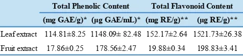
of R2 = 0.9999). The total phenolic and flavonoid content
for LE and FE was shown in Table 1. LE was found to contain a much higher phenolic content when compared
with FE; 114.81 and 17.86 mg GAE/g dry weight for LE
and FE, respectively. Similarly, LE was observed to have a
considerably higher flavonoid content than FE, which were 152.17 and 19.88 mg RE/gram dry weight for LE and FE,
respectively.
(mg GAE/g)* (μg GAE/mL)* (mg RE/g)** (µg RE/g)**
Leaf extract 114.81±8.25 1148.09± 82.48 152.17±2.64 1521.73±26.38
Fruit extract 17.86±0.25 178.56±2.47 19.88±0.34 198.83±3.41 Total Phenolic Content Total Flavonoid Content Table 1. Total phenolic and flavonoid content of aqueous
extracts from P. guajava.
*Total phenolic content was analysed as mg GAE/g dry weight of plant material and μg GAE/mL; values are mean±SD (n=3). GAE: gallic acid equivalent.
**Total flavonoid content was analysed as mg RE/g dry weight of plant material and μg RE/ml; values are mean±SD (n=3). RE: rutin equivalent.
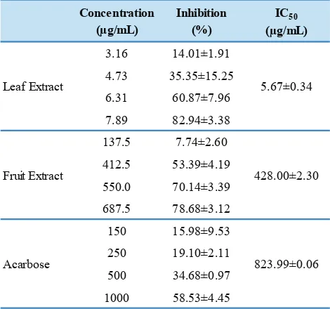
Inhibition of α-Glucosidase
As shown in Table 2, both the extracts inhibited
α-glucosidase in a concentration-dependent manner. Both extracts exhibited marked inhibition against α-glucosidase,
with IC50 values much lower than acarbose; 5.67 and 428.00
μg/mL for LE and FE, respectively, and acarbose 823.99 μg/
Table 2. α-Glucosidase inhibitory activities of aqueous extracts
from P. guajava.
Concentration (µg/mL)
Inhibition (%)
IC50
(µg/mL)
3.16 14.01±1.91 4.73 35.35±15.25 6.31 60.87±7.96 7.89 82.94±3.38 137.5 7.74±2.60 412.5 53.39±4.19 550.0 70.14±3.39 687.5 78.68±3.12 150 15.98±9.53 250 19.10±2.11 500 34.68±0.97 1000 58.53±4.45
823.99±0.06 5.67±0.34
Acarbose
Fruit Extract 428.00±2.30 Leaf Extract
Figure 1. Kinetics of inhibition on α-glucosidase by aqueous leaf extract of P. guajava (a) and acarbose (b).
Antioxidant Activities
Four methods were used in this study to evaluate the antioxidant activities. DPPH assay was the method used to evaluate the radical scavenging activity of the extracts. Both LE and FE were able to scavenge the DPPH radicals, with activities weaker than the standards (Table 3); IC50 of 74.77
and 843.84 μg/mL for LE and FE, compared with 53.24 and 21.36 μg/mL for ascorbic acid and BHT, respectively.
However, it is worth pointing that LE exhibited more effective scavenging activity than FE.
The ferrous ion chelating activities of extracts and standard are presented in Table 3. Both LE and FE showed low chelating activity for ferrous ion compared with the standard chelator Na2EDTA; IC50 of 147.07 and 2105.05 μg/ mL for LE and FE, compared with 66.50 for Na2EDTA.
DPPH radical scavenging
Ferrous ion chelating
Leaf extract 74.77±5.26 147.07±13.95 Fruit extract 843.84±9.52 2105.05±58.57 Ascorbic acid 53.24±0.82 NA BHT 21.36±0.80 NA EDTA NA 66.5±1.02
IC50 (µg/mL)
Table 3. DPPH radical scavenging and ferrous ion chelating activities of aqueous extracts from P. guajava and standards.
Values are mean±SD (n=3) NA: not assayed
The reducing power assay assesses antioxidant potential by measuring the reduction of Fe(III) to Fe(II) in the presence of antioxidant compounds in the extract. The potassium ferricyanide reducing assay was used to this end, and the results are shown in Figure 2a. The absorbance of LE and standards at 700 nm increased with increasing
concentrations of the samples (50 to 200 µg/mL), suggested an increased in reducing ability. At 50 and 100 µg/mL,
LE showed an effective reducing power, similar with the
standard BHT (the difference is not statistically significant).
However, weaker activity was observed when compared with ascorbic acid. LE reducing activity is observed to be
stronger than BHT at 200 µg/mL (p<0.05). In contrast, FE
did not show significant reducing activity.
The phosphomolybdate assay assesses the total antioxidant capacity in term of the reductive activity of both phenolics and non-phenolics compounds, such as ascorbic acid, tocopherol, etc. As can be seen in Figure 2b, at the range
of 50 to 400 µg/mL, the extracts and standards exhibited an
Figure 2. Reducing activities of aqueous extracts from P. guajava and standards as measured by reducing power method (a) and phosphomolybdate method (b). AA: ascorbic acid, BHT: butylated hydroxytoluene, LE: leaf extract, FE: fruit extract. Values are mean of three measurements.
of Mo(VI) to Mo(V) by the antioxidant compounds in the tested materials. Overall, LE has lower activity than the standards ascorbic acid and BHT (p<0.05). However, at the
concentration of 100 µg/mL, LE exhibited stronger reducing
ability than ascorbic acid exhibited (p<0.05). The reducing ability for LE was lower than standards at concentrations
200 and 400 µg/mL (p<0.05). Similarly to those found in the reducing power assay, FE did not show substantial reducing activity, observing no modulation in absorbance with increasing concentrations.
Discussion
As can be seen from the results above, LE had stronger biological activities when compared with FE, in terms of its
inhibitory activity on α-glucosidase and antioxidant activity.
These differences were probably due to that LE containing more concentrated bioactive compounds that FE. Many studies have shown that bioactivities of plant materials are closely related to their phenolic type compounds.(5,17) To this end, we have evaluated the polyphenolics content in the aqueous extracts from P. guajava by measuring their
phenolic content, expressed as GAE, and flavonoid content, expressed as RE. LE had higher total phenolic and flavonoid
content than those observed for FE. The higher levels of polyphenolics found in LE than FE is consistent with other previous reports.(7,18) High biological activities may be attributed to polyphenolic compounds in the extracts. Results obtained from this study offer a valid starting point for the exploitation of P. guajava for use as antidiabetic and
antioxidant natural source. Isolation and identification of
active chemical constituents could be the direction of future studies.
The study herein was designed to investigate the antidiabetic potentials from the natural derived products with increased potency and less side effects than that of the
synthetic inhibitors. In the present work, the α-glucosidase
inhibitory activity of aqueous extracts from P. guajava
has been investigated. Results reveal that both LE and FE
exerted remarkable inhibitory effect on α-glucosidase when
compared with acarbose. The LE, however, was observed to have a stronger inhibitory activity than FE. Marked difference in inhibitory activities between the two extracts is probably related to the difference in their polyphenolic compound contents, as shown from their total phenolic
and flavonoid content. Previous studies on α-glucosidase
inhibitors isolated from medicinal plants suggest that some potential inhibitors belong to phenolic type group, including
flavonoid class, have features inhibiting a-glucosidase activity.(19, 20)
A number of previous studies have reported more
potent α-glucosidase inhibitory activities from plant
extracts compared to acarbose. Water extract from
Brickellia cavanillesii was reported to have a stronger activity compared to acarbose (IC50 extract 0.169 mg/mL
and acarbose 1.12 mg/mL).(21) In another study, several
Echeveria species extracted with methanol were found to be more active than acarbose (IC50 of E. subrigida, E. kimnachii and E. craiginia were 0.025, 0.057 and 0.051
mg/mL, respectively, whereas acarbose 3.59 mg/mL).(22)
and Pseudocedrela kotschyi exhibited better activities than acarbose (IC50 extracts 1.5 µg/mL and 5 µg/mL, respectively,
whereas acarbose 0.726 mg/ml).(23)
Kinetic study was conducted to understand the mode
of inhibition on α-glucosidase by P. guajava extract. The study has been carried out for LE, which has the strongest inhibitory activity. The result was compared with the standard drug, acarbose. Figure 1 shows the double reciprocal plot of the inhibition. The plot generated straight lines which had different intersections in the x-axis. This
indicates that the mode of α-glucosidase inhibition by LE is
of competitive mode, similar to that of acarbose. This could
suggest that the extract inhibited α-glucosidase by binding
with the free enzyme in a manner that prevents substrate binding.
In the living system, hyperglycaemic condition often leads to stress oxidative. Antioxidant in the diets may help protect against oxidative damages. In this direction, antioxidant potential of the extracts was also investigated. Due to the multiple ways in which antioxidant protect biological systems, various methods were employed in assessing antioxidant activity, as to determine the reactions that may contribute to the antioxidant potential of the extracts. These methods include assays evaluating the radical scavenging activity, metal chelating ion ability, and reducing ability. One of the pathways of antioxidant mechanism of action is by removing free radicals. This can be achieved by donating hydrogen to free radicals, leading to the formation of unreactive species. For the evaluation of radical scavenging activity, we have used DPPH radical scavenging assay, as the DPPH molecule is considered to be a model of lipophilic radicals formed by lipid auto oxidation.(24) The results reveal that both LE and FE exhibited activities in scavenging DPPH radicals, in
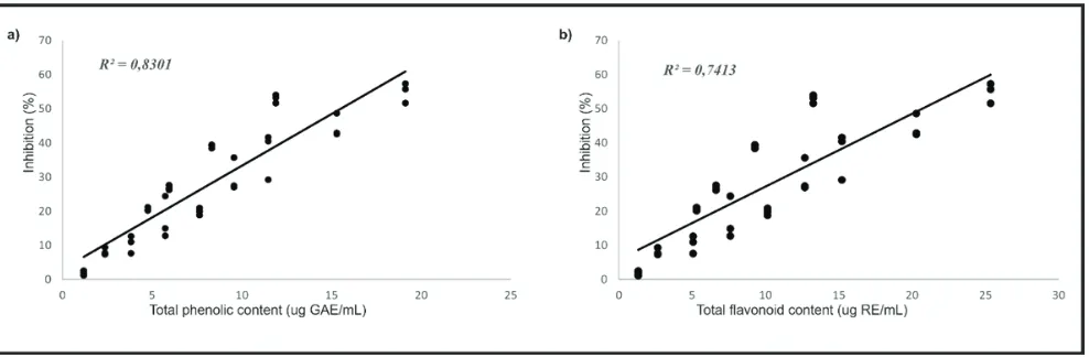
Figure 3. Correlation between DPPH inhibition (%) and total phenolic compound (µg GAE/mL) (a) and between DPPH inhibition (%) total flavonoid content (µg RE/mL) of aqueous extracts from P. guajava (b).
particular LE, observing noticeable activity when compared with the standard ascorbic acid. The scavenging activity
may be related to their phenolic and flavonoid content. The
stronger radical scavenging activity was observed for LE
which has higher phenolic and flavonoid content compared with FE. In this sense, we attempted to find a correlation
between antioxidant activities with the phytochemical
contents. It can be seen in Figure 3 that there is a significant
linear correlation between the DPPH radical scavenging
activity (% of inhibition) and total phenolic (µg GAE/ mL) and flavonoid (µg RE/mL) compounds (R2 = 0.8301 and 0.7413 for phenolic and flavonoid, respectively). This finding provides evidence that polyphenols in the extract
are likely to contribute to the radical scavenging activities in LE. This result is in agreement with the previous report for the strong relationship between antioxidant activities and polyphenolics in medicinal plants.(17) The structural features of the phenolic compounds may be responsible for the activity, due to the ease of proton donation from phenolic structure and the nucleophilic character of the benzene ring. Radical reactions can be initiated by the presence of transition metal ions, such as Cu(I) and Fe(II) ions. These metal ions is able to participate in a one-electron transfer reaction that
generate ROS, such as •OH from H2O2 through the Fenton
reaction.(25) Thus, ability to chelate transition metal ions is an important antioxidant property and measuring chelation of ferrous ion is one method to evaluate this property. It was evident that both extracts were able to chelate Fe(II) ion, however their binding activities were weak compared with the standard EDTA. Between these two extracts, LE was found to have much higher activity than FE. As in the radical scavenging assay, this result was in accordance with the phytochemical content of the extracts, i.e., phenolic and
with the reported studies for the chelating activities of the polyphenolic class compounds.(26,27) The mechanism that is operative may be through the formation of bidentate complexes between Fe(II) ion and the poly hydroxyl structure in the polyphenolics.
Studies have reported that the antioxidant effect is concomitant with development of reducing power and that the antioxidant properties is associated with the presence of reductones.(28) In this direction, we have investigated the reducing capacity of our extracts. Herein, LE was again showed stronger reductive activity than FE using the ferricthiocyanate method. The result is similar to that found for total antioxidant activity based on the phosphomolybdate method. The observed differences between LE and FE may be attributed to the content of polyphenolic compounds. This may suggest that polyphenols in the extract may act as electron donor, reacting with free radicals to terminate the chain reaction.
Conclusion
Aqueous extracts of leaf and fruit from P. guajava are good sources for neutraceutical material. Their antidiabetic activities are excellent. The study found that LE is better than FE in their antioxidant activities. Results obtained from this study offer a valid starting point for the exploitation of
P. guajava for use as antidiabetic and antioxidant natural source. However, further studies are necessary in order to
confirm their biological activities in different in vivo system, along with their mode of action.
Acknowledgment
We acknowledged funding from the Research Institution in the Faculty of Medicine Krida Wacana Christian University. We thank Mrs. Tjhia Lie Hwa from Pagarawan village, Bangka Island, Indonesia for kindly providing us with the plant materials.
References
1. Cefalu WT. Diagnosis and classification of diabetes mellitus. Diabetes
Care. 2004; 27(Suppl 1): s5-10.
2. Kim JG, Jo SH, Ha KS, Kim SC, Kim YC, Apostolidis E, et al. Effect of long-term supplementation of low molecular weight chitosan oligosaccharide (GO2KA1) on fasting blood glucose and HbA1c
in db/db mice model and elucidation of mechanism of action. BMC Complement Altern Med. 2014; 14: 272. doi:
10.1186/1472-6882-14-272.
3. Li N, Frigerio F, Maechler P. The sensitivity of pancreatic β-cells to
mitochondrial injuries triggered by lipotoxicity and oxidative stress. Biochem Soc Trans. 2008; 36: 930-4.
4. Hung HY, Qian K, Morris-Natschke SL, Hsu CS, Lee KH. Recent discovery of plant-derived anti-diabetic natural products. Nat Prod Rep. 2012; 29: 580-606.
5. Katalinic V, Milos M, Kulisic T, Jukic M. Screening of 70 medicinal plant extracts for antioxidant capacity and total phenols. Food Chem. 2006; 94: 550-7.
6. Matsuda H, Asao Y, Nakamura S, Hamao M, Sugimoto S, Hongo M, et al. Antidiabetogenic constituents from the Thai traditional medicine Cotylelobium melanoxylon. Chem Pharm Bull. 2009; 57: 487-94. 7. Gutiérrez RMP, Mitchell S, Solis RV. Psidium guajava: a review
of its traditional uses, phytochemistry and pharmacology. J Ethnopharmacol. 2008; 117: 1-27.
8. Giovannini P, Howes MJR, Edwards SE. Medicinal plants used in the traditional management of diabetes and its sequelae in Central America: A review. J Ethnopharmacol. 2016; 184: 58-71.
9. Díaz-de-Cerio E, Verardo V, Gómez-Caravaca AM, Fernández-Gutiérrez A, Segura-Carretero A. Health effects of Psidium guajava L. Leaves: An overview of the last decade. Int J Mol Sci. 2017; 18:
pii: E897. doi: 10.3390/ijms18040897.
10. Ahmed D, Fatima K, Saeed R. Analysis of phenolic and flavonoid
contents, and the anti-oxidative potential and lipid peroxidation inhibitory activity of methanolic extract of Carissa opaca roots and its fractions in different solvents. Antioxidants. 2014; 3: 671-83.
11. Kamtekar S, Keer V, Patil V. Estimation of phenolic content, flavonoid
content, antioxidant and alpha amylase inhibitory activity of marketed polyherbal formulation. J Appl Pharm Sci. 2014; 4: 61-5.
12. Dehghan H, Sarrafi Y, Salehi P. Antioxidant and antidiabetic activities
of 11 herbal plants from Hyrcania region, Iran. J Food Drug Anal. 2016; 24:179-88.
13. Lineweaver H, Burk D. The determination of enzyme dissociation constants. J Am Chem Soc. 1934; 56: 658-66.
14. Kilic I, Yesiloglu Y, Bayrak Y. Spectroscopic studies on the antioxidant activity of ellagic acid. Spectrochim Acta A Mol Biomol Spectrosc. 2014; 130: 447-52.
15. Jakovljević VD, Milićević JM, Stojanović JD, Solujić SR, Vrvić MM.
Antioxidant activity of ethanolic extract of Penicillium chrysogenum and Penicillium fumiculosum. Hem Ind. 2014; 68: 43-9.
16. Alam MN, Bristi NJ, Rafiquzzaman M. Review on in vivo and in vitro
methods evaluation of antioxidant activity. Saudi Pharm J. 2013; 21: 143-52.
17. Paixão N, Perestrelo R, Marques JC, Câmara JS. Relationship between antioxidant capacity and total phenolic content of red, rosé and white wines. Food Chem. 2007; 105: 204-14.
18. El Bedawey A, Mansour E, Zaky M, Hassan AA. Characteristics of antioxidant isolated from some plant sources. Food Nutr Sci. 2010; 1: 5-12.
19. Matsui T, Ogunwande I, Abesundara K, Matsumoto K. Anti-hyperglycemic potential of natural products. Mini-Rev Med Chem. 2006; 6: 349-56.
20. Oboh G, Agunloye OM, Adefegha SA, Akinyemi AJ, Ademiluyi AO. Caffeic and chlorogenic acids inhibit key enzymes linked to type 2 diabetes (in vitro): a comparative study. J Basic Clin Physiol Pharmacol. 2015; 26: 165-70.
21. Escandón-Rivera S, González-Andrade M, Bye R, Linares E,
Navarrete As, Mata R. α-Glucosidase inhibitors from Brickellia
22. López-Angulo G, Montes-Avila J, Díaz-Camacho SP, Vega-Aviña R, Ahumada-Santos YP, Delgado-Vargas F. Chemical composition and
antioxidant, α-glucosidase inhibitory and antibacterial activities of
three Echeveria DC. species from Mexico. Arab J Chem. 2014; n.v:
n.p. doi: 10.1016/j.arabjc.2014.11.050.
23. Moradi-Afrapoli F, Asghari B, Saeidnia S, Ajani Y, Mirjani M, Malmir M, et al. In vitro α-glucosidase inhibitory activity of phenolic constituents from aerial parts of Polygonum hyrcanicum. DARU.
2012; 20: 37. doi: 10.1186/2008-2231-20-37.
24. Shukla S, Park J, Kim DH, Hong SY, Lee JS, Kim M. Total phenolic
content, antioxidant, tyrosinase and α-glucosidase inhibitory
activities of water soluble extracts of noble starter culture Doenjang,
a Korean fermented soybean sauce variety. Food Control. 2016; 59: 854-61.
25. Adiani V, Gupta S, Chatterjee S, Variyar PS, Sharma A. Activity guided characterization of antioxidant components from essential oil of Nutmeg (Myristica fragrans). J Food Sci Technol. 2015; 52: 221-30.
26. Mladěnka P, Macáková K, Filipský T, Zatloukalová L, Jahodář L,
Bovicelli P, et al. In vitro analysis of iron chelating activity of
flavonoids. J Inorg Biochem. 2011; 105: 693-701.
27. Adjimani JP, Asare P. Antioxidant and free radical scavenging activity of iron chelators. Toxicol Rep. 2015; 2: 721-8.