CONSTRUCTION AND RETROSPECTIVE VALIDATION OF
STRUCTURE-BASED VIRTUAL SCREENING PROTOCOLS TO
IDENTIFY POTENT LIGANDS FOR HUMAN ADRENERGIC BETA-2
RECEPTOR
Enade Perdana Istyastono1,2,*, Dewi Setyaningsih3
1Division of Drug Discovery, Faculty of Pharmacy, Sanata available. However, the original retrospective structure-based virtual screening campaign accompanying the dataset showed relatively poor quality with enrichment factor of true positives at 1% false positives (EF1%) value of 3.9. In this article, the construction and retrospective validation of a structure-based virtual screening protocol by employing PLANTS1.2 as the molecular docking software and PyPLIF as an alternative post docking scoring functions are presented. The results show that the developed protocols have better quality compared the original structure-based virtual screening with EF1% values of 24.24 and 8.22 by using ChemPLP from PLANTS1.2 and by using Tc-PLIF from PyPLIF, respectively. Further investigation by performing systematic filtering resulted in the identification of D113, S203, and N293 as molecular determinants in ADRB2-ligand binding.
Key words: Structure-based virtual screening, molecular docking, adrenergic beta-2 receptor, protein-ligand interaction fingerprinting, molecular determinants.
INTRODUCTION
Adrenergic Beta-2 Receptor (ADRB2) plays an important role as the molecular target for drugs in the therapy for diseases as diverse as heart failure, hypertension and asthma
(Cherezov et al., 2007; Taylor, 2007). ADBR2 is
a member of G-Protein Coupled Receptors (GPCRs) family, to which more than 30% of top-selling drugs in the market bind (Klabunde
and Hessler, 2002; Surgand et al., 2006).
Notably, human ADRB2 was also the first human GPCR that could be crystallized and publicly available to provide insight on how
ligands bind to GPCRs (Cherezov et al., 2007).
The ADRB2 crystal structure has been subsequently employed in some prospective Structure-Based Virtual Screening (SBVS) campaigns and successfully discovered novel
potent ADRB2 ligands (Kolb et al., 2009; Yakar
and Akten, 2014).
The successful three dimensional (3D)
structure characterization through
crystallography of ADRB2 bound to its
antagonist carazolol (Cherezov et al., 2007) was
followed by 3D characterization of some other
GPCRs (Chien et al., 2010; Jaakola et al., 2008;
Shimamura et al., 2011; Wacker et al., 2010; Wu
et al., 2010), which have offered opportunities
to construct, validate and perform SBVS to discover novel potent ligands for a particular GPCR both on the crystal structures (Carlsson
et al., 2010; de Graaf et al., 2011a; Katritch et al.,
2010; Kolb et al., 2009; Yakar and Akten, 2014)
and homology models (Carlsson et al., 2011; de
Graaf et al., 2011b; Istyastono et al., 2011b; Sirci
et al., 2012; Tarcsay et al., 2013). Solely used of
SBVS approaches on Histamine H1 Receptor
(HRH1) crystal structure in the recent virtual screening campaigns showed extraordinary results, both retrospectively and prospectively
(de Graaf et al., 2011a). One of the key
strategies of the virtual screening was filtering the Protein-Ligand Interaction Fingerprint
(PLIF) (Marcou and Rognan, 2007; Radifar et
al., 2013a): Only docking poses that form a
Enade Perdana Istyastono
interaction with D107 were considered (de
Graaf et al., 2011a). This strategy can be
recognized as “using prior knowledge” in SBVS
campaigns (Seifert, 2009; Yuniarti et al., 2011).
The customization of the SBVS protocols by filtering on key interactions has increased the
SBVS quality significantly (de Graaf et al.,
2011a; Sirci et al., 2012; Yuniarti et al., 2011).
Unfortunately, the information of key
interactions is available only for a few drug targets. The key interaction used in the SBVS campaigns on crystal structure of HRH1 was
identified from previous Site-Directed
Mutagenesis (SDM) studies and chemogenomic
analysis (de Graaf et al., 2011a; Shin et al., 2002;
Surgand et al., 2006). Besides SDM studies and
chemogenomic analysis, some computer-aided strategies could be employed in order to obtain information on key interactions that can assist
the improvement of SBVS quality. Istyastono et
al. (2011a) employed QSAR, 3D-QSAR,
homology modeling and molecular dynamics to identify the molecular determinants of ligand
binding modes in the Histamine H3 and H4
Receptors (HRH3 and HRH4, respectively). The obtained information was subsequently used in SBVS campaigns on homology models
the receptors (Istyastono et al., 2011a; Sirci et al.,
2012). However, the use of multiple or combined approaches reflects time and resource consuming methods. Therefore, development of more effective and efficient computational methods to identify key interact-tions as well as the molecular determinants in protein-ligand binding to increase the SBVS quality is of considerable interest.
The research presented in this paper was
aimed to perform retrospective SBVS
campaigns on a newly published enhanced dataset of ligands and decoys (DUD-e) for
ADRB2 (Mysinger et al., 2012) and to identify
the PLIF of each compound to ADRB2 by
employing PyPLIF (Radifar et al., 2013a; Radifar
et al., 2013b; Setyaningsih et al., 2013). The
SBVS qualities were subsequently assessed (de Graaf and Rognan, 2008) and compared to the original SBVS accompanying the release of
DUD-e (Mysinger et al., 2012). The
results showed that both scoring strategies
employed in this research, i.e. ChemPLP score
andTc-PLIFresulted ina better SBVS. Notably
the SBVS quality using ChemPLP scores outperformed the SBVS quality using Tc-PLIF values. The PLIFs of ADRB2-ligands identified in this research were subsequently employed in the key interactions identification in a further investigation by systematic filtering. These approaches have led to the identification of
test compounds to perform retrospective SBVS. All calculations and computational simulations were performed on a Linux (Ubuntu 10.04 LTS Lucid Lynx) machine with Intel(R) Xeon(R) CPU E31220 (@ 3.10 GHz) as the processors and 8.00 GB of RAM. Computational medicinal chemistry applications employed in this research were SPORES (ten Brink and Exner, 2009),
PLANTS1.2 (Korb et al., 2009), Open Babel
2.2.3 (O’Boyle et al., 2011), PyPLIF 0.1.1
(Radifar, 2013a), and PyMOL 1.2r1 (Lill and
Danielson, 2011). Statistical analysis was performed by using R 3.1.0 (R Development Core Team, 2008). A shell script to take into account only poses that have the predefined
interaction bitstring after the PLIF
identification using PyPLIF (Table I).
Computational methods
molecular docking simulation employing PLANTS1.2 docking software. This procedure
produced the virtual target protein.mol2 and the
Ligands preparation for retrospective
downloaded and stored locally as actives_final.ism
and decoys_final.ism. The files were subsequently
concatenated into a file named all.smi. Each
compound in the file was then subjected to Open Babel 2.2.3 conversion software to be converted in its three dimensional (3D) format
at pH 7.4 as a mol2 file. The settypes module in
SPORES was subsequently employed to
properly check and assign the mol2 file into a
proper mol2 file ready to dock by using
PLANTS1.2 docking software.
Automated molecular docking and
virtual screening
All virtual screenings were performed by docking program PLANTS1.2. For each compound, 50 poses were calculated and scored by the ChemPLP scoring function at speed setting 2. The binding pocket of ADRB2 was defined by the coordinates of the center of the reference ligand and a radius of 5 Å (which is the maximum distance from the center defined by a 5 Å radius around the reference ligand). All other options of PLANTS1.2 were left at their default setting. Every compound
was virtually screened three times.
Rescoring using protein-ligand interaction fingerprints calculated by PyPLIF
The co-crystal ligand binding mode in the ADRB2 crystal structure was used to generate reference PLIF by using PyPLIF. Seven different interaction types (negatively charged, positively charged, hydrogen bond (H-bond) acceptor, H-bond donor, aromatic face-to-edge, aromatic face-to-face and hydrophobic interactions) were used to define the PLIF. The cavity used for the PLIF analysis is consisted of a set of residues in the binding pocket of ADRB2 defined in subsection Automated molecular docking and virtual screening. Note that for each PLANTS docking pose, a unique subset of protein coordinates with rotated hydroxyl hydrogen atoms were used to define the PLIF. Standard PLIF scoring parameters, and a Tanimoto coefficient (Tc-PLIF) measuring PLIF similarity with the reference molecule pose was used to re-rank the docking poses of the known active ADRB2 ligands and their decoys.
SBVS quality assessment
The docking pose with the best ChemPLP score or the best Tc-PLIF value was selected for each virtually screened compound. Virtual screening accuracies were determined in terms of Area Under the Curve (AUC) of the Receiver-Operator Characteristic (ROC) plots computed with R statistical computing software
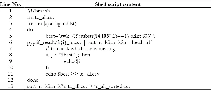
Table I. Shell script to filter based on the predefined interaction bitstring (Radifar, 2013a).
Line No. Shell script content
1
for i in $(cat ligand.lst) do
best=`awk '{if (substr($4,103*),1)==1) print $0}' \
pyplif_result/${i}_tc.csv | sort -n -k3rn -k2n | head -n1` # to check which csv is missing
if [ -z "$best" ]; then echo $i fi
echo $best >> tc_all.csv done
sort -n -k3rn -k2n tc_all.csv > tc_all_sorted.csv
*) The shell script should was adopted according to the relevant predefined bitstring. In this example by
Enade Perdana Istyastono filtering on PLIFs resulted in subsection Rescoring using protein-ligand interaction fingerprints calculated by PyPLIF was created by adopting the one (Table I) provided by
Radifar et al. (2013a). For every filtering result, a
new rank based on the ChemPLP values was
created and the EF1% values were then
calculated (de Graaf et al., 2011a; Sirci et al.,
2012). The molecular determinants were identified by correlating the bitstring interaction
that give significantly better EF1% values
compared to the default ones (without PLIF filtering) to the relevant binding pocket
residues (Wacker et al., 2010). The results were
then retrospectively validated by examining available mutation data in the literatures stored
in GPCRDB (Vroling et al., 2010).
Visual inspection
Visual inspection using PyMOL 1.2r1 (Lill and Danielson, 2011) was performed to investigate manually some representative docking poses to examine the plausible molecular determinants of the ADRB2-ligands
binding.
RESULTS AND DISCUSSION
This research was aimed to construct a valid SBVS protocol to identify potent human
ADRB2 ligands by employing PLIF
identification using PyPLIF as an alternative
rescoring strategy (Radifar et al., 2013b).
The additional rescoring procedures offer
possibilities to identify the molecular
determinant in the ADRB2-ligand binding by
providing PLIF bitstrings from every
interaction types of every docking poses to all
amino acids in the binding pocket (de Graaf et
al., 2011a; Marcou and Rognan, 2007; Radifar et
al., 2013a). Subsequent investigation by
performing systematic filtering on the bitstrings could lead to the identification of the critical bitstrings that affect the SBVS quality. The identified critical bitstrings were suggested to
be correlated to the potential molecular determinant in the ADRB2-ligand binding (de
Graaf and Rognan, 2008; Istyastono et al.,
2011b).
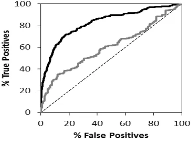
Figure 1. ROC curves resulted in the retrospective SBVS campaign. The black lines represent the ROC curves when the results were ranked by ChemPLP scores, while the grey lines represent the ROC curves when the results were ranked by Tc-PLIF values. The dashed lines represent random selection.
The virtual screening campaigns has
resulted 2,284,650 docking poses and
799,627,500 bitstrings for all 15,231 screened ADRB2 ligands or decoys downloaded from DUD-e. By employing either ChemPLP score originated from PLANTS1.2 or Tc-PLIF value
of PyPLIF (Korb et al., 2009; Radifar et al., plotted were accordingly (Figure 1). The results showed that the developed protocols had better qualities compared the original SBVS with
EF1% values of 24.24 by using ChemPLP from
Compared to the original SBVS accompanying
Interestingly, the results indicated also that the SBVS on ADRB2 employing ChemPLP as the scoring functions outperformed the SBVS employing Tc-PLIF as the scoring functions. However, the PLIFs resulted in the PLIF identification using PyPLIF could serve as starting points in the identification of the critical bitstrings, which in turn could be correlated to the important residues in the ADRB2-ligand binding.
The PLIFs of docking poses resulted in this research have subsequently served as useful tools to identify the molecular determinants in
ADRB2-ligands binding. The systematic
filtering on all PLIF bitstrings for both “on” (represents favorable interaction) and “off”
(represents unfavorable interaction) resulted in
some bitstrings that gave better EF1% values
compared to the unfiltered SBVS campaigns (the default ones). These important bitstrings
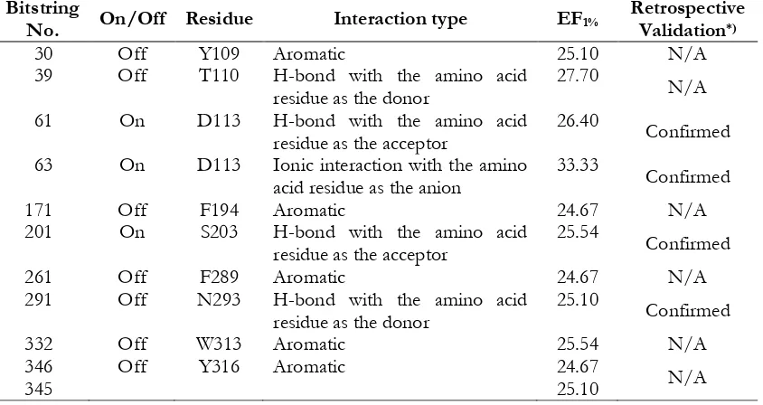
and their related amino acid residues are presented in Table 2. By employing mutation
data stored in GPCRDB (Vroling et al., 2011),
the following were the identified
and retrospectively validated molecular
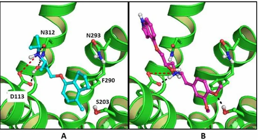
determinants in ADRB2-ligand binding: D113, S203 and N293. These could serve as the key information in the future ADRB2-ligand design (Figure 2): Potent ligands for ADRB2 should form ionic interaction to D113 and H-bond to
both D113 (Strader et al., 1987; Elling et al.,
compound recognized in the EF1% value in the
retrospective SBVS campaigns CHEMBL38205
Tabel II. Filtering on PLIF bitstrings for both “on” (represents favorable interaction) and “off”
(represents unfavorable interaction) that gave better EF1% values compared to the unfiltered SBVS
campaigns (EF1% = 24.24).
63 On D113 Ionic interaction with the amino
acid residue as the anion
33.33
Enade Perdana Istyastono
(Ki at ADRB2 = 3-7nM (Tejani-Butt and
Brunswick, 1986; Mysinger et al., 2012);
ChemPLP score = -101.916) has 33.33 times better opportunities to be confirmed as a ADRB2 ligand compared to any random selected compounds. Figure 2 is presented to examine how the representative ligand CHEMBL38205 interacts to the ADRB2 binding pocket in comparison to the co-crystal
ligands ICI 118,551 (Wacker et al., 2010). As
can be seen in Figure 2, the co-crystal ligand ICI 118,551 forms an ionic bond to D113, H-bonds to D113 and N312, and an aromatic
interaction to F290 (Wacker et al., 2010)
(Figure 2A), while the representative ligand forms ionic bond to D113, H-bonds to D113 and S203, and an aromatic interaction to F290 (Figure 2B). Notably, both compounds do not form H-bond to N293. Similar to histamine receptors, ADRB2 as an aminergic GPCR has a
conserved D113 residue as an ionic bond
anchor (Cherezov et al., 2007; de Graaf et al.,
2011a; Istyastono et al., 2011a; Istyastono et al.,
2011b; Shimamura et al., 2011; Wacker et al.,
2010). The SBVS protocols developed in this research showed that by adding knowledge of molecular determinants in ADRB2-ligand binding in the protocols could increase the virtual screening quality as well as to identify the most plausible binding pose of ligands in the ADRB2 binding pocket. The similar strategy has successfully shown in the SBVS on a HRH1 crystal structure and HRH3 homology
models (de Graaf et al., 2011a; Sirci et al., 2012).
CONCLUSIONS
The constructed SBVS protocol
employing PLANTS1.2 and PyPLIF to identify ligands for ADRB2 has been retrospectively validated using newly published database Figure 2. The co-crystal ligands ICI 118.551 (cyan carbon atoms; balls and sticks mode) pose in the
ADRB2 binding pocket (Wacker et al., 2010) (A) and the docking pose of the representative ligand
CHEMBL38205 (magenta carbon atoms; balls and sticks mode; Ki at ADRB2 = 3-7nM
(Tejani-Butt and Brunswick, 1986; Mysinger et al., 2012); ChemPLP score = -101.916) in the ADRB2
DUD-e. The protocol showed better virtual screening qualities in ligand identification
compared to the original protocol
accompanying the release of DUD-e. An improvement on virtual screening quality was subsequently achieved by adding information of molecular determinants in ADRB2-ligand binding into the protocol. The produced PLIFs have served as useful tools in the recognition of the molecular determinants in ADRB2-ligand binding: D113, S203, and N293.
ACKNOWLEDGEMENT
This research was financially supported by Indonesian Directorate General of Higher Education (Fundamental Research Block Grant No. 1320/K5/KM/2014 to E.P.I. and D.S.) and Faculty of Pharmacy, Sanata Dharma University (Collaborative Research Travel Grant No. Far/091/XI/2014/ST/D to E.P.I.).
REFERENCES
Ballesteros, JA., Shi, L., Javitch, JA., 2001, Structural mimicry in G protein-coupled receptors: Implications of the high-resolution structure of rhodopsin for structure-function analysis of
rhodopsin-like receptors, Mol. Pharmacol., 60,1-19.
Carlsson J., Coleman RG., Setola V., Irwin JJ., Fan H., Schlessinger A., Sali A., Roth BL., Shoichet BK., 2011, Ligand
discovery from a dopamine D3 receptor
homology model and crystal structure.
Nat. Chem. Biol., 7, 769–778.
Carlsson J., Yoo L., Gao, Z., Irwin JJ., Shoichet BK., Jacobson KA., 2010, Structure-resolution crystal structure of an
engineered human 2-adrenergic G
protein-coupled receptor. Science, 318,
1258–1265.
Chien EYT., Liu W., Zhao Q., Katritch V., Han GW., Hanson MA., Shi L., Newman AH., Javitch JA., Cherezov V., Stevens RC., 2010, Structure of the
human dopamine D3 receptor in
complex with a D2/D3 selective
antagonist. Science, 330, 1091–1095.
De Graaf C., Kooistra AJ., Vischer HF., Katritch V., Kuijer M., Shiroishi M., Iwata S., Shimamura T., Stevens RC., de
Esch IJP., Leurs R., 2011a, Crystal
structure-based virtual screening for fragment-like ligands of the human
histamine H1 receptor. J. Med. Chem., 54,
8195–8206.
De Graaf C., Rein C., Piwnica D., Giordanetto
F., Rognan D., 2011b, Structure-based
discovery of allosteric modulators of two
related class B G-protein-coupled
receptors. ChemMedChem, 6, 2159–2169.
De Graaf C., Rognan, D., 2008, Selective structure-based virtual screening for full
and partial agonists of the 2 adrenergic
receptor, J. Med. Chem., 51, 4978–4985.
Elling, CE., Thirstrup, K., Holst, B., Schwartz, TW., 1999, Conversion of agonist site to
metal-ion chelator site in the 2
-adrenergic receptor, Proc. Natl. Acad. Sci.
U S A., 96, 12322-12327.
Gouldson, PR., Snell, CR., Reynolds, CA., 1997, A new approach to docking in the
2-adrenergic receptor that exploits the
domain structure of G-protein-coupled
receptors, J. Med. Chem., 40, 3871-3886.
Istyastono EP., de Graaf C., de Esch IJP.,
Leurs R., 2011a, Molecular determinants
of selective agonist and antagonist
binding to the histamine H4 receptor.
Curr. Top. Med. Chem., 11, 661–679.
Istyastono EP., Nijmeijer S., Lim HD., van de Stolpe A., Roumen L., Kooistra AJ., Vischer HF., de Esch IJP., Leurs R., de
Graaf, C., 2011b, Molecular determinants
of ligand binding modes in the histamine
H4 receptor: Linking ligand-based
three-dimensional quantitative
structure−activity relationship (3D
-QSAR) models to in silico guided
receptor mutagenesis studies. J. Med.
Chem., 54, 8136–8147.
Jaakola V., Griffith MT., Hanson MA., Cherezov V., Chien EYT., Lane JR., Ijzerman AP., Stevens RC., 2008, The 2.6 angstrom crystal structure of a
human A2A adenosine receptor bound to
Enade Perdana Istyastono
Katritch V., Jaakola V., Lane JR., Lin J., Ijzerman AP., Yeager M., Kufareva I., Stevens RC., Abagyan R., 2010, Structure-based discovery of novel
chemotypes for adenosine A2A receptor
antagonists. J. Med. Chem. 53, 1799–1809.
Klabunde T., Hessler G., 2002, Drug design
strategies for targeting
G-protein-coupled receptors, ChemBioChem, 3, 928–
944.
Kolb P., Rosenbaum DM., Irwin JJ., Fung JJ., Kobilka BK., Shoichet BK., 2009,
Structure-based discovery of 2
-adrenergic receptor ligands. Proc. Natl.
Acad. Sci. U. S. A., 106, 6843–6848.
Korb O., Stützle T., Exner TE., 2009,
Empirical scoring functions for
advanced protein-ligand docking with
PLANTS. J. Chem. Inf. Model., 49, 84–96.
Liapakis, G., Ballesteros, JA., Papachristou, S., Chan, WC., Chen, X., Javitch, JA., 2000, The forgotten serine. A critical role for Ser-2035.42 in ligand binding to and
activation of the 2-adrenergic receptor,
J. Biol. Chem., 275, 37779-37788.
Lill, MA., Danielson, ML., 2011, Computer-aided drug design platform using
PyMOL, J. Comput. Aided Mol. Des., 25,
13–19.
Marcou G., Rognan D., 2007, Optimizing fragment and scaffold docking by use of
molecular interaction fingerprints. J.
Chem. Inf. Model., 47, 195–207.
O’Boyle, NM., Banck, M., James, CA., Morley,
C., Vandermeersch, T., Hutchison, GR., 2011, Open Babel: An open chemical
toolbox, J. Cheminform., 3, 33.
R Development Core Team, 2008, R: A
Language and Environment for Statistical
Computing, Vienna,
http://www.r-project.org.
Radifar M., Yuniarti N., Istyastono EP., 2013a,
PyPLIF: Python-based protein-ligand
interaction fingerprinting, Bioinformation,
9, 325–328.
Radifar M., Yuniarti N., Istyastono EP., 2013b,
PyPLIF-assisted redocking
indomethacin-(R)- -ethyl-ethanolamide
into cyclooxygenase-1. Indo. J. Chem. 13,
283–286.
Rasmussen, SG., Choi, HJ., Fung, JJ., Pardon, E., Casarosa, P., Chae, PS., Devree, BT., Rosenbaum, DM., Thian, FS., Kobilka, TS., Schnapp, A., Konetzki, I., Sunahara, RK., Gellman, SH., Pautsch, A., Steyaert, J., Weis, WI., Kobilka, BK., 2011, Structure of a nanobody-stabilized active
state of the 2 adrenoceptor, Nature, 469,
175-180.
Sato, T., Kobayashi, H., Nagao, T., Kurose, H., 1999, Ser203 as well as Ser204 and Ser207 in fifth transmembrane domain
of the human 2-adrenoceptor
contributes to agonist binding and
receptor activation, Br. J. Pharmacol., 128,
272-274.
Seifert MHJ., 2009, Targeted scoring functions
for virtual screening. Drug Discov. Today,
14, 562–569.
Setyaningsih D., Radifar M., Murti YB.,
Istyastono EP., 2013, Construction of in
silico structure-based screening tools to
study the oxidative metabolites
formation of curcumin by human
cytochrome 450 3A4. Indonesian J. Pharm.,
24, 75–85. 2002, Molecular modeling and site-specific mutagenesis of the
histamine-binding site of the histamine H4
screening: Discovery of histamine H3
receptor ligands using ligand-based and
protein-based molecular fingerprints. J.
Chem. Inf. Model., 52, 3308–3324.
1987, Mutations that uncouple the beta-adrenergic receptor from Gs and
increase agonist affinity, J. Biol. Chem.,
262, 16439-16443.
Surgand J., Rodrigo J., Kellenberger E., Rognan D., 2006, A chemogenomic analysis of the transmembrane binding cavity of human G-protein-coupled receptors.
Proteins, 62, 509–538.
Suryanarayana, S. and Kobilka, BK., 1993, Amino acid substitutions at position 312 in the seventh hydrophobic segment of
the 2-adrenergic receptor modify
ligand-binding specificity, Mol. Pharmacol., 44,
111-114.
Tarcsay A., Paragi G., Vass M., Jójárt B., Bogár
F., Keserű GM., 2013, The impact of
molecular dynamics sampling on the performance of virtual screening against
GPCRs. J. Chem. Inf. Model., 53, 2990–
2999.
Taylor MRG., 2007, Pharmacogenetics of the
human -adrenergic receptors.
Pharmacogenomics J., 7, 29–37.
Tejani-Butt, SM., Brunswick, DJ., 1986, Synthesis and beta-adrenergic receptor blocking potency of 1-(substituted
amino)-3-(4-indolyloxy)propan-2-ols, J.
Med. Chem., 29, 1524-1527.
Ten Brink, T., Exner, TE., 2009, Influence of
protonation, tautomeric, and
stereoisomeric states on protein-ligand
docking results, J. Chem. Inf. Model., 49,
1535–1546.
Vroling, B., Sanders, M., Baakman, C., Borrmann, A., Verhoeven, S., Klomp, J., Oliveira, L., de Vlieg, J., Vriend, G.,
2011, GPCRDB: Information system for
G protein-coupled receptors, Nucleic
Acids Res., 39, D309-D319.
Wacker D., Fenalti G., Brown MA., Katritch V., Abagyan R., Cherezov V., Stevens RC., 2010, Conserved binding mode of
human 2 adrenergic receptor inverse
agonists and antagonist revealed by
X-ray crystallography. J. Am. Chem. Soc.,
132, 11443–11445.
Wieland, K., Zuurmond, HM., Krasel, C., Ijzerman, AP., Lohse, MJ., 1996,
Involvement of Asn-293 in
stereospecific agonist recognition and in
activation of the 2-adrenergic receptor,
Proc. Natl. Acad. Sci. U S A., 93,
9276-9281.
Wu B., Chien EYT., Mol CD., Fenalti G., Liu W., Katritch V., Abagyan R., Brooun A., Wells P., Bi FC., Hamel DJ., Kuhn P., Handel TM., Cherezov V., Stevens RC., 2010, Structures of the CXCR4 chemokine GPCR with small-molecule
and cyclic peptide antagonists. Science,
330, 1066–1071.
Yakar R., Akten ED., 2014, Discovery of high
affinity ligands for 2-adrenergic receptor
through pharmacophore-based
high-throughput virtual screening and
docking. J. Mol. Graph. Model. 53, 148–
160.
Yuniarti N., Ikawati Z., Istyastono EP., 2011, The importance of ARG513 as a hydrogen bond anchor to discover COX-2 inhibitors in a virtual screening