1
PATHOLOGICAL CASE STUDY OF RABBIT HEPATIC COCCIDIOSIS IN TABANAN, BALI
I Made Kardena
Laboratory of Animals Pathology, Faculty of Veterinary Medicine Udayana University, 80232 Denpasar, Indonesia
Email: [email protected]; Phone: +6281353353399
I Ketut Berata
Laboratory of Animals Pathology, Faculty of Veterinary Medicine Udayana University, 80232 Denpasar, Indonesia
Email: [email protected]; Phone: +628123645132
Ida Bagus Oka Winaya
Laboratory of Animals Pathology, Faculty of Veterinary Medicine Udayana University, 80232 Denpasar, Indonesia
Email: [email protected]; Phone: +628123680143
Ida Bagus Made Oka
Laboratory of Animal Parasitology, Faculty of Veterinary Medicine Udayana University, 80232 Denpasar, Indonesia dissected to examine the liver changes microscopically and macroscopically. Examination of color, size, and consistency were done to evaluate the liver samples. The samples were then processed for tissue processing by using Hematoxillin-Eosin method before performing histophatological examination by using 10x and 40x magnificence under a binocular microscope. The result demonstrates all of the rabbit samples showed pathological changes in their livers, such as: the livers were relatively dark-reddish and bigger in size. In addition, the surface of the liver mainly contained white nodules with diameter between 1-5 mm. Microscopically, degeneration and necrotic in the liver cells were
examined in all of the liver samples. Additionally, hypertrophy of the bile duct’s epithelial cells was observed. It is high likely that these macroscopic and microscopic changes were due to Eimeria stidae parasite infestation. It is also assumed that the infection could cause death of the infected rabbits.
2
INTRODUCTION
Coccidiosis is a parasitic disease that can cause economic losses of the rabbit breeders. The disease can spread rapidly, especially when the rabbits were placed in a cage with a low hygiene level and high quantity of livestock (Gonzales-Redondo, et al., 2008). Coccidiosis is caused by parasites of the genus Eimeria. This disease is quite infectious to rabbits, especially the young rabbits (Wang and Tsai, 1991). Infection in adult rabbits generally does not show clinical symptoms, therefore adult rabbits called as career because they can spread the parasite to the young rabbit or other susceptible animals (Al-Rukibat, et al., 2001).
Based on the clinical symptoms, the coccidiosis in rabbits is relatively mild to severe. Rabbits that been infected, both hepatic and intestinal types, sometimes show less manifest clinical symptoms. However, not rare coccidiosis can cause death (Gendron and Earle-Briges, 2000). This is often confusing, especially for rabbit breeders who tend to think that nothing happened on their livestock. This is also supported by the very limited data on morbidity and mortality of this disease. On the other hand, some researchers suggest that the morbidity and mortality, in particular the hepatic type of coccidiosis in rabbits, are relatively high. Wang and Tsia (1991) reported the coccidiosis morbidity rate could reach 100%. Similar research was reported by El Akabawy, et al., (2004), on the high mortality rate of hepatic coccidiosis in rabbits.
There are several risk factors that allegedly affect the increase number of coccidiosis cases in rabbits, including the enclosure management, hygiene level, season, and age of rabbits. Enclosure with low level of hygiene and feeding with decaying leaves can increase the incidence of coccidiosis in rabbits. The prevalence of Eimeria sp. infection in rabbits also tends to increase during the rainy season when compared to the dry season (Bhat, et al., 1996). Other factors, such as age of rabbits also greatly influence the level of coccidiosis infection. The risk of hepatic coccidiosis markedly decreases with advancing age (Wang and Tsai, 1991).
There are various species of Eimeria sp, but the most common species that infects rabbits and causes the death is E. stidae. The predilection of Eimeria stidae is in bile duct epithelium of the rabbit liver. The other species, such as E. perforans, E. magna, E. media, E. irresidua, and E. intestinalis, generally infect gastrointestinal tract (Cam, et al., 2008). This other species of Eimeria are relatively harmless compared to Eimeria stidae.
Rabbit can be infected by coccidiosis in many different ways. Rabbits infected by ingesting foliage containing sporulated oocyst of Eimeria sp. Infection can also occur when rabbit cleaning the contaminated fur with tongue. The fur usually contaminated by feces containing oocyst of Eimeria sp. (Berata, 2009).
The rabbit which infected by coccidiosis is likely associated with clinical symptoms such as growth inhibition, weaknesses, anorexia, icterus, dull hair, constipation, and diarrhea (Darzi, et al., 2007). Post-mortem examination of the infected rabbit could show organ changes. The changes are strongly influenced by the species of Eimeria that infects. In this study, we focused on anatomic pathological changes in rabbit liver infected by coccidiosis.
METHODS
A total of 10 rabbits was used in this study, with an average age of 1-3 months and were given feed pellets and grass. Samples were taken from a rabbit farm in Bedugul area, the Candi Kuning Village, Tabanan Regency, the Province of Bali. Rabbits that used as sample in this study were naturally dead rabbits within less than 2 hours prior to necropsy. All died rabbits that used in this study showed no clinical symptoms, except the presence of a yellowish color in the conjunctiva. Necropsy was performed at the Pathology Laboratory, Faculty of Veterinary Medicine, Udayana University for pathognomonic observation. Identification of the parasites was conducted at the Parasitology Laboratory, Faculty of Veterinary Medicine, Udayana University by examining bile duct material.
Necropsy
3
size of about 1x1x1 cm3 and immersed in a fixative solution of 10% neutral buffered formalin, prior to further processing for making preparations for histopathological examination.Histopathology Preparation Method
The steps in making preparations for histopathology adapted from Kiernan (2001) method. Liver tissue was immersed in a fixative solution for 24 hours, and then sliced and put into a box of cassette to be further processed in a tissue processor machine. In this box, the tissue were immersed in a solution of 70%, 80%, 90%, and 96% alcohol, and two toluene solutions for each 2 hours. The tissue was then inserted into liquid paraffin with a temperature of 56°C for 2 hours. Furthermore, the tissues were taken by tweezers and were blocked by using paraffin. Tissue slicing was done by using microtome machine with a thickness of around 4-5 microns in paraffin blocks. The excised tissue developed on the water in a water bath at a temperature of 37°C which was then captured with an object glass. After drying at room temperature, the preparation was ready to be colored.
Hematoxilin-Eosin staining was used in this study. After staining and mounting process was done, the preparation was ready to be observed with a microscope. Examinations of the liver tissue and bile duct changes were done by using 10x and 40x magnificence under a binocular microscope.
RESULTS AND DISCUSSION
Anatomical pathology examination showed main changes in the liver. The liver changes morphologically indicating the diagnosis of hepatic coccidiosis infection. Indeed, the bile’s material examination showed the presence of E. stidae oocyst.
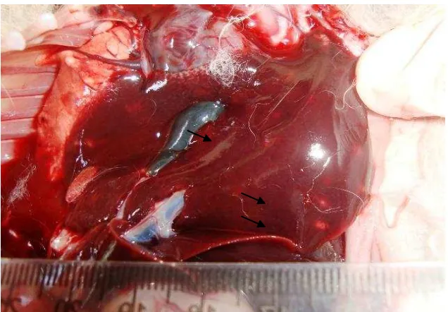
In anatomic pathology examination, most of rabbit liver experienced swelling, congestion, and edema. In addition, it was also observed abnormalities such as the presence of yellowish white nodules with various diameters between 1-5 mm, spread either on the surface or in the surrounding liver parenchyma (Figure 1). Caseus materials were observed when the nodules incised. Clinically, these changes were believed to be associated with symptoms caused yellowish in infected rabbit cornea, as it associated with a disturbance of liver function damaged by E. stidae parasitic infections. The infection is generally associated with general symptoms, such as; anorexia, depression, rough hair coat, weakness, pendulous, and diarrhea that occurred on the infected rabbits (Singla, et al., 2008). Although in these samples, the farmers mainly said that no specific symptoms were showed before their rabbits dead.
4
Coccidiosis is a parasitic disease caused by Eimeria sp.. In general there are two types of coccidiosis in rabbits which have typical pathognomonic symptoms, namely hepatic and intestinal types (Bhat, et al., 1996). Coccidiosis which left specific lesions in the liver is caused by Eimeria stidae infection. The other species, such as E. magna, E. intestinalis and E. irresidua generally infected the intestinal tract without any significant effect (Gardiner, et al., 1998).More specifically, E. stidae is a protozoa which has predilection in the epithelial cells of the bile duct in rabbits. This parasite will cause damage to the cells or tissue. Eimeria can also cause severe pathological disorders in the liver of infected rabbits, causing metabolic disorders in these organs. This situation is believed to have contributed to the host deaths (Wang & Tsai 1991).
Rabbits can be infected when ingesting sporulated oocysts. Sporulated sporozoites from oocysts penetrate the intestinal mucosa of the gastrointestinal tract passes through the hepatic portal system to the liver. Eimeria stidae sporozoites enters the epithelial cells of the bile ducts and also infect the parenchyma cells of the liver. In the epithelial cells of the bile duct, sporozoites develop into schizon, and produce merozoites. Within 18 days, the oocysts can be observed in the feces of infected animals. According to El-Akabawy, et al, (2004), the presence of Eimeria stidae can cause lesions in the liver of infected rabbits.
Microscopic observation of the E. stidae was not clear, whereas observation of its oocyst was clearly visible. On histopathologic observation, Eimeria oocyst observed in the liver, especially in the bile duct (Figure 2). In severe infections, most of oocysts were found in the lumen of the bile duct, even in parenchyma tissue of the liver (Al-Rukibat, et al., 2001). This caused bile duct epithelium undergoes swelling and thickening. Specifically, villous epithelium of the bile duct showed hypertrophy (Figure 3). This may occurs due to inflammatory reaction against oocyst of Eimeria, and lead to bile duct epithelial villi changes. Fibrous connective tissue can be observed in the inflammation area of chronic inflammatory conditions. It is supported by a research stating that hepatic coccidiosis caused congestion, severe dilation, rupture of the vascular endothelium in the liver, and also hypertrophy on villous epithelium of the bile ducts in portal area (Al Mathal, 2008). As a result, infected livers were bigger with tend to be darker in the color.
5
Figure 3. Histopathological examination of the coccidiosis infection. Arrow show bile ducts undergo hypertrophy.
(Stained with Hematoxylin and Eosin, 100 times magnification)
It is high likely that the damage that occured in the liver and bile ducts was due to the penetration of the sporozoites to the hepatic portal system, entered the bile duct epithelium, subsequently developed into trophozoites and continued to form schizont (Figure 4). The development of thesporozoites had contributed to the degeneration and necrosis of the hepatocytes. All of those microscopic changes affected the macroscopic liver changes. The livers observed bigger with darker in the color and brittle for their consistency.
Figure 4. Microscopic observation of material obtained from the bile duct showed various stage development of the Eimeria stidae (arrow).
(Stained with Hematoxylin and Eosin, 400 times magnification)
CONCLUSION
6
SUGGESTION
Experimental prospective studies are needed regarding Eimeria stidae infection in rabbits so that stage of the pathogenesis of this parasite infection can be more clearly known. Similarly, research on particular treatment to infected rabbits needs to be done to improve quality and quantity of the livestock.
ACKNOWLEDGEMENT
7
REFERENCE
Al-Mathal, E.M. (2008). Hepatic Coccidiosis of The Domestic Rabbit (Oryctolagus cinniculus) in Saudi Arabia. World Journal of Zoology, 3(1), 30-35.
Al-Rukibat, R.K., Irizarry, A.R., Lacey, J.K., Kazacos, K.R., Storandt, S.T., & deNicola, D.B. (2001). Impression smear of liver tissue from a rabbit. Veterinary Clonical Pathology, 30(2), 57-61.
Bhat, T.K., Jithendran, K.P., & Kurade, N.P. (1996). Rabbit Coccidiosis and its control: a review. World Rabbit Science, 4(1), 37-41.
Berata, I.K. (2009). Kelinci Sebagai Hewan Coba, Sumber Daging, dan Kesayangan. Denpasar: Swasta Nulus.
Cam, Y., Alasever, A., Eraslan, G., Kibar, N., Atyalay, O., Beyaz, L., Inci, A., & Liman, B.C. (2008). Eimeria stidae: Experimental infection in rabbits and the effect of treatment with toltrazuril and ivermectin. Experimental Parasitology, 119, 164-172.
Darzi, M.M., Mir, M.S., Kamil, S.A., Nashiruddullah, N., & Munshi, Z.H. (2007). Pathological changes and local defense reaction occurring in spontaneous hepatic coccidiosis in rabbits (Oryctolagus cuniculus). World Rabbit Sci, 15, 23-28.
El Akabawy, L.M., Zayan, K.A., Tantawy, A.A., & Omar, R.E.M. (2004). Anticoccidial Efficacy of Propolis and Toltrazuril Against Eimeria stidae in New Zealand White Rabbit's. Zag. Vet. J., 32(1), 129-145.
Gardiner, G.H., Fayer, R., & Dubey, J.P. (1998). Apicomplexa. In: An Atlas of Protozoan Parasites in Animal Tissues. Washington: Armed Forces Institute of Pathology.
Gonzales-Redondo, P., Finzi, A., Negretti, P., & Micci, M. (2008). Incidence of coccidiosis in different rabbit keeping systems. Arg. Pras. Med. Vet. Zootec., 60(5), 1267-1270.
Gendron, K., & Earle-Briges, M. (2000). The Rabbit Handbook. New York: Barron's Educational Series, Inc.
Hussein, F.N. (2008). Anasthesia and Euthanasia in Laboratory Animals. Workshop on the Care and Use of Lab An Res. Collaboration Fac.Vet.Med Airlangga Univ. and Fac.Vet.Med. UPM. Surabaya.
Kiernan, J.A. 2001. Histological and Histochemical Methods. 3r d Edition. Toronto: Arnold Pub. pp. 330-354.
Singla, L.D., Juyal, P.D., & Sandhu, B.S. (2000). Pathology and Theraphy in Naturally Eimeria stidae - infected Rabbit. The Journal of Protozoology Research, 10(4), 185-191.
Toulah FH. and Al-Rawi MM. 2007. Efficacy of garlic extract on hepatic coccidiosis in infected rabbit (Orictolagus cuniculus): histological and biochemical studies. J.Egypt Soc Parasitol 37(3): 957-968.