Molecular cloning and characterization of soybean peroxidase gene
families
Huabang Chen, Richard A. Vierling *
Indiana Crop Impro6ement Association and Department of Agronomy,Purdue Uni6ersity,West Lafayette,IN47907,USA
Received 2 July 1999; received in revised form 25 August 1999; accepted 25 August 1999
Abstract
Plant peroxidases play major roles in many physiological processes. A soybean seedbud (21 days after flowering) Uni-ZAP XR cDNA library was screened with a peroxidase-specific probe. The probe was generated by 3%rapid amplification of cDNA ends with soybean seedbud total RNA and a degenerate primer derived from a plant peroxidase conserved amino acid region (distal heme ligand). Positive clones were recovered by PCR using the degenerate peroxidase-specific primer and the vector primer T7
flanking the cloning site. Four cDNAs, designatedGmEpa1,GmEpa2,GmEpb1, andGmEpb2, contained 1298, 1326, 1171, and 1145 nucleotides, excluding poly(A) tail, and encoded mature proteins of 303, 303, 292, and 292 amino acids, respectively. The four predicted amino acid sequences showed homology to other peroxidases. GmEpa1 andGmEpa2 exhibited 97% amino acid identity,GmEpb1 andGmEpb2 exhibited 93% amino acid identity, andGmEpa1 andGmEpb1 exhibited 47% amino acid identity. GmEPa1 andGmEPb1 were expressed as fusion proteins inEscherichia coli. The recombinant fusion proteins were sequestered in inclusion bodies and active forms of the two denatured proteins were recovered after in vitro folding in a medium containing hemin, urea and Ca2+.GmEpa1 andGmEpa2 messages were detected in developing seed and root, whileGmEpb1 andGmEpb2
messages were present in root, leaf, stem and seed pod. These cDNAs and cDNA-specific primers will allow investigations into peroxidase’s role in development, stress response and in other physiological processes. © 2000 Elsevier Science Ireland Ltd. All rights reserved.
Keywords:Peroxidase; cDNA; Differential expression; Soybean
www.elsevier.com/locate/plantsci
1. Introduction
Peroxidases, a ubiquitous class of protein, are enzymes whose primary function is to oxidize a variety of hydrogen donors. Peroxidase has been studied extensively in higher plants and has been implicated in a variety of physiological processes including lignin biosynthesis [1], extensin polymer-ization [2], disease and pathogen response [3],
wound healing [4], and response to air pollutant stress [5].
Most higher plants possess a number of differ-ent peroxidase isozymes. In the tobacco plant there are at least 12 distinguishable isozymes [1]. The soybean peroxidase family consists of 20 isozymes [6]. The large number of peroxidase isozymes could represent gene copy numbers through polymorphism or post-translational mod-ification. These many possibilities of so-called isozyme expression make it difficult to study the actual function of peroxidase. Peroxidase expres-sion has been reported to be tissue-specific, devel-opmentally regulated, and influenced by environmental factors [6].
Considering the vast number of studies in this field, the physiological function of individual members within this class of enzymes is only
The nucleotide sequences have been registered in the EMBL Nucleotide Database under the accession numbers U51191 (GmEpa1), U51192 (GmEpa2), U51193 (GmEpb1) and U51194 (GmEpb2).
* Corresponding author. Present address: 1150 Lilly Hall, West Lafayette, IN 47907-1150, USA. Tel.: +1-765-5232535; fax: +1-765-5232536.
E-mail address:[email protected] (R.A. Vierling)
tially understood. The lack of knowledge is due, in part, to the complexity of peroxidase isozymes expressed within a given plant tissue. Cloning of plant peroxidase genes could help in elucidating the physiological role of individual peroxidase iso-forms during plant normal development and in response to various environmental factors. Under-standing the expression pattern of individual per-oxidase isozymes may lead to understanding their function. Without gene-specific probes, separation of the expression patterns of closely related perox-idases may be difficult due to similarity in se-quence and transcript size. Cloning of plant peroxidase genes also could open the possibility of transgenic studies and in vitro site-mutagenesis analysis of various peroxidase isoforms. In this report, we described the isolation and characteri-zation of four closely related peroxidase cDNAs, the development of four sets of gene-specific per-oxidase primers, and the expression ofGmEPa1 as a fusion protein inEscherichia coliand its in vitro folding to yield an active enzyme.
2. Materials and methods
2.1. cDNA library construction and screening
Total RNA was extracted from soybean
(Glycine max cul. Resnik) seedbuds 21 days after flowering as previously described [7]. Poly(A)-en-riched RNA was prepared from total RNA using PolyATract (Promega) and the cDNA library was constructed in the unidirectional vector Uni-ZAP XR (Stratagene).
A plant peroxidase specific primer (PSP) was designed from a plant peroxidase conserved amino acid region (distal heme ligand, HFHDCFV) (5%
CA(C/T)TT(T/C)CA(C/T)GA(C/T)TG(C/T)TT(C/ T)GT 3%) [8]. The plant peroxidase-specific probe
was generated using the 3% RACE system with soybean seed bud total RNA and PSP as described by the manufacturer (GIBCO/BRL) except that hot-start PCR was performed. The PCR-RACE products were cloned into pCR™ II plasmid (In-vitrogen). DNA from 20 clones was purified and digested withEcoRI, fractionated by electrophore-sis on a 1% agarose gel, and blotted on a nylon membrane and probed with [g-32
P]dATP-end-la-beled PSP. A single positive clone was random-prime-labeled with [a-32P]dCTP and used for
primary screening of the cDNA library. Prehy-bridization was conducted in 6× SSPE, 5× Den-hardt’s, 0.5% (w/v) SDS, 100 mg/ml denatured salmon sperm DNA, and 50% (v/v) formamide at 42°C for 2 h. Hybridizations were done overnight and the conditions were the same as those in prehybridization, except that 1× Denhardt’s solu-tion was used. Membranes were washed three times for 30 min each with 0.1× SSPE and 0.1% (w/v) SDS at 65°C.
PCR using PSP and the vector primer T7 was
used to purify single phage clones. Phage particles were eluted by incubating primary picks and/or single plagues in 500 ml of SM buffer (100 mM NaCl, 10 mM MgSO4, 0.01% (w/v) gelatin in 50
mM Tris pH 7.5) at room temperature for 2 h. The PCR cycling parameters were 94°C for 4 min followed by 30 cycles of 1 min at 94°C, 1 min at 57°C, and 1 min at 72°C, and then followed by a final extension at 72°C for 5 min. PCR reaction conditions were 1× reaction buffer (500 mM KCl, 100 mM Tris – HCl, pH 9.0 and 1.0% (v/v) Triton X-100), 1.5 mM MgCl2, 200 mM each
dNTP, two units of Taq DNA polymerase, 1mM each primer and 2 ml of phage particle elution in 50 ml total volume.
2.2. DNA sequencing and analysis
DNA sequencing of both strands was performed using Sequenase Kit 2.0 (USB) and SK and KS primers. Synthetic primers corresponding to inter-nal sequences of cDNA were made to complete sequencing. Sequences were optimally aligned us-ing the PILEUP program of the Wisconsin GCG software package, and the DISTANCES program was used to determine the similarity percentage. The dendrogram was generated using the cluster-ing algorithm in PILEUP, which aligns the most similar sequences prior to alignment with more distantly related sequences.
2.3. Re6erse transcription PCR (RT-PCR)
cDNA specific primers designed from 3%
un-translated regions of each peroxidase cDNA and PSP were used in reverse transcript PCR (RT-PCR) to study expression patterns. For GmEpa1,
GmEpa2, GmEpb1, andGmEpb2 the primers were
5% AAATTAACTCAGCTGTGGG 3%, 5%
CC-CAAGACATGCTTGAGAT 3%, and 5%
AAGTTCATACTTCTAAC 3%, respectively. A total of 5 mg of total RNA from different soybean tissues was used for synthesizing the first strand of cDNA using SUPERSCRIPT™ II Rnase H−REVERSE TRANSCRIPTASE as
sug-gested by the manufacturer (GIBCO/BRL). RT-PCR conditions were the same as those in 3%
RACE except that the annealing temperature for
GmEpb2 was 45°C. A total of 20 ml of PCR
products was electrophoresed on a 1% (w/v) agarose gel and visualized with ethidium bromide.
2.4. In 6itro folding of bacterially expressed
GmEPa1 and GmEPb1 proteins
The open reading frames including the 5%leader sequences of GmEPa1 and GmEPb1 were PCR-amplified and cloned into the pET-34b(+)
expres-sion vector (Novagen). The primers
complementary to the lower strands were designed with a BamH I site at the 5% ends and the primers complementary to the upper strands with a Xho I
site (GmEPa1: 5%
GACGGATCCATGGGAAG-CAACTTGAGGTTTTTG 3%, 5%
GACCTC-GAGTTAGCTATTTATAAATGCACAATG 3%;
GmEPb1: 5%
GACGGATCCATGGCTGT-CATGGGTGCATTCTTG 3%, 5%
ACCTCGAG-TAATTCTGCAGCCCTTCTTTCCTCCTG 3%).
PCR products were digested with BamH I and Xho I and ligated to pET-34b(+) digested with the same two enzymes. The constructs were then transformed into BL21 (DE3) competent E. coli cells.
An overnight culture of 5 ml of BL21 was inoculated into a 100-ml culture containing 50 mg/ml kanamycin. The inoculated culture was grown at 37°C with vigorous shaking until an OD600of 1.0. The 100-ml culture was then split
into 2×50-ml cultures. IPTG was added to one of the 50-ml cultures to a final concentration of 1 mM. The other culture was used as an uninduced control. The 2×50-ml cultures were further grown for 5 h after IPTG induction. Different temperatures and media additives were used dur-ing the bacterial growth. Isolation of total cell proteins, inclusion bodies and cellulose binding domain (CBD) fused peroxidase were performed according to the manufacturer (Novagen). Inclu-sion bodies were washed twice with buffer (200 mM Tris – Cl pH 8.0, 100 mM EDTA, 100 mM
DTT, 10% (v/v) Triton X-100) before being fully solubilized in 10 vol. of 6 M urea, 1 mM DTT in 50 mM Tris – Cl (pH 8.0).
A single-step dilution was used for the dena-tured protein refolding. A total of 10 mg (1mg/ml) of the inclusion body prep was slowly diluted in 190 ml of PBS, which once diluted, contained 2 M urea, 5 mM CaCl2, 10 mM hemin and 0.1 mM
DTT (PBS: 137 mM NaCl, 1.47 mM KH2PO4,
8.10 mM Na2HPO4, and 2.68 mM KCl, pH 8.0).
After overnight incubation at room temperature, 20-ml aliquots of the folding mixtures were trans-ferred to the wells of a microtiter plate, and perox-idase activity was monitored using substrate tetramethylbenzadine as described by Vierling and Wilcox [9].
3. Results
3.1. Isolation of soybean peroxidase cDNA
The vast number of plant peroxidase sequences documented and the rapid amplification of cDNA ends (RACE) technique made possible the genera-tion of a plant peroxidase-specific probe. A degen-erate plant peroxidase-specific primer (PSP) corresponding to a highly conserved region, distal heme ligand (HFHDCFV) was synthesized. Using PSP and anchor primer complementary to the poly(dT) end of the cDNA, the 3% RACE experi-ment resulted in amplification of a major DNA band of 900 bp (data not shown). The fragment was cloned and one of the clones was used as probe to screen the cDNA library.
Approximately 2×10⁵ recombinant phages
from the soybean seedbud cDNA primary library were screened using the plant peroxidase-specific probe. A total of 25 clones were obtained by primary screening, and 11 positive clones were recovered after two rounds of PCR using PSP and T7 vector primers. The four longest clones,
desig-nated GmEPa1,GmEPa2,GmEPb1 and GmEPb2, were further analyzed.
3.2. Nucleotide and deduced protein sequences of
the soybean peroxidase cDNA
GmEPa1, GmEPa2, GmEPb1, and GmEPb2
U51192, U51193 and U51194. They contained 1298, 1326, 1171 and 1145, nucleotides, excluding poly(A) tail, with 86, 82, 59 and 38-bp 5%
untrans-lated leaders, and 240, 272, 173 and 168-bp 3%
untranslated regions, respectively. Two copies of the putative polyadenylation signals AATAAG are present at nucleotides 25 and 81 upstream of the poly(A) tail in GmEpa1, and 45 and 112 bases upstream of the poly(A) tail in GmEpa2. There was only one copy of the putative polyadenylation signal AATAAA 42 bases upstream of the poly(A) tail inGmEpb1 and 20 bases upstream inGmEpb2. The open reading frames (ORFs) of GmEPa1,
GmEPa2, GmEPb1 and GmEPb2 were 972, 972,
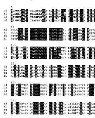
942 and 942 bp long. The deduced amino acid sequences encoded by the four ORFs are shown in Fig. 1. It was predicted from these sequences that
the proteins were synthesized as preproteins of 324, 324, 314 and 314 amino acids with hydropho-bic putative signal sequences of 21, 21, 22 and 22 residues, respectively. The mature proteins from
GmEPa1, GmEPa2, GmEPb1 and GmEPb2 were
designated as a1, a2, b1 and b2. Cleavage of putative signal sequences releases mature proteins of 303, 303, 292, and 292 residues with theoretical Mr of 33 333, 33 333, 32 412 and 32 412 Da. The
theoretical pIs of mature a1, a2, b1 and b2 were 6.96, 7.41, 10.04 and 9.05, respectively. There were six putative glycosylation sites specified by N-X-T/ S at amino acid residues 56, 69, 128, 142, 183 and 214 in a1 and a2, and four putative glycosylation sites at residues 70, 142, 185 and 195 inb1 and b2. Peroxidases a1 and a2 had the [Q L X X X F Y] motif at the NH2 terminus that was a feature
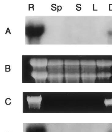
Fig. 2. (A) Northern blot analysis ofGmEPa1 expression in root (R), seedpod (Sp), stem (S), leaf (L), and developing seed coat (Ds). (B) Ethidium bromide stained RNA gel indicating roughly equal loading of total RNA. (C) Ethidium bromide stained RT-PCR products amplified from cDNA synthesized from total RNA of R, Sp, S, L, and Ds. The two primers were PSP and GmEPa1-specific primer. (D) Southern blot analysis of RT-PCR products amplified using PSP and
GmEPa1-specific primers. The probe was 32P-labelled
GmEPa1.
region of each peroxidase cDNA and PSP were used in RT-PCR to study expression patterns. The RT-PCR results (Fig. 2) for GmEPa1 were consis-tent with the above Northern blotting analysis for GmEPa1, and the RT-PCR products were also confirmed by probing the products with GmEPa1 (Fig. 2). Based on the results of cDNA-specific primers, transcripts from GmEpa2 were also de-tected in root and developing seed, and transcripts
from GmEpb1 and GmEpb2 were detected in root,
stem, leaf, and seedpod (Fig. 4).
3.4. In 6itro folding of bacterially expressed
GmEPa1 proteins
In order to obtain expression of soybean perox-idases, the ORFs of GmEPa1 and GmEPb1 in-cluding the leader sequences were cloned into the expression vector pET-34b(+). Since the two se-quences showed the same features in both expres-sion and in vitro folding, onlyGmEPa1 is reported here. Peroxidase a1 was found toxic to E. coli growth upon early IPTG induction, even though the peroxidase was produced as a CBD-peroxidase fusion protein (Figs. 5 and 6). The E. coli culture containing the vector construct was then induced by IPTG at OD600=1.0 to maximize the fusion
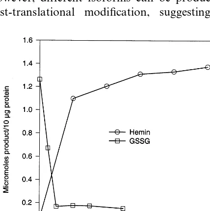
protein production. As shown in Fig. 6, a fusion protein of 60 kDa was produced and the fusion protein was accumulated in inclusion bodies under all conditions tested. After four rounds of sonica-tion and washing, the urea-solublized inclusion body prep gave \90% pure fusion proteins as judged by SDS – PAGE (Fig. 6). As can be seen in Figs. 7 and 8, the recovery of peroxidase activity was critically dependent on the addition of hemin and on the concentration of urea, with 2 M being optimal at pH 8.0. The addition of oxidized glutathione (GSSG) inhibited correct folding (Fig. 8). No difference in folding efficiency existed among solubilized inclusion bodies obtained under different bacteria growing conditions (data not shown).
4. Discussion
We have cloned four soybean peroxidase cD-NAs. The similarity of the predicted amino acid
from GmEPa1 andGmEPa2 to other plant
perox-idases ranges from 54% (cotton, L08199) to 78% found in most plant peroxidases, but no [Q L X X
X F Y] motif existed in b1 or b2. Each mature protein, except b2, had eight cysteines that may form disulfide bridges. Peroxidases a1 and a2 ex-hibited 99% amino acid similarity and 97% iden-tity, b1 and b2 had 95% similarity and 93% identity, and a1 and b1 shared 60% similarity and 47% identity.
3.3. Differential expressions of peroxidase mRNA
Total RNA from leaf, stem, root, seedpod, and developing seed were probed with a 300-bp gene-specific Kpn-Tif I fragment from the 3% end of
GmEpa1. Transcripts of 1400 nucleotides from
GmEpa1 were present in developing seed and root (Fig. 2). Since both the coding regions and the non-coding regions of the four cDNAs are highly homologous, RT-PCR experiments were con-ducted to study the differential expressions of peroxidase mRNA. The divergence at the 3%end of
GmEPa1 and GmEPa2, GmEPb1 and GmEPb2,
(tomato, L13653). Mohan et al. [10] reported the induction of L13653 by wounding and pathogen attack, which suggested these peroxidases may play a role in wound healing and/or plant defense response. Although GmEPa1 and GmEPa2 also are expressed in seed coat, they share only about 46% amino acid identity with two other soybean seed coat peroxidases (U41657 and AF014502) [11]. The similarity of the predicted amino acid
from GmEPb1 and GmEPb2 to other plant
perox-idases ranges from 47% (soybean, U41657) to 64% (turnip, B23116). Further studies are needed to address the physiological roles of these genes.
It is predicted from the cDNA sequences that all four proteins are initially synthesized as pre-proteins with predominantly hydrophobic amino
acid signal sequences, suggesting that the mature proteins could be secreted through cell mem-branes. The four soybean peroxidases differ in the number of glycosylation sites, their pIs, and their expression patterns. Other plant peroxidases vary greatly in the number of glycosylation sites, from one in peanut and turnip to eight in horseradish [12]. The heterogeneity of glycosylation indicates that peroxidases exist in different glycosylated forms or glycoforms. Lerouge et al. [13] reported
that N-glycosylation is a major modification of
plant proteins, and glycosylation is necessary for an efficient secretion of plant glycoproteins. Vari-ability in N-linked oligosaccharide chain number and location also may be adaptively important for fine tuning catalytic properties of the functional
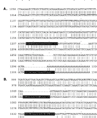
Fig. 4. Ethidium bromide stained RT-PCR products amplified from cDNA synthesized from total RNA of root (e, j, p), seedpod (d, i, o), stem (c, h, n), leaf (b, g, l), and developing seed coat (a, f, k). The primer pairs used were PSP and cDNA-specific primers derived from the 3% untranslated re-gions ofGmEpa2 (I),GmEpb1 (II) andGmEpb2 (III), respec-tively. Lane M was the 1-kb DNA marker.
Fig. 6. Expression of CBD-soybean peroxidase inE.coliand SDS – PAGE of different fractions. (1) Protein molecular weight marker. (2) Total protein of BL21. (3) Total protein of BL21 with plasmid containing insert but no IPTG induction. (4) Total protein of BL21 with plasmid containing insert and IPTG induction. (5) Bacteria growth temperature of 25°C. (6) Bacteria growth temperature of 30°C. (7) Bacteria growth temperature of 37°C. (8) Bacteria growth temperature of 37°C and addition of d-ALA. (9) Bacteria growth temperature of 37°C and addition of hemin.
could be that the E. coli expression system lacks glycosylation machinery, and non-glycosylation of peroxidase may result in loss of the epitope or the antibody recognized a conformational epitope.
Peroxidase genes comprise multigene families in the plant genomes. In order to analyze each mem-ber of a gene family, the availability of gene-spe-cific probes is essential. The divergence at the 3%
ends of the highly homologous soybean peroxidase coding regions enabled us to design gene-specific primers and study the expression of highly ho-enzyme molecule. However, a glycosylation site
(Asp-185) is common to most peroxidases. Wan and Huystee [14] reported that oligosaccharides were the major antigenic sites of glycoproteins during the production of antibodies. We initially screened the cDNA expression library with an anti soybean seed coat monoclonal antibody and failed to obtain any positive clones. One possible reason
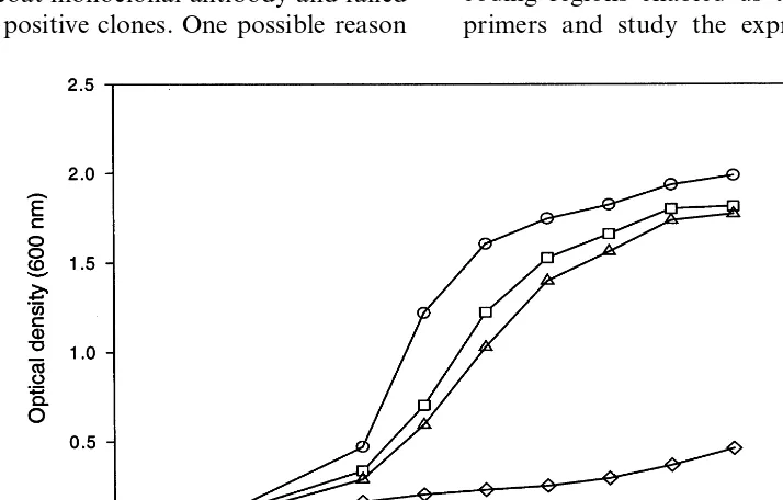
Fig. 7. Optimum urea concentrations for folding of recombi-nant soybean peroxidase. , BL21+plasmid+insert+
IPTG;, BL21+plasmid+insert.
different genes might not be necessary to provide different functions. A second possibility is that multiple genes can allow for greater regulatory flexibility. Some genes may be expressed in tissue-specific organs or at tissue-specific stages, and the expres-sion of the genes may be determined by different signals. This appears to be the case in soybean as we have demonstrated that the four soybean per-oxidases are expressed in different tissues. The use of gene-specific probes will allow us to analyze individual gene expression independent of the other highly related gene sequences. And the availability of these genes also will allow us to further study their catalytic mechanisms by site-mutagenesis, and their physiological roles by transgenic studies.
The cDNA open reading frame encoding soy-bean peroxidase was found to be growth inhibiting upon expression inE. coli, even when expressed in an inactive form. Thus, it is unlikely that the growth inhibiting activity is mediated by peroxi-dase activity generating oxygen radicals. Bar-tonek-Roxa and Eriksson [15] found that the first 64 N-terminal amino acids of a mature, neutral horseradish peroxidase was responsible for its tox-icity to E. coli growth. This region was highly conserved among peroxidases, but no homology was found when this region was compared to other protein sequences. Kempf et al. [16] reported that formation of heme-containing holoenzyme cy-tochrome P450 2D6 was strictly dependent on addition of the heme precursor d-aminolevulinic acid to the E. coli culture. The addition of d-aminolevulinic acid and hemin to the E. coli cul-ture, however, did not result in functional expression of soybean peroxidase. Unlike previous studies [17,18], GSSG was inhibitory in this study. One possible explanation is that soybean peroxi-dases have seven or eight cysteine residues that form disulfide bridges. The addition of GSSG may facilitate the formation of incorrect, mispaired in-tra and/or intermolecular disulfide bridges among the cysteines of the reduced, inactive polypeptide. Whether refolding of the completely reduced plant enzyme would occur more readily is uncertain. The native enzyme differs from the recombinant enzyme in being glycosylated and in having a blocked N-terminus, whereas the N-terminal amino acid of the recombinant enzyme is pre-sumably methionine. Glycosylation may serve to prevent irreversible aggregation of partially un-mologous genes. The expression patterns of these
genes based on both Northern analysis and RT-PCR indicated differential expression of soybean peroxidase genes, which was expected based on expression patterns of other plant peroxidases. It is not apparent why some organisms have several expressed peroxidase genes, but one possibility is that different isoforms have different functions. However, different isoforms can be produced by post-translational modification, suggesting that
Fig. 8. Effects of hemin and oxidized glutathione (GSSG) on folding of recombinant soybean peroxidase. , Hemin; ,
folded intermediates, and the lack of glycosylation in E. coli system may explain, in part, the low efficiency of refolding. Meanwhile, the addition of an anti-seed-coat peroxidase monoclonal antibody did not affect the in vitro protein refolding (data not shown). Further refinement of the expression/ folding/activation of soybean seed coat peroxidase is needed to provide sufficient active protein for characterization of both the wild type and site-di-rected mutants.
All of the purified sequences contained the ex-pected peroxidase conserved regions and the cDNA expressed in E. coli was confirmed to have peroxidase activity. The use of gene-specific probes will allow the analysis of individual gene expres-sion independent of highly related gene sequences. This will allow for greater discrimination in study-ing tissue specificity, inducibility and regulation of related soybean peroxidase genes.
References
[1] L.M. Lagrimini, W. Burkhart, M. Moyer, S. Rothstein, Molecular cloning of complementary DNA encoding the lignin-forming peroxidase from tobacco: molecular anal-ysis and tissue-specific expression, Proc. Natl. Acad. Sci. USA 84 (1987) 7542 – 7546.
[2] S.C. Fry, Cross-linking of matrix of polymers in the growing cell walls of angiosperms, Annu. Rev. Plant Physiol. 37 (1986) 165 – 186.
[3] L.M. Lagrimini, S. Rothsteins, Tissue specificity of to-bacco peroxidase isozymes and their induction by wounding and tobacco mosaic virus infection, Plant Physiol. 84 (1987) 438 – 442.
[4] K.E. Espelie, V.R. Franceschi, P.E. Kolattukudy, Im-munocytochemical localization and time course of ap-pearance of an anionic peroxidase associated with suberization in wound-healing potato tuber tissue, Plant Physiol. 81 (1986) 487 – 492.
[5] C. Breda, D. Buffard, R.B. Huystee, R. Esnault, Differ-ential expression of two peanut peroxidase cDNA clones in peanut plants and cells in suspension culture in re-sponse to stress, Plant Cell 12 (1993) 268 – 272.
[6] J.W. Gillikin, J.S. Graham, Purification and develop-mental analysis of the major anionic peroxidase from the
seed coat ofGlycine max, Plant Physiol. 96 (1991) 214 – 220.
[7] R.A. Vierling, H.T. Nguyen, Heat-shock protein gene expression in diploid wheat genotypes differing in ther-mal tolerance, Crop Sci. 32 (2) (1992) 370 – 377. [8] H. Tyson, Relationships, derived from optimum
align-ments, among amino acid sequences of plant peroxi-dases, Can. J. Bot. 70 (1991) 543 – 556.
[9] R.A. Vierling, J.R. Wilcox, Microplate assay for soybean seed coat peroxidase activity, Seed Sci. Technol. 24 (1996) 485 – 494.
[10] R. Mohan, A.M. Bajar, P.E. Kolattukudy, Induction of a tomato anionic peroxidase gene (tap1) by wounding in transgenic tobacco and activation of tap1/GUS and tap2/GUS chimeric gene fusions in transgenic tobacco by wounding and pathogen attack, Plant Mol. Biol. 21 (1993) 341 – 354.
[11] M. Gijzen, A deletion mutation at the ep locus causes low seed coat peroxidase activity in soybean, Plant J. 12 (5) (1997) 991 – 998.
[12] D. Buffard, C. Breda, R.B. Van Huystee, O. Asemota, M. Pierre, D.B.D. Ha, R. Esnault, Molecular cloning of complementary DNAs encoding two cationic peroxidases from cultured peanut cells, Proc. Natl. Acad. Sci. USA 87 (1990) 8874 – 8877.
[13] P. Lerouge, M. Cabanes-Macheteau, C. Rayon, A.C. Fischette-Laine, V. Gomord, L. Faye, N-Glycoprotein biosynthesis in plants: recent developments and future trends, Plant Mol. Biol. 38 (1998) 31 – 48.
[14] L. Wan, R.B. Huystee, Immunogenicity of the N-glycans of peanut peroxidase, Phytochemistry 37 (1994) 933 – 940.
[15] E. Bartonek-Roxa, H. Eriksson, Expression of a neutral horseradish peroxidase inE.coli, J. Biotechnol. 37 (1994) 133 – 142.
[16] A.C. Kempf, U.M. Zanger, U.A. Meyer, Truncated hu-man P450 2D6: expression in E. coli, Ni2+-chelate
affinity purification, and characterization of solubility and aggregation, Arch. Biochem. Biophys. 321 (2) (1995) 277 – 288.
[17] W.A. Doyle, A.T. Smith, Expression of lignin peroxidase H8 inE.coli: folding and activation of the recombinant enzyme with Ca2+ and haem, Biochem. J. 315 (1996)
15 – 19.
[18] A.T. Smith, N. Santama, S. Dacey, M. Edwards, R.C. Bray, R.N.F. Thorneley, J.F. Burke, Expression of a synthetic gene for horseradish peroxidase C in E. coli and folding and activation of the recombinant enzyme with Ca2+ and heme, J. Biol. Chem. 265 (22) (1990)
13335 – 13343.