F
our types of protein kinases constitute the calcium-dependent protein kinase or calmodulin-like domain protein kinase (CDPK) superfamily. These kinases differ in whether they are regulated by binding Ca21(CDPKs), Ca21
/calmodulin [calmodulin-dependent protein kinases (CaMKs)], a combination of both [calcium and calmodulin-dependent protein kinases (CCaMKs)], or neither [CDPK-related protein kinases (CRKs)].
The abundant calcium-stimulated protein kinase activity found in plant extracts is associated with CDPKs. These enzymes contain three functional domains1–4
: catalytic, autoinhibitory and calcium-binding (Fig. 1). The calcium-binding domain of the archetypal CDPK is similar to calmodulin in sequence (~40% identity) and contains four EF-hand calcium-binding motifs. In addition to plants, CDPKs are found in protozoans such as para-mecium and Plasmodium falciparum (the causative agent of malaria). Notably, CDPKs are absent from the completed genome sequence of yeast (Saccharomyces cerevisiae) and of nematode
(Caenorhabditis elegans). Thus, it is tempting to speculate that CDPKs might be present in plants and protozoans only.
CCaMKs are rarer than CDPKs, and might be expressed in a few plant tissues only5
. Like CDPKs, they contain a calcium-binding domain6
(Fig. 1), but this domain contains only three EF-hands and is more similar to visinin (another EF-hand protein) than to cal-modulin. The autoinhibitory domain contains a binding site for calmodulin, and calmodulin stimulates the activity of these kinases. A third type of calcium-regulated protein kinases, the CaMKs, is well characterized from animals and yeast, but only one puta-tive representaputa-tive is known in plants7
. The plant CaMK is more similar in sequence to CCaMKs than to animal CaMKs, having an identical calmodulin-binding site, but lacking the C-terminal domain containing EF-hands (Fig. 1). The biochemical properties of this enzyme have not been characterized fully.
The fourth type of protein kinase in the superfamily is the CDPK-related protein kinases (CRKs). They have catalytic domains closely related to those of CDPKs, and their C-terminal domains have some sequence similarity to calmodulin (20% iden-tity), but their EF-hands are poorly conserved. Representative members of this group appear to be unresponsive to calcium8–10
. It is not known how these protein kinases are regulated or what their physiological roles are. Another type of CRK was reported recently: phosphoenolpyruvate carboxylase kinase has a catalytic domain related to those of CRKs, but no C-terminal domain. This protein kinase phosphorylates and regulates phosphoenolpyruvate carboxylase in vivoand is regulated at the level of transcription11
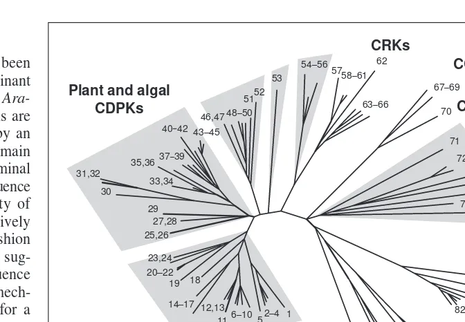
. In phylogenetic analyses (Fig. 2), the clustering of the plant CDPKs and CRKs away from the non-plant CaMKs and the SNF1-like kinases suggests a single common origin for plant CDPK and CRK genes. However, an important evolutionary question remains unresolved. Did different branches of the super-family have a common origin or did the fusion of genes encoding a protein kinase and a calcium-binding domain occur more than once in evolution?
Based on the analysis of the currently available (~70% com-plete) genomic sequence from Arabidopsis, we estimate that there will be a total of 40 CDPKs and seven CRKs. An Arabidopsis CCaMK sequence has not yet been identified, but southern blot analysis suggests that there is a single gene12
. Proliferation of fam-ily members might be related to expression of some of these genes in specific tissues, physiological conditions or developmental stages (reviewed in Ref. 13). Also there might be specialization of cellular roles, which might be related to differences in substrate specificity, subcellular location and calcium sensitivity.
CDPKs – a kinase for every
Ca
2
1
signal?
Alice C. Harmon, Michael Gribskov and Jeffrey F. Harper
Numerous stimuli can alter the Ca21concentration in the cytoplasm, a factor common to many
physiological responses in plant and animal cells. Calcium-binding proteins decode infor-mation contained in the temporal and spatial patterns of these Ca21signals and bring about
changes in metabolism and gene expression. In addition to calmodulin, a calcium-binding protein found in all eukaryotes, plants contain a large family of calcium-binding regulatory protein kinases. Evidence is accumulating that these protein kinases participate in numerous aspects of plant growth and development.
Fig. 1.Domain structure of calcium-dependent protein kinase or calmodulin-like domain protein kinases (CDPKs) and three related protein kinases. The N-terminal domain is highly variable in length and sequence. An autoinhibitor is predicted in the region immediately following the kinase domain. A distinguishing fea-ture of CDPKs, CDPK-related protein kinases (CRKs) and cal-cium and calmodulin-dependent protein kinases (CCaMKs) is the number of functional EF-hands in a C-terminal regulatory domain: EF-hands that can bind calcium are denoted by black boxes, whereas degenerated EF-hands are denoted by gray boxes. In a conventional CDPK the regulatory domain has four EF-hands and an overall sequence similarity to calmodulin (CaM). CaMK, calmodulin-dependent protein kinase.
Trends in Plant Science CaM-like CDPK
Kinase
CRK
CCaMK
CaMK
Autoinhibitor
N-terminal
Association domain Degenerated
Activity and regulation
Regulation of CDPKs by Ca21
How calcium regulates CDPKs has been the subject of studies with recombinant soybean (Glycine max) CDPKaand Ara-bidopsisCPK1 (Refs 1–4,14). CDPKs are kept in a low basal state of activity by an autoinhibitor located in a junction domain that connects the kinase to its C-terminal calmodulin-like domain. A peptide sequence from the junction inhibits the activity of wild-type enzymes and of a constitutively active mutant, in a competitive fashion with respect to the peptide substrate, sug-gesting that the autoinhibitory sequence functions through a pseudosubstrate mech-anism3
, analogous to that proposed for a typical CaMK from animals.
The simplest model for the activation of CDPK by Ca21
is provided by analogy to the mechanism for stimulation of animal CaMKs. For CaMKs, calcium promotes a bimolecular binding of calmodulin to a region immediately downstream of an autoinhibitory sequence. This binding event somehow disrupts the autoinhibitor and results in a ‘release of inhibition’. The dis-tinction for a CDPK is that this ‘release of inhibition’ involves intramolecular binding with its calmodulin-like domain (Fig. 3). One line of evidence supporting a close analogy to a CaMK is the observation that the activity of a truncated CDPK (DC), in which the calmodulin-like domain is deleted, can be partially stimulated by either calmodulin or an isolated cal-modulin-like domain, with half maximal activation at ~3 mM for both activators1,2.
Although this indicates that a CDPK can be reconstituted as a bimolecular interaction with calmodulin (i.e. like a CaMK), it is possible that the natural mechanism of intramolecular activation (i.e. the whole) is distinct from its reconstitution as two sep-arate fragments (i.e. the sum of the parts). An important challenge is to understand the structural basis for activation of CDPK (and CaMKs), and to determine whether the presence of a tethered calmodulin-like domain endows CDPKs with unique bio-chemical and physiological properties.
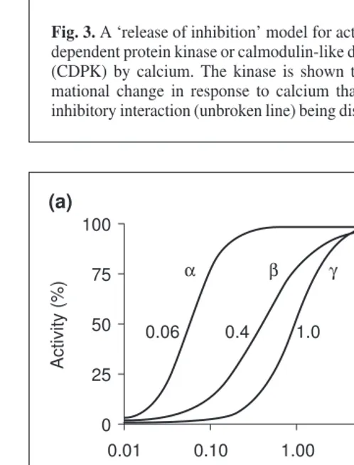
One reason for the multiplicity of CDPKs in a given plant species might be related to the specialization of different iso-forms with respect to calcium binding and activation. Dose–response curves (Fig. 4) for three CDPK isoforms from soybean show that they are responsive to different ranges of calcium concentrations15
. The concentration of calcium required for half-maximal activity (K0.5) for CDPKs a, band g varies over two orders of magnitude when using the synthetic peptide substrate syn-tide-2. Other CDPKs, such as P. falciparum
Fig. 2.Unrooted phylogenetic tree showing the relationship between the CDPK superfamily, plant SNF1-like kinases, and the most closely related animal kinases, the calmodulin-dependent protein kinases (CaMKs). Calcium and calmodulin-dependent protein kinases (CcaMKs) and the single plant CaMK (No. 70) share a branch close to the protozoan CDPKs. Plant and proto-zoan CDPKs form two distinct groups on the tree, with the plant isoforms being found on four branches. The majority of CDPKs from vascular (Nos 1–17, 19–26 and 29–52) and nonvascu-lar plants (18, 27 and 28) are interspersed on three branches. The only available CDPK sequence from an alga (No. 53) might represent a first example for a fifth branch of algal homologs. Few of these CDPKs have been characterized at the biochemical level, so distinc-tions cannot be made between the funcdistinc-tions of the enzymes on these branches. Sequences of plant/algal (1–56) and protozoan (71–78) CDPKs (highlighted in gray) and related kinases in the NCBI nr database were identified by BLAST 2.09 (Ref. 43) – the evolutionary profile method44
. Alignments were constructed by ClustalW45
, corrected manually, and used to gener-ate a neighbor-joining tree46
, which was subjected to 1000 rounds of bootstrap analysis. The tree shown was constructed from sequences of kinase catalytic domains. Each entry contains a two-letter code indicating genus and species, common name (if available) and Accession no. Entries for CDPKs from Arabidopsisinclude names (underlined) assigned in Ref. 13 followed by names given in the literature. See http://plantsP.sdsc.edu for additional information on meth-ods and sequences. (1) AtCPK20, 3928078; (2) AtCPK1, AK1, 304105; (3) AtCPK2, 1399271; (4) CpCPK1, 1899175: (5) zm3320104; (6) AtCPK26, 4467129; (7) VrCDPK-1, 967125; (8) AtCPK6, AtCDPK3, 603473; (9) AtCPK6, 1399275; (10) AtCPK5, 1399273; (11) Zm506413; (12) ZmCDPK1, 1632768; (13) ZmCDPK7, 1504052; (14) OsCDPK1, 435466; (15) Os6063536; (16) Oscdpk12, 2944385; (17) Oscpk11, 587500; (18) TrCPK1, 2315983; (19) AtCPK12, AtCDPK9, 836946; (20) AtCPK10, AtCDPK1, 604880; (21) AtCPK11, AtCDPK2, 604881; (22) AtCPK4, 1399267; (23) GmCDPKb, 2501764; (24) GmSK5, GmCDPKa, 116054; (25) AtCPK3, AtCDPK6, 2129550; (26) AtCPK3, AtCDPK6alt, 2129553; (27) MpCDPK-a, 4874268; (28) MpCDPK-b, 5162878; (29) Zm639722; (30) AtCPK22, 4115942; (31) AtCPK27, 5706728; (32) AtCPK31, 5732059; (33) AtCPK15, 2961339; (34) AtCPK21, 4115943; (35) AtCPK23, At4115945; (36) AtCPK19, 3367525; (37) Dc1765912; (38) NtCDPK1, 3283996; (39) GmCDPKg, 2501766; (40) AtCPK9, 1399265; (41) Ib1552214; (42) Mc4336426; (43) OsCPK2, 587498; (44) ZmCDPK2,886821; (45) ZmCDPK9,1330254; (46) AtCPK13, 1314711; (47) AtCPK14, 1871195; (48) AtCPK8, AtCDPK19, 2129551; (49) AtCPK7, 1399277; (50) Fa2665890; (51) AtCPK30, At5882721; (52) AtCPK24, 4589951; (53) Ce806542; (54) StCPK1,3779218; (55) AtCPK16, 2708745; (56) AtCPK18, 3036811; (57) AtCRK3, 3831444; (58) Zm2443388; (59) ZmCRK1, 1313907; (60) ZmMCK1, 1839597; (61) ZmCRK3, 1313909; (62) AtCRK4, AtCP4, 4741927; (63) DcPK421,1103386; (64) AtCRK5, 2154715; (65) AtCRK2, 5020368; (66) AtCRK1, 5020366; (67) LlCCAMK, 860676; (68) NtCCAMK, 4741991; (69) NtCAMK1, 5814023; (70) MdKCCS, 1170626; (71) PfCDPK2, 2315243; (72) TgTPK4, 2854042; (73) EtCDPK, 1279425; (74) PfCPK, 422320; (75) PtCPKb, 2271461; (76) PtDPKa, 2271459; (77) TgTPK6, 4325074; (78) TgTPK5, 4325072; (79) HvKIN12a, 3341452; (80) At4099088; (81) StPKIN1, 1216280; (82) HvBKIN2, 575292; (83) Gm4567091; (84) AtAKIN10, 322596; (85) Cs1743009; (86) NtNPK5, 1076633; (87) CgCMK, 2654181; (88) EmCMKB, 5053101; (89) SpCAMKI, 3309070; (90) DmCAMKI, 3893099; (91) CeCMK-1, 5672678; (92) RnCAMK1, 3122310; (93) RnCAMKII, 125288; (94) CeCAMKII, 5834390; (95) DmCAMKII, 84904.
Trends in Plant Science
PfCPK1 (Ref. 16), require concentrations of calcium that are another order of magnitude higher than those observed for the soybean enzymes.
Calcium-binding properties have been experimentally deter-mined for only a few CDPKs, and it is difficult to predict from sequence information what the calcium-binding properties of each isoform will be. Some CDPKs appear to have defects in one or more of their EF hands that would affect their calcium-binding properties17
. Studies with PfCPK1 (Ref. 16), in which each EF hand was disabled by the mutation of a critical glutamate residue, showed that for the enzyme to be stimulated by calcium only the first EF-hand must be functional. Therefore, CDPKs with defects in EF hands can still be regulated by calcium.
Another nuance in the regulation of CDPKs is that their sensi-tivity to calcium can be influenced by the type of protein substrate (Fig. 4). In the absence of any substrates, CDPKa binds Ca21
with a Kdof 50mM. However, in the presence of substrates, calcium sensitivity can increase tenfold or more (Fig. 4).
These differences in sensitivity to calcium might mean that each isoform of CDPK responds to a specific set of calcium signals, which differ in frequency of oscillation, magnitude and duration depending on the stimulus (reviewed in Refs 18,19). The difficult question of how to test in vivofor differential activities of specific isoforms remains unanswered.
Regulation by calmodulin
CCaMK binds both calcium ions and Ca21
/calmodulin20 . Ca21 stimulates autophosphorylation, but not phosphorylation of the in vitrosubstrate histone IIAS. By contrast Ca21
/calmodulin stimu-lates histone IIAS phosphorylation, but inhibits autophosphoryl-ation. Activation is proposed to occur through the binding of Ca21
/calmodulin to a site in the autoinhibitory domain, similar in position and sequence to the intramolecular binding site for the calmodulin-like domain in CDPKs.
The isolated autoinhibitory domains of soybean CDPKaand ArabidopsisAtCPK1 bind calmodulin2,21
, but these enzymes are not greatly stimulated by calmodulin. Because the calmodulin-binding sequence in these CDPKs interacts intramolecularly with the calmodulin-like domain in the holoenzyme, it is probably unavailable for binding calmodulin. Carrot CRK is also un-affected by the addition of calmodulin (J. Choi, pers. commun.) in spite of the presence of a potential calmodulin-binding site in its putative autoinhibitory domain. Nevertheless, the question of whether calmodulin might regulate some isoforms is still open, as apo-calmodulin can bind to the variable amino-terminal domain of AtCPK1 (Ref. 1). Whether this binding site might have a role in docking the kinase into a protein complex or in modifying a more subtle feature of regulation is not known.
Regulation by myristoylation and lipids
The question of regulation by lipids is an important one because this could represent a possible point of crosstalk between signal-ing pathways, or a means of targetsignal-ing activity to specific locations by reversible membrane association. CDPKs are found in several subcellular locations including membranes. The structural basis for membrane-association is not known because CDPKs have nei-ther predicted membrane spanning regions nor C2 domains, which are responsible for the interaction of proteins such as protein kinase C with lipids and Ca21
. However, it is possible that myristoylation, which is known to affect the membrane localization of several pro-teins, might underlie membrane-association. Many CDPKs have predicted myristoylation sites at their N-termini13,17
. A carrot CDPK can be myristoylated during co-expression with appropriate modi-fying enzymes in E.coli22
, and a zucchini (Cucurbita pepo) CDPK Fig. 3.A ‘release of inhibition’ model for activation of a
calcium-dependent protein kinase or calmodulin-like domain protein kinase (CDPK) by calcium. The kinase is shown to undergo a confor-mational change in response to calcium that results in an auto-inhibitory interaction (unbroken line) being displaced (broken line).
Trends in Plant Science
Basal Ca Activated
2+
K K
Autoinhibitor
Tether Calmodulin-like domain +
Fig. 4. Sensitivity of calcium-dependent protein kinase or calmodulin-like domain protein kinase (CDPK) isoforms to Ca21.
(a) Isoform-specific thresholds for Ca21-activation showing the Ca21
dose-response curves for phosphorylation of syntide-2 by soybean CDPKs a, band g(data from Ref. 15), and phosphorylation of casein by Plasmodium falciparumCDPK1 (data from Ref. 16). To emphasize differences in calcium sensitivity, the maximal activity for each enzyme was set to 100%. The K0.5 for each CDPK is
indicated. (b) Substrate-dependent thresholds for Ca21-activation,
showing the influence of substrate on the Ca21 sensitivity of
soybean CDPKa(data from Ref. 15). In the absence of any sub-strates, CDPKabinds Ca21(broken line) with a K
dof 50mM. In
activity assays, the K0.5s were 0.4 and 4.0 mMsyntide-2 and histone
IIIS, respectively.
Trends in Plant Science
100
(a)
75 50 25 0
0.01 0.10 1.00
Activity (%)
10.00 100.00
α β γ PF
0.06 0.4 1.0 15
100
(b)
75 50 25 0
0.01 0.10 1.00 Free calcium (µM)
Activity or
calcium binding (%)
10.00 100.00 Syntide-2
has been found to be myristoylated in vivo23
. Because CDPK activity is found in the cytosol as well as in membranes, the ques-tion arises as to whether their associaques-tion with membranes might be regulated by a Ca21
-dependent myristoyl switch, in a manner similar to that of recoverin24
.
The activity of some CDPK isoforms is stimulated by phos-pholipids in addition to activation by calcium. Activation of AtCPK1 (Ref. 21) and carrot (Daucus carota) DcCPK1 (Ref. 10) by phospholipids such as phosphatidylserine and phosphatidyl-inositol is synergistic. However, because these lipids are not con-sidered to be signaling molecules, it is arguable whether they serve as regulators or as structural components of membrane-associated CDPKs. One possible scenario is that certain CDPKs have low activity when located in the cytosol, but are activated upon translocation to the membrane.
It is possible that some CDPKs are regulated in vivoby signal-ing molecules derived from lipids, but only a few have been tested. Recombinant DcCPK1 is stimulated by phosphatidic acid10
, a component in phospholipase D signaling pathways. It is not stimulated by diacylglycerol, which is produced by the cleav-age of phosphatidylinositol bisphosphate by phospholipase C and is a regulator of protein kinase C in animals. It will be interesting to see if CDPKs are subject to cross-regulation by components of other pathways such as jasmonic acid or brassinosteroids.
Regulation by phosphorylation
Both native and recombinant CDPKs exhibit intramolecular autophosphorylation, but the sites of autophosphorylation have not been identified and there is no consensus as to the role of autophosphorylation. Many CDPK isoforms contain a potential autophosphorylation site (Lys-Gln-Phe-Ser) in their auto-inhibitory domains. Because autophosphorylation of CaMKII at a similar site yields an active enzyme that no longer requires Ca21
/calmodulin, it is tempting to speculate that these CDPKs are potentially activated by a similar mechanism. However, available evidence does not support this possibility. Soybean CDPKadoes not phosphorylate peptides matching the sequence of the auto-inhibitory domain3
, and autophosphorylation does not affect the calcium requirement of either groundnut (Arachis hypogaea)25
or soybean CDPKa(B.C. Yoo and A.C. Harmon, unpublished). In addition, the activity of a CDPK purified from spinach (Spinacia oleracea) was not altered by incubation in conditions that favor phosphorylation or by treatment with phosphatase26
. In the case of CCaMK, autophosphorylation of the lily (Lilium) isoform stimu-lates activity fivefold, but it still requires Ca21
/calmodulin20 . However, other reports have shown that autophosphorylation affects the activity of some CDPKs. Autophosphorylation of groundnut CDPK is required for its activity, but it occurs at low concentrations of Ca21
and might not have a regulatory role in vivo25 . By contrast, autophosphorylation of CDPK purified from winged bean (Psophocarpus tetragonolobus) is inhibitory27
. Thus, clear evidence showing phosphorylation-dependent activation of a CDPK has not yet emerged.
The 14-3-3 connection
The 14-3-3 proteins serve as both regulatory and docking proteins (reviewed in Ref. 28). Several CDPKs bind 14-3-3 proteins, and the activity of at least one is stimulated through binding 14-3-3 (Refs 29–31). The 14-3-3s also bind to sites in proteins, such as nitrate reductase, that have been phosphorylated by CDPK (Ref. 28). Several other proteins including sucrose-phosphate syn-thase, trehalose-6-phosphate synsyn-thase, glutamine synthetases and LIM17 bind 14-3-3 proteins in a phosphorylation-dependent man-ner30
. It has been proposed that 14-3-3s might dock enzymes
together that are involved in two consecutive metabolic steps32 . Roles for 14-3-3s in the activation and targeting of CDPKs need to be explored further.
Substrates and physiological roles
Insight into the physiological roles of CDPKs has come from identification of substrates and from experiments using constitu-tively active CDPKs to activate a pathway in the absence of a calcium signal33
. For example, it has been shown that expression of a constitutively active version of isoform AtCPK10 [but not AtCPK1 or AtCPK11 (numbering as in Ref. 17), or four protein kinases from another family] led to the expression of a stress-, Ca21
- and ABA- responsive reporter gene. This is the first stimulus-response pathway shown to be activated by a specific isoform. It will be interesting to see what protein(s) in this path-way are phosphorylated by this CDPK.
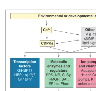
Biochemical approaches have identified a variety of CDPK substrates that suggest potential regulatory roles in gene expres-sion, metabolism and signaling pathway components, traffic of ions and water across membranes, and the dynamics of the cytoskeleton (Fig. 5). More information on these substrates can be found at the protein kinase and phosphatase Web site (Box 1), and in a recent review13
. Here, we highlight two substrates for which there is strong experimental support for regulation by CDPK.
Sucrose phosphate synthase (SPS) is a key enzyme in the sucrose synthesis pathway, and nitrate reductase is the rate-limit-ing enzyme in the assimilation of nitrogen from nitrate (reviewed in Refs 34,35). Both enzymes are phosphorylated in the dark, resulting in their inhibition. SPS is directly inhibited by phospho-rylation of Ser153, and nitrate reductase is inhibited by a two-step mechanism involving phosphorylation of Ser543 and binding of a 14-3-3 protein to the phosphorylated site (reviewed in Ref. 28). Inhibition of these enzymes in the dark when carbon fixation is not occurring diminishes the partitioning of carbon skeletons into exported sucrose and amino acids, and conserves glucose and fructose for use in other pathways in leaf cells, such as glycolysis or starch synthesis. Evidence that supports this in vivofunction includes co-purification of a CDPK that has been identified as a homolog of AtCPK3 (Ref. 26), which phosphorylates both of these enzymes at the regulatory sites26,36–38
. These observations raise the possibility that a single CDPK can coordinately regulate both activities.
The hypothesis that CDPK down-regulates nitrate reductase and SPS in response to the dark is consistent with the observation that cytoplasmic calcium concentrations are higher at night than during the day39
. However, it should be noted that a calcium-inde-pendent kinase with properties of SNF1-related kinases also phos-phorylates Ser153 of SPS and Ser543 of nitrate reductase38,40,41
. Thus, it is possible that both types of kinase down-regulate SPS and nitrate reductase in vivo, but in response to a separate signaling pathway.
A CDPK might also be involved in activating SPS by phospho-rylation at a site (Ser424) distinct from the inhibitory site42
. This phospho-dependent activation occurs in response to hypo-osmotic stress and presumably increases cytosolic sucrose and thereby decreases the water potential of the cell to help retain water. These intriguing results raise the possibility that two different CDPK pathways that are differentially activated by separate stimuli oppositely regulate SPS activity.
The future
• Plants have many CDPK isoforms (.40 in Arabidopsis), which are predicted to have kinase activities directly activated by cal-cium and potentially modified by cross-talk with other signal-ing systems.
• CDPKs are multifunctional, with individual isoforms providing specific pathways to control transcription, metabolic enzymes, membrane transport and cell structure.
• Plants also have at least three types of CDPK-related kinases, with some members regulated by calmodulin and some totally unresponsive to calcium signals.
A major challenge for the future is to create an integrated picture of how members of this kinase family are used in plant develop-ment and physiology. To this end, we offer the following basic strategy for investigating every isoform in a model plant such as Arabidopsis.
• Use biochemistry to define isoform-specific calcium activation thresholds and substrate specificities.
• Use cell biology tools to delineate subcellular locations.
• Use genetics to identify biological func-tions, as indicated by the phenotypes that result from the disruption of genes or the expression of de-regulated mutants (e.g. calcium independent).
• Use bioinformatics to provide a picture of how CDPK signaling pathways are in-tegrated into the dynamic interactions of all signaling pathways in a cell.
CDPKs represent a potential gold mine of opportunity for biotechnology applications. Given the involvement of calcium signals in so many aspects of plant biology, including biotic and abiotic stress responses, the differ-ent CDPK pathways provide many potdiffer-ential points of intervention to suppress or activate a specific response. Structure–function stud-ies on CDPKs have provided an important paradigm by showing how a CDPK can be converted into an active, calcium-indepen-dent kinase to be used as a dominant, posi-tive transgene1
. This approach has provided a precedent by establishing a role for one CDPK isoform in selectively activating a cold, dark and osmotic stress response pathway in the absence of other calcium signaling pathways33
.
Acknowledgements
We are supported by funding from the National Science Foundation, DBI-9975808.
References
1Huang, J.F. et al.(1996) Activation of a Ca21-dependent protein kinase involves intramolecular binding of a calmodulin-like regulatory domain. Biochemistry35, 13222–13230
2Yoo, B.C. and Harmon, A.C. (1996) Intramolecular binding contributes to the activation of CDPK, a protein kinase with a calmodulin-like domain. Biochemistry35, 12029–12037
3Harmon, A.C. et al.(1994) Pseudosubstrate inhibition of CDPK, a protein kinase with a calmodulin-like domain. Biochemistry33, 7278–7287
4Harper, J.F. et al.(1994) Genetic identification of an autoinhibitor in CDPK, a protein kinase with a calmodulin-like domain. Biochemistry33, 7267–7277
Box 1. Plant protein kinase and phosphatase Web site
This Web site (http://plantsP.sdsc.edu) focuses on Arabidopsis genes encoding protein kinases and phosphatases. When completed, this site will have an annotated database of all families of protein kinases and phosphatases, and will announce the availability of protein kinase and phos-phatase mutants as they are identified and made available to the research community. Information on the CPDK family can be found at http://plantsP.sdsc.edu/cdpk, in-cluding fully annotated figures from this review, expanded information on phylo-genetic trees and sequence alignments, and a categorized list of literature related to all the CDPKs that have been found in plant and protist species to date.
Fig. 5.Calcium-dependent protein kinase or calmodulin-like domain protein kinase (CDPK) substrates and physiological roles. A hypothetical model for the roles of CDPKs in plant growth and development and cellular defense responses. CDPK is stimulated by an increase in Ca21that results from various stimuli. These stimuli might also activate additional
signal-ing pathways and Ca21 might also activate calmodulin-dependent pathways. Activated
CDPK(s) phosphorylate certain substrates, depending on the nature of the stimulus. Other signaling pathways might also be involved in the stimulation of CDPKs or the substrates. Phosphorylation of the substrates alters their activities and this contributes to the physiologi-cal response. Abbreviations: G/HBF1, soybean G- and H-box binding factor 1; HBP-1a(17), wheat H-box binding factor 1a(17); GT1BP, ArabidopsisGT1-binding protein; SPS, sucrose phosphate synthase; NR, nitrate reductase; SuSy, sucrose synthase; HMGR, 3-hydroxy-3-methylglutaryl-coenzyme A reductase; SAT, serine O-acetyltransferase; EF1-a, elongation factor 1-a; Phox, p67-Phox and p47-Phox are components of NADPH oxidase; aquaporins, water channels including Nodulin 26, a soybean root nodule water and weak anion channel, and TIP from tobacco vacuoles.
Trends in Plant Science Environmental or developmental signals
Cell growth, development and defense Ca2+
CDPKs
Other pathways
e.g. calmodulin, cGMP, G proteins, lipid signals, MAPKs
Transcription factors
G/HBF1? HBP-1a(17)?
GT1BP?
Metabolic enzymes and
regulators
SPS, NR, SuSy, HMGR, SAT, EF1-α, Phox
Cytoskeletal proteins
Actin depolymerizing
factor
Ion pumps and channels
Aquaporins H+ and Ca2+
pumps, K+ and
anion channels
Gene expression
Sugar and amino acid metabolism,
signaling
Cell architecture Membrane
5Poovaiah, B.W. et al. (1999) Developmental regulation of the gene for chimeric calcium/calmodulin-dependent protein kinase in anthers. Planta209, 161–171
6Ramachandiran, S. et al. (1997) Functional domains of plant chimeric calcium/calmodulin-dependent protein kinase: regulation by autoinhibitory and visinin-like domains. J. Biochem.121, 984–990
7Watillon, B. et al. (1995) Structure of a calmodulin-binding protein kinase gene from apple. Plant Physiol.108, 847–848
8Furumoto, T. et al. (1996) Plant calcium-dependent protein kinase-related kinases (CRKs) do not require calcium for their activities. FEBS Lett. 396, 147–151
9Lindzen, E. and Choi, J.H. (1995) A carrot cDNA encoding an atypical protein kinase homologous to plant calcium-dependent protein kinases. Plant Mol. Biol.28, 785–797
10Farmer, P.K. and Choi, J.H. (1999) Calcium and phospholipid activation of a recombinant calcium-dependent protein kinase (DcCPK1) from carrot (Daucus carotaL.). Biochim. Biophys. Acta1434, 6–17
11Hartwell, J. et al. (1999) Phosphoenolpyruvate carboxylase kinase is a novel protein kinase regulated at the level of expression. Plant J.20, 333–342
12Wang, W. and Poovaiah, B.W. (1999) Interaction of plant chimeric calcium/calmodulin-dependent protein kinase with a homolog of eukaryotic elongation factor-1a. J. Biol. Chem.274, 12001–12008
13Hrabak, E.M. Calcium-dependent protein kinases and their relatives. In Plant Protein Kinases (Advances in Botanical Sciences) (Kreis, M. and Walker, J.C., eds), Academic Press (in press)
14Vitart, V. et al. Intramolecular activation of a Ca21-dependent protein kinase is disrupted by insertions in the tether that connects the calmodulin-like domain to the kinase. Biochemistry (in press)
15Lee, J.Y. et al.(1998) Kinetic and calcium-binding properties of three calcium-dependent protein kinase isoenzymes from soybean. Biochemistry 37, 6801–6809
16Zhao, Y. et al. (1994) Calcium-binding properties of a calcium-dependent protein kinase from Plasmodium falciparumand the significance of individual calcium-binding sites for kinase activation. Biochemistry33, 3714–3721
17Hrabak, E.M. et al. (1996) Characterization of eight new members of the calmodulin-like domain protein kinase gene family from Arabidopsis thaliana. Plant Mol. Biol.31, 405–412
18McAinsh, M. and Hetherington, A. (1998) Encoding specificity in Ca21 signalling systems. Trends Plant Sci.3, 32–36
19Sanders, D. et al.(1999) Communicating with calcium. Plant Cell11, 691–706
20Takezawa, D. et al. (1996) Dual regulation of a chimeric plant
serine/threonine kinase by calcium and calcium/calmodulin. J. Biol. Chem. 271, 8126–8132
21Binder, B.M. et al.(1994) Characterization of an Arabidopsiscalmodulin-like domain protein kinase purified from Escherichia coliusing an affinity sandwich technique. Biochemistry33, 2033–2041
22Farmer, P.K. and Choi, J.H. (1995) Expression and potential myristoylation of a calcium-dependent protein-kinase. J. Cell Biochem.(Suppl.) 21A, 507
23Ellard-Ivey, M. et al. (1999) Cloning, expression and N-terminal
myristoylation of CpCPK1, a calcium-dependent protein kinase from zucchini (Cucurbita pepoL.). Plant Mol. Biol.39, 199–208
24Tanaka, T. et al. (1995) Sequestration of the membrane-targeting myristoyl group of recoverin in the calcium-free state. Nature376, 444–447
25Chaudhuri, S. et al. (1999) Autophosphorylation-dependent activation of a calcium-dependent protein kinase from groundnut. Plant Physiol.120, 859–866
26Douglas, P. et al. (1998) Purification of a nitrate reductase kinase from Spinacea oleracea leaves, and its identification as a calmodulin-domain protein kinase. Planta206, 435–442
27Saha, P. and Singh, M. (1995) Characterization of a winged bean
(Psophocarpus tetragonolobus) protein-kinase with calmodulin-like domain: regulation by autophosphorylation. Biochem. J.305, 205–210
28Chung, H.J. et al.(1999) The 14-3-3 proteins: cellular regulators of plant metabolism. Trends Plant Sci4, 367–371
29Camoni, L. et al. (1998) 14-3-3 proteins activate a plant calcium-dependent protein kinase (CDPK). FEBS Lett.430, 381–384
30Moorhead, G. et al. (1999) Phosphorylation-dependent interactions between enzymes of plant metabolism and 14-3-3 proteins. Plant J.18, 1–12
31van der Hoeven, P.C. et al. (1996) A calcium and free fatty acid-modulated protein kinase as putative effector of the fusicoccin 14-3-3 receptor. Plant Physiol.111, 857–865
32Toroser, D. et al. (1998) Site-specific regulatory interaction between spinach leaf sucrose-phosphate synthase and 14-3-3 proteins. FEBS Lett. 11, 110–114
33Sheen, J. (1996) Ca21-dependent protein kinases and stress signal transduction in plants. Science274, 1900–1902
34Huber, S.C. et al. (1996) Post-translational regulation of nitrate reductase activity: a role for Ca21and 14-3-3 proteins. Trends Plant Sci1, 432–438
35Huber, J. and Huber, S. (1996) Role and regulation of sucrose phosphate synthase in higher plants. Annu. Rev. Plant Physiol. Plant Mol. Biol. 47, 431–444
36Bachmann, M. et al. (1996) Identification of the major regulatory phosphorylation site as Ser-543 in spinach leaf nitrate reductase and its phosphorylation by a calcium-dependent protein kinase in vitro. Plant Cell 8, 505–517
37Bachmann, M. et al. (1995) Partial purification and characterization of a calcium-dependent protein kinase and an inhibitor protein required for inactivation of spinach leaf nitrate reductase. Plant Physiol.108, 1083–1092
38McMichael, R.W., Jr et al. (1995) Spinach leaf sucrose-phosphate synthase and nitrate reductase are phosphorylated/inactivated by multiple protein kinases in vitro. Plant Physiol.108, 1077–1082
39Johnson, C.H. et al. (1995) Circadian oscillations of cytosolic and chloroplastic free calcium in plants. Science269, 1863–1865
40Douglas, P. et al. (1997) Three spinach leaf nitrate reductase-3-hydroxy-3-methylglutaryl-CoA reductase kinases that are required by reversible phosphorylation and/or Ca21ions. Biochem. J.325, 101–109
41Sugden, C. et al. (1999) Two SNF1-related protein kinases from spinach leaf phosphorylate and inactivate 3-hydroxy-3-methylglutaryl-coenzyme A reductase, nitrate reductase, and sucrose phosphate synthase in vitro. Plant Physiol.120, 257–274
42Toroser, D. and Huber, S.C. (1997) Protein phosphorylation as a mechanism for osmotic-stress activation of sucrose-phosphate synthase in spinach leaves. Plant Physiol.114, 947–955
43Altschul, S.F. et al. (1997) Gapped BLAST and PSI-BLAST: a new generation of protein database search programs. Nucleic Acids Res. 25, 3389–3402
44Gribskov, M. and Veretnik, S. (1996) Identification of sequence patterns with profile analysis. Methods Enzymol.266, 198–212
45Thompson, J.D. et al. (1994) CLUSTAL W: improving the sensitivity of progressive multiple sequence alignment through sequence weighting, position-specific gap penalties and weight matrix choice. Nucleic Acids Res. 22, 4673–4680
46Saitou, N. and Nei, M. (1987) The neighbor-joining method: a new method for reconstructing phylogenetic trees. Mol. Biol. Evol.4, 406–425
*Alice C. Harmon is at the Program in Plant Molecular and Cellular Biology, Dept of Botany, University of Florida, PO Box 118526, Gainesville, FL 32611-8526, USA; Michael Gribskov is at the San Diego Supercomputer Center, University of California San Diego, 9500 Gilman Drive, La Jolla, CA 92093-0537, USA (tel 11 858 534 8312; fax 11 858 822 0873;
e-mail [email protected]); Jeffrey F. Harper is at the Dept of Cell Biology, The Scripps Research Institute, 10550 N. Torrey Pines Rd, La Jolla, CA 92037, USA (tel 11 858 784 2862;
fax 11 858 784 9840; e-mail [email protected]).


