Embryogenic Callus Induction from Leaf Tip Explants and Protocorm-Like Body
Formation and Shoot Proliferation of
Dimorphorchis lowii
: Borneon Endemic Orchid
Juddy E. Jainol and Jualang Azlan Gansau*)
Tissue Culture Laboratory, Faculty of Science and Natural Resources, University Malaysia Sabah, 88999 Kota Kinabalu, Sabah, Malaysia
*) Corresponding author E-mail: [email protected]
Received: April 17, 2016/ Accepted: September 13, 2016
ABSTRACT
Dimorphorchis lowii is a rare epiphytic orchid endemic to Borneo and very popular due to its ornamental value. For the purpose of mass propagation, procedures for in vitro propagation through callus were developed. The objectives of this study were to determine the effects of plant growth regulators (PGRs) on callus induction from leaf tip explant and the effects of complex additives on PLBs formation and shoot develop-ment. The best combination of PGR was 3.0 mg L-1 Thidiazuron (TDZ) with 0.046 mg L-1 NAA supplemented in half-strength medium (½ MS) (Murashige & Skoog, 1962). The percentage of survival and callus formation obtained were 96.0 % ± 19.8 and 52.0 % ± 16.5 respectively. Maximum shoot proliferation from PLBs was observed in Knudson C (KC) medium enriched with 15 % (v/v) coconut water. In this treatment, 10.2 ± 6.2 shoots were produced from one callus explant. Shoots were rooted on KC medium containing 15 % (v/v) coconut water and transferred in a medium mixture containing sphagnum moss, charcoal, brick and coco peat with the ratio of 1:1:1:1. After 2 months being acclimatized in the glasshouse, 78 % of plantlets survived. Histology observations showed that embryogenic callus derived from leaf tip explants might originate from epidermis and mesophyll cells and was capable of developing into complete plantlets.
Keywords: callus, in vitro, orchid, protocorm like-bodies (PLBs)
INTRODUCTION
Dimorphorchis lowii is an endemic orchid of Borneo and is one of the rarest epiphytic orchids, which commonly inhibiting the area with the elevation of 600-1800 meter above sea level (m asl) on Kinabalu, Tombuyukon and Trus Madi mountains in Sabah (Cribb & Bell, 2008). This
orchid possess unique and beautiful dimorphic feature, which means it has two distinct flowers on one inflorescent and has the prospect to be marketed as a valuable basket or potted flowers. The increasing popularity, high degree of rarity and its difficulty-to-locate have put this species among the most highly sought after species. Therefore, it is imperative to develop a method for multiplication to reproduce a great number of plantlets in order to fulfill the floriculture demand as well as for conservation purposes.
Dimorphorchis orchids, just like other monopodial orchids are asexually propagated using the top cuttings of the mother plants. Nevertheless, reproduction through this conven-tional technique can be detrimental to the stock plants as it poses threat due to the removal of meristematic tips (Gantait, Bustam, & Sinniah (2012). Conventional method through seed propagation is sometimes problematic as seed germination in natural environment is a slow process and it requires mycorrhizal or fungal
partners’ association for its germination (Pant & Thapa, 2012) and any disturbance in the habitat or physical environment during germination will destroy the whole population (Mathews & Rao, 1980). In vitro propagation of orchids by shoot tip culture is a well-developed method. However, by using shoot tip as explant means the most significant part that is actively growing has to be sacrificed. Therefore, a technique of reproduction using other insignificant parts such as leaves of the plant would be beneficial (Chugh, Guha, & Rao, 2009).
Successful culture of plantlet from leaf culture of many orchid species has been reported. Among them are Aerides (Devi, Devi, & Singh, 2013), Aranda (Gantait, Bustam, & Sinniah, 2012), Phalaenopsis (Kuo, Chen, & Chang, 2005; Khoddamzadeh et al., 2011), Vanda and Dendrobium (Martin & Madassery, 2006; Tee, Wong, Lam, & Maziah, 2010), and Oncidium Cite this as: Jainol, J. E., & Gansau, J. A. (2017). Embryogenic callus induction from leaf tip explants and protocorm-like body formation and shoot proliferation of Dimorphorchis lowii: Borneon endemic orchid. AGRIVITA Journal of Agricultural Science, 39(1), 1-10. http://doi.org/10.17503/agrivita.v39i1.895
Accredited: SK No. 81/DIKTI/Kep/2011
(Mayer, Stancato, & Appezzato-Da-Glória, 2010). In leaf cultures, for example in Renanthera (Wu et al., 2012) and Vanilla (Janarthanam & Seshadri, 2008), wound surface of leaf explants had been reported to produce callus. Wu et al., (2012) reported that 99.67 % plantlet formation was obtained from callus originated from leaf segments of Renanthera orchid. Protocorm-like bodies (PLBs) generally have high potency to proliferate rapidly and regenerate into a whole plantlet (Bustam, Sinniah, Kadir, Zaman, & Subramaniam, 2013). Therefore, they are the most common target tissues used for mass propagation or producing a large number of uniform plantlets for commercial purposes.
This study investigated (I) the effects of PGR on callus induction from leaf tip explants and (II) the effect of complex additives on the PLBs formation and shoot development of D. lowii. Histology and scanning electron microscope observations were also conducted to study the callus induction from leaf tip explants.
MATERIALS AND METHODS
The mother plant was collected in early 2009 from a local nursery in Sabah, Malaysia. This plant was brought to the Orchid Unit nursery at University Malaysia Sabah, grown and maintained in the glasshouse conditions. In December 2009, seeds were obtained and cultured in Vacin & Went (VW) (Vacin & Went, 1949) medium added with 10 % (v/v) potato homogenate. Protocorms developed from these seed cultures were then transferred to half-strength MS (½ MS) (Murashige & Skoog, 1962) medium containing 2.0 % (w/v) sucrose and 0.2 % (w/v) yeast extract. This served as the staging medium for explants source. During series of subcultures, protocorms grew into multiple shoots. Multiple shoots bearing 3-4 leaves each were chosen as source for leaf explants. The first two leaves from the top with the diameter of 2-5 mm were chosen and dissected with a sterile sharp scalpel and the tips were cut approximately 5-7 mm in length.
Medium Preparation and Culture Condition
In this study, three media formulations were used as basal media: Vacin and Went (VW), modified MS containing half-strength macro and micro nutrients (½ MS), and Knudson C (KC) (Yam & Arditti, 2009). VW enriched with 10 % (v/v) potato homogenate was used for seed
germination, ½ MS was used as basal medium for protocorm development into seedlings in which the leaves were used as the source of explants in experiment I. ½ MS was also used for basal medium in experiment I, and KC was used as basal medium in experiment II. All media were supplemented with 2 % sucrose and solidified with 0.8 % agar powder (Sigma, USA). ½ MS was also enriched with 100 mg L-1 myo-inositol, 0.5 mg L-1 pyridoxine-HCl, 0.1 mg L-1 thiamine-HCl, 0.5 mg L-1 nicotinic acid and 2 mg L-1 glycine. Different PGRs (TDZ or NAA) and concentrations were added to ½ MS medium in as treatments experiment I, and different complex additives and concentrations (15 % (v/v) coconut water, 0.2 % (w/v) peptone or 0.2 % (w/v) yeast extracts) were added to KC medium as treat-ments in experiment II.
The pH of the media was adjusted to 5.2-5.3 and agar powder was weighed and added prior to autoclaving for 20 minutes at 121 oC and 15 kPa. The autoclaved medium was poured aseptically into sterile plastic petri dishes (60 mm x 15 mm, Sterilin, UK) at 20 ml for each petri dish. These petri dishes were allowed to cool down, covered and then sealed with parafilm M®
(American National Cam™, Menasha, WI)before used. For callus induction experiment, cultures were maintained at 25± 2 oC under continuous darkness. For PLB formation and shoot development experiment, cultures were maintained at 25± 2 oC under continuous cool-white fluorescent light with photon flux density (PFD) of 15-25 μmol m-2 s-1.
Effect of PGRs on Embryogenic Callus Induc-tion from Leaf Tip Explants
PGR tested in this investigation included TDZ at 0.22, 0.33, 1.0, 3.0 and 4.0 mg L-1 and NAA at 0.046, 0.5 and 1.0 mg L-1. Both TDZ and NAA were applied individually or in combination. ½ MS medium supplemented with 2.0 % (w/v) sucrose was used as basal medium.
Effects of Complex Additives on PLBs Development and Shoot Proliferation from Embryogenic Callus Derived from Leaf Tip Explants
(w/v) and yeast extract at 0.2 % (w/v) were used to investigate the PLBs development and sub-sequent shoot formation from embryogenic callus of D. lowii. KC medium supplemented with 2.0 % (w/v) sucrose was used as basal medium. Once shoots were established, they were transferred into 100 ml Erlenmeyer flask containing 20 ml of KC medium supplemented with 15 % (v/v) coconut water for shoot elongation and root formation.
Data Analysis
All experiments were performed in a Complete Randomized Design (CRD) with five replicates. For embryogenic callus induction experiment, one petri dish represented one replicate and in each replicate contained 20 leaf tip explants. Data on percentage of survival, percentage of callus formation, callus density and color were recorded after 60 days of culture. For PLBs shoot proliferation from embryogenic callus derived from leaf tip explants, one petri dish represented one replicate and in each replicate contained 20 callus clumps. Data on percentage of survival rate, percentage of PLB formation, number of shoots, and number of leaves were recorded after 60 days and 120 days of culture. Significance of treatment effects on these data were analyzed using ANOVA, p<0.05 and comparison between mean values of treatments were made by Duncan Multiple Range Test using SPSS version 20 (IBM Corporation, New York, USA)
Histology and Scanning Electron Microscope Observation on Embryogenic Callus Derived from Leaf Tip Explants
Light Microscopy
Tissues for histological observation were secured in PAA solution (15 Picric acid: 1 Acetic acid), dehydrated in an ethanol series, and lodged in paraffin wax. The materials embedded in paraffin
were sectioned at 5 μm and stained with
hematoxylin-eosin. The observation and photography were done using light microscope (Leica ICC50 HD, UK).
Scanning Electron Microscopy (SEM)
The samples were fixed in 2 % (v/v) glutaraldehyde in 0.1 M phosphate buffer (pH 7.0) and dehydrated in an ethanol series. The samples were dried to the critical point with liquid CO2 in a critical point dryer (CPD) (Leica EM300,
Austria), affixed to aluminium stubs and coated with gold palladium in a sputter coater (EMITECH, K550X, England). The mounted specimens were examined with scanning electron microscope (Zeiss EVO MA10, U.K).
RESULTS AND DISCUSSION
Effect of PGRs on Embryogenic Callus Induc-tion from Leaf Tip Explants
The effects of PGRs is shown in Table 1. Incorporation of TDZ and NAA at a concen-tration ranges from 1.0 to 3.0 mg L-1 and 0.046 to 1.0 mg L-1, respectivelyin ½ MS medium was crucial in promoting the percentages of survival of explants and embryogenic callus formation. The highest percentage of explant survival was observed in medium containing 3.0 mg L-1 TDZ and 0.046 mg L-1 NAA at 96.0 % ± 19.8 and 1.0 mg L-1 TDZ and 0.046 mg L-1 NAA at 92.0 % ± 15.3. There were no significant differences between these two treatments. However, there were significant differences observed when compared to the other concentrations of TDZ and NAA as shown in Table 1. For embryogenic callus induction, it was observed that medium containing 3.0 mg L-1 TDZ and 0.046 mg L-1 NAA produced the highest percentage of callus formation (52.0 % ± 16.5), while other PGR treatments produced only 18-34 % of the explants forming callus.
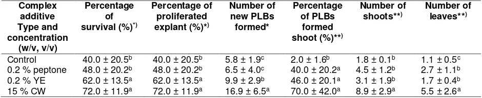
Table 1. Effects of plant growth regulator types and concentrations on embryogenic callus induction from leaf tip explants of D. lowii cultured in ½ MS medium supplemented with 2.0 % (w/v) sucrose under continuous darkness at temperature of 25 ± 2 oC after 60 days of culture
Cytokinin/Auxin
(mg L-1) Percentage of
Survival (%)
Percentage of callus formation (%)
Density of
callus Color of callus
TDZ NAA
Control 24.0±8.0g 2.0±1.47d* + Greenish-white
0.22 0.00 64.0±28.5cd 18.0±10.4bc + Greenish-white 0.33
1.0 0.00 0.00
60.0±24.5cde 48.0±20.5def
22.0±11.5bc 24.0±13.1bc
+ +
Yellowish-white Greenish-white 3.0 0.00 90.0±30.3ab 28.0±17.4bc ++ Greenish-white 0.0
0.0
0.0 0.046 0.5
1.0
75.0±43.9bc 65.0±48.3cd 42.0±29.8efg
28.0±17.4bc 25.0±13.9bc 0.0±0.0e
++ +
-Yellowish-white Greenish-white
-
1.0 0.046 92.0±15.3a 34.1±25.8bc ++ White
3.0 3.0
0.046 0.5
96.0±19.8a 34.0±8.9fg
52.0±16.5a 12.0±8.8bc
+++ +
White Yellowish-white
3.0 1.0 86.0±21.1ab 28.0±17.9bc ++ White
4.0 0.5 62.0±25.0cd 18.0±10.4bc + Greenish-white
4.0 1.0 50.0±26.0def 14.0±9.1bc + Greenish-white
Remarks: *Mean values within a column followed by the same letters are not significantly different at p < 0.05 according
to Duncan’s Multiple Range Test. Note. +: callus at tip, ++: callus at tip and midsection, +++: callus covered all surfaces, - : no callus
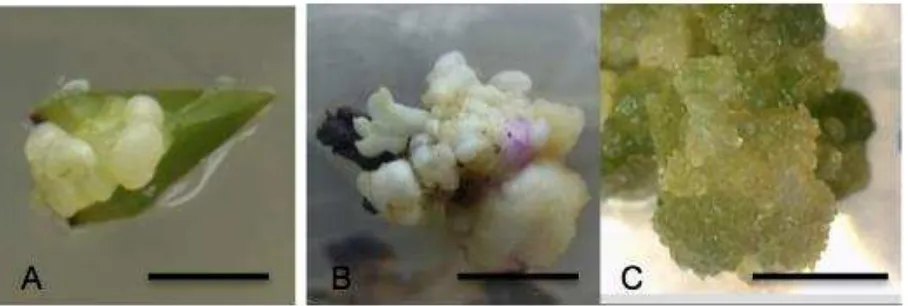
Figure 1. Embryogenic callus induction from leaf explant. A. Callus induction from leaf tip explant cultured on ½ MS medium supplemented with 3.0 mg L-1 TDZ and 0.046 mg L-1 NAA after 30 days of culture (DOC). B. Callus induction from leaf tip explant cultured on ½ MS medium supple-mented with 3.0 mg L-1 TDZ and 0.046 mg L-1 NAA after 60 DOC and C. after 90-100 DOC when transferred under 24hr light.(Scale bars = 5 mm).
The highest frequency of SE formations were found in media containing 3 mg L-1 TDZ without NAA, or in combination with 0.1 or 1.0 mg L-1 NAA, i,e., 93.8 %, 87.5 % and 81.3 %, respectively. In line with these results, Tariq, Ali, & Abbasi (2014) also reported that 2.0 mg L-1 TDZ in combination with 1.0 mg L-1 NAA produced optimum result in callus formation from leaf explant of Artemisia absinthium. In this study, greenish-white callus development was seen on
cultured in ½ MS medium supplemented with 3.0 mg L-1 TDZ alone.
PGR(s) proportions have been known to have species-specific effect on callus and PLB induction from leaf explants (Shimura & Koda, 2004; Gantait, Bustam, & Sinniah, 2012). TDZ is a substituted phenyl urea with cytokinin-like activity (van Staden, Zažímalová, & George, 2008; Jitsopakul, Thammasiri, & Ishikawa, 2013). The ascendancy of TDZ in inducing somatic embryos (infant PLBs) from leaf explants was recounted in numerous orchid species including Oncidium (Mayer, Stancato, & Appezzato-Da-Glória, 2010), Doritaenopsis (Park, Yeung, Chakrabarty, & Paek, 2002), Dendrobium and Phalaenopsis (Kuo, Chen, & Chang, 2005). TDZ is also useful for rapid plant regeneration through organogenesis (Kishor & Devi, 2009; Jitsopakul, Thammasiri, & Ishikawa, 2013), somatic em-bryogenesis (Jheng, Do, Liauh, Chung, & Huang, 2006; Gow, Chen, & Chang, 2009), or PLB formation of orchids (Mayer, Stancato, & Appezzato-Da-Glória, 2010; Khoddamzadeh et al., 2011; Roy, Sajeev, Pattanayak, & Deka, 2012). NAA is a synthetic auxin plant growth regulator that is routinely used in in vitro propagation of orchid to promote callus induction such as in Dendrobium (Mei, Danial, Mahmood, & Subramaniam, 2012; Salam, Salam, & Mohanty, 2013), Vanilla (Tan, Chin, & Alderson, 2011), Oncidium (Mayer, Stancato, & Appezzato-Da-Glória, 2010), Phalaenopsis (Khoddamzadeh et al., 2011), and Renanthera (Wu et al., 2012).
Callus from leaf tip explant cultured on ½ strength MS medium turned green when transferred under 24hr light after 90-100 DOC (Figure 1C).
Effect of complex additive on PLB develop-ment and shoot proliferation from embryo-genic callus derived from leaf tip explants
Table 2 shows the result obtained after 120 days of cultures. 15 % (v/v) coconut water presented the highest percentage of survival and PLBs formation both at 72.0 % ± 11.9 followed by 0.2 % (w/v) yeast extract at 62.0 % ± 13.5 in both percentages of survival and PLBs formation.
There were no significant differences between the effects of these two complex additives. However, significant differences were observed between these two complex additives in terms of number of PLBs per explant with 15 % (v/v) coconut water produced the highest 16.9±6.5 and 0.2 % (w/v) yeast extract produced 9.9±2.9. In both media, the emergence of nodular structures from the callus was seen after 20-30 days of culture (Figure 2A). These nodular tissues later progressed further and developed into aggregates of PLBs after 60 days of culture (Figure 2B). After 120 days of culture, 15 % (v/v) coconut water produced a significantly higher percentage of PLBs forming shoot at 70.0 % ± 42.0 with 8.9±2.9 shoots and 5.5±2.6 leaves (Figure 2C) followed by 0.2 % (w/v) peptone at 40.0 % ± 20.2 with 4.5±1.2 shoots and 2.7±1.1 leaves.
Table 2. Effect of complex additives types and concentrations on PLBs development and shoot proliferation from callus culture of D. lowii cultured on KC medium supplemented with 2.0 % (w/v) sucrose under 24-hour light photoperiod with PFD of 15-25 μmol m-2 s-1 at 25 ± 2 oC after 60 and 120 days of culture
Complex additive Type and concentration
(w/v, v/v)
Percentage of survival (%)*)
Percentage of proliferated explant (%)*)
Number of new PLBs formed*
Percentage of PLBs
formed shoot (%)**)
Number of shoots**)
Number of leaves**)
Control 40.0 ± 20.5b 40.0 ± 20.5b 5.8 ± 1.9c 2.0 ± 1.6b 1.8 ± 0.1b 1.1 ± 0.5c 0.2 % peptone 48.0 ± 20.2b 48.0 ± 20.2b 6.5 ± 4.0c 40.0 ± 20.2a 4.5 ± 1.2b 2.7 ± 1.1b 0.2 % YE 62.0 ± 13.5a 62.0 ± 13.5a 9.9 ± 2.9b 46.0 ± 20.1a 3.1 ± 1.9b 1.7 ± 0.4b 15 % CW 72.0 ± 11.9a 72.0 ± 11.9a 16.9 ± 6.5a 70.0 ± 42.0a 8.9 ± 2.9a 5.5 ± 2.6a
Remarks: Mean values within a column followed by the same letters are not significantly different at p<0.05 according
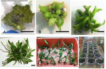
Figure 2.Plantlet development from callus: A. Callus cultured on KC medium added with 15 % (v/v) coconut water. B. PLBs with shoots after 50-60 DOC and C. Shootlets after 120 DOC. (Scale bars = 5 mm). D. Plantlets formed when cultured on KC medium supplemented with 15 % (v/v) coconut water after 150-180 DOC. E. Plantlets were separated from clumps and prepared for acclimatization. F. Plantlets acclimatization after 2 months. (Scale bars = 10 mm)
Overall, coconut water at 15 % (v/v) showed the superiority compared to the other two complex additives (peptone & yeast extract). Coconut water is extensively used in the plant micropro-pagation due to its growth enhancer substance and cytokinin-type action that assist cell division and stimulate fast growth (Arditti, 2008; Yong, Ge, Ng, & Tan, 2009; Prando, Chiavazza, Faggio, & Contessa, 2014). Coconut water contains high levels of Z-type cytokinin (trans-zeatin riboside, trans-zeatin O-glucoside, dihydrozeatin O-gluco-side, trans-zeatin, dihydrozeatin, trans-zeatin riboside-5 -monophosphate), iP-type cytokinin (N6-isopentenyladenine), kinetin, kinetin riboside and ortho-topolin in addition to sugars, vitamins, minerals and amino acids (Yong, Ge, Ng, & Tan, 2009; Tan, Cheng, Bhat, Rusul, & Easa, 2014). Murdad et al., (2006) reported that the production
of PLBs in Phalaenopsis gigantean is improved with the addition of coconut water in the culture medium. The PLB and callus induction in Cymbidium hybrids is also promoted by the coconut water in the culture media (Teixeira da Silva & Tanaka, 2006). Coconut water has also been reported to enhance shoot multiplication in Vanilla planifolia (Kalimuthu, Senthilkumar, & Murugalatha, 2006).
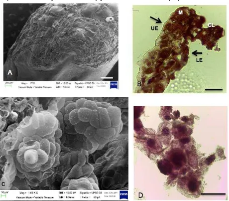
Figure 3. Scanning electron microscope (SEM) and histology view on A. SEM view on embryogenic callus formed on the abaxial part of leaf tip explant, scale bar = 400 μm, 77 x. B. Light microscopic views of leaf tip showed longitudinal section of explant showing cell dedifferentiation in callus that produced unorganized cell mass, upper & lower epidermis (UE) & (LE), mesophyll cell (M), callus (CL), scale bar = 50 μm, 40x. C. Single cells divide and developed into PLBs of different sizes, scale bars = 40 μm, 1.00K x.D. Cross section of calli with multi-globular compact callus cells, scale bar = 50 μm, 40x (Jainol, 2016).
Histology and Scanning Electron Microscope Observations on Embryogenic Callus Derived from Leaf Tip Explants
Embryogenic callus derived from leaf tip explants shows cells dedifferentiation (Figure 3A-B). Begum, Tamaki, Tahara, & Kako (1994) reported that callus formation from orchids used explants that contained meristematic cells or epidermal cells, suggesting that callus cells arise from these types of cells. Figure 3B shows the cross-section through the leaf tip explants which
showed densely lower epidermis and mesophyll cells underwent dedifferentiation. Most of the time, callus developed at the cut ends of leaf tip explants, suggesting that the wounding might have triggered dedifferentiation of cells to form callus. Kuo, Chen, & Chang (2005) reported that the highest ability of Phalaenopsis leaf explants to form embryos was found at adaxial surfaces near cut ends on TDZ-containing medium. Figure 3C-D show that the callus masses composed of spherical, compact and loose aggregates of cells.
Spherical single cells can be seen embedded within the friable callus. These cells started to divide and gave rise to somatic embryos or PLBs of different sizes. Globular-stage somatic embryo or PLBs might originate either from the cells within the callus or from the callus surface. These PLBs developed into shoots that would formed complete plantlets when cultured on KC medium supplemented with 15 % (v/v) coconut water.
CONCLUSION
The present study demonstrated that the success of embryogenic callus induction from leaf tip explants of Dimorphorchis lowii is greatly influenced by plant growth regulators. Leaf tip explants were able to produce embryogenic callus in medium free of PGR but the frequency was very low (2 %). In this study, the best result in embryogenic callus formation on leaf tip explants was obtained at combination of 3.0 mg L-1 TDZ and 0.046 mg L-1 NAA Furthermore, 15 % (v/v) coconut water was found to be the best complex additive to promote PLBs development and subsequent shoot formation from embryo-genic callus of D. lowii. Histology observations revealed that, embryogenic callus might have originated from epidermis and mesophyll cells and these calli have the capability to multiply and formed complete plantlets. However, further study needs to be conducted to study cell de-differentiation, and the structural and biological functions of cells involved in the initiation and formation of embryogenic callus.
ACKNOWLEDGMENT
We thank Tissue culture laboratory, Uni-versity Malaysia Sabah for providing the facilities and equipment for this research.
REFERENCES
Arditti, J. (2008). Micropropagation of orchids (2nd ed., Vol. 1). Oxford, UK: Wiley-Blackwell.
Begum, A. A., Tamaki, M., Tahara, M., & Kako, S. (1994). Somatic embryogenesis in Cymbidium through In vitro culture of inner tissue of protocorm-like bodies. Journal of the Japanese Society for Horticultural Science, 63(2), 419-427. http://doi.org/10.2503/jjshs.63.419 Bustam, S., Sinniah, U. R., Kadir, M. A., Zaman,
F. Q., & Subramaniam, S. (2013). Selection of optimal stage for protocorm-like bodies and production of artificial seeds for direct regeneration on different media and short term storage of Dendrobium Shavin White. Plant Growth Regulation, 69(3), 215–224. http://doi. org/10.1007/s10725-012-9763-6
Chen, J. T., & Chang, W. C. (2006). Direct somatic embryogenesis and plant regeneration from leaf explants of Phalaenopsis amabilis. Biologia Plantarum, 50(2), 169-173. http://doi. org/10.1007/s10535-006-0002-8
Chugh, S., Guha, S., & Rao, I. U. (2009). Micropropagation of orchids: A review on the potential of different explants. Scientia Horticulturae, 122(4), 507–520. http://doi.org/10.1016/j.scienta.2009.07. 016
Cribb, P., & Bell, A. (2008). The Bornean endemic genus Dimorphorchis (Vandeae: Aeridinae). Malesian Orchid Journal, 2, 77-92. Devi, H. S., Devi, S. I., & Singh, T. D. (2013). High
frequency plant regeneration system of Aerides odorata Lour. through foliar and shoot tip culture. Notulae Botanicae Horti Agrobotanici Cluj-Napoca, 41(1), 169– 176. Retrieved from https://www. researchgate.net/publication/236949935 _High_Frequency_Plant_Regeneration_ System_of_Aerides_odorata_Lour_Thro ugh_Foliar_and_Shoot_Tip_Culture Gantait, S., Bustam, S., & Sinniah, U. R. (2012).
Alginate-encapsulation, short-term storage and plant regeneration from protocorm-like bodies of Aranda Wan Chark Kuan
“Blue” Vanda coerulea Grifft. ex. Lindl. (Orchidaceae). Plant Growth Regulation, 68(2), 303–311. http://doi.org/10.1007/ s10725-012-9699-x
Gow, W. P., Chen, J. T., & Chang, W. C. (2009). Effects of genotype, light regime, explant position and orientation on direct somatic embryogenesis from leaf explants of Phalaenopsis orchids. Acta Physiologiae Plantarum, 31(2), 363–369. http://doi. org/10.1007/s11738-008-0243-6
doctoral dissertation). University Malaysia Sabah, Kota Kinabalu, Sabah.
Janarthanam, B., & Seshadri, S. (2008). Plantlet regeneration from leaf derived callus of Vanilla planifolia Andr. In Vitro Cellular and Developmental Biology - Plant, 44(2), 84–89. http://doi.org/10.1007/ s11627-008-9123-4
Jheng, F. Y., Do, Y. Y., Liauh, Y. W., Chung, J. P., & Huang, P. L. (2006). Enhancement of growth and regeneration efficiency from embryogenic callus cultures of Oncidium “Gower Ramsey” by adjusting carbohydrate sources. Plant Science, 170(6), 1133–1140. http://doi.org/10. 1016/j.plantsci.2006.01.016
Jitsopakul, N., Thammasiri, K., & Ishikawa, K. (2013). Efficient adventitious shoot regeneration from shoot tip culture of Vanda coerulea, a Thai orchid. ScienceAsia, 39(5), 449–455. http://doi.
org/10.2306/scienceasia1513-1874.2013.39.449
Kalimuthu, K., Senthilkumar, R., & Murugalatha, N. (2006). Regeneration and mass multiplication of Vanilla planifolia Andr. - A tropical orchid. Current Science, 91(10), 1401–1403. Retrieved from http://www.iisc.ernet.in/~currsci/nov2520 06/1401.pdf
Khoddamzadeh, A. A., Sinniah, U. R., Kadir, M. A., Kadzimin, S. B., Mahmood, M., & Sreeramanan, S. (2011). In vitro induction and proliferation of protocorm-like bodies (PLBs) from leaf segments of Phalaenopsis bellina (Rchb.f.) Christenson. Plant Growth Regulation, 65, 381. http://doi.org/10.1007/s10725-011-9611-0
Kishor, R., & Devi, H. S. (2009). Induction of multiple shoots in a monopodial orchid hybrid (Aerides vandarum Reichb.f × Vanda stangeana Reichb.f) using thidiazuron and analysis of their genetic stability. Plant Cell, Tissue and Organ Culture (PCTOC), 97(2), 121-129. http:// doi.org/10.1007/s11240-009-9506-1 Kuo, H. L., Chen, J. T., & Chang, W. C. (2005).
Efficient plant regeneration through direct somatic embryogenesis from leaf explants of Phalaenopsis “Little Steve.” In Vitro Cellular & Developmental Biology - Plant, 41(4), 453–456. http://doi.org/10.
1079/IVP2005644
Martin, K. P., & Madassery, J. (2006). Rapid in vitro propagation of Dendrobium hybrids through direct shoot formation from foliar explants, and protocorm-like bodies. Scientia Horticulturae, 108(1), 95–99. http://doi.org/10.1016/j.scienta.2005.10. 006
Mathews, V. H., & Rao, P. S., (1980). In vitro multiplication of Vanda hybrids through tissue culture technique. Plant Science Letters, 17(3), 383-389. http://doi.org/ 10.1016/0304-4211(80)90171-6
Mayer, J. L. S., Stancato, G. C., & Appezzato-Da-Glória, B. (2010). Direct regeneration of protocorm-like bodies (PLBs) from leaf apices of Oncidium flexuosum Sims (Orchidaceae). Plant Cell, Tissue and Organ Culture, 103(3), 411–416. http:// doi.org/10.1007/s11240-010-9782-9 Mei, T. A., Danial, M., Mahmood, M., &
Subramaniam, S. (2012). Exquisite protocol of callus induction and protocorm-like bodies (PLBs) regeneration of Dendrobium sonia-28. Australian Journal of Crop Science, 6(5), 793–800. Retrieved from http://www. cropj.com/sabrumanian_6_5_2012_793 _800.pdf
Murashige, T., & Skoog, F. (1962). A revised medium for rapid growth and bioassays with tobacco tissue cultures. Physiologia Plantarum, 15, 473-497. http://doi.org/ 10.1111/j.1399-3054.1962.tb08052.x Murdad, R., Hwa, K. S., Seng, C. K., Latip, M. A.,
Aziz, Z. A., & Ripin, R. (2006). High frequency multiplication of Phalaenopsis gigantea using trimmed bases protocorms technique. Scientia Horticulturae, 111(1), 73–79. http://doi.org/10.1016/j.scienta. 2006.08.008s
Pant, B., & Thapa, D. (2012). In vitro mass propagation of an epiphytic orchid, Dendrobium primulinum Lindl. through shoot tip culture. African Journal of Biotechnology, 11(42), 9970–9974. http://doi.org/10.5897/AJB11.3106 Park, S., Yeung, E., Chakrabarty, D., & Paek, K.
Reports, 21(1), 46–51. http://doi.org/10. 1007/s00299-002-0480-x
Prando, M. A. S., Chiavazza, P., Faggio, A., & Contessa, C. (2014). Effect of coconut water and growth regulator supplements on in vitro propagation of Corylus avellana L. Scientia Horticulturae, 171, 91–94. http://doi.org/10.1016/j.scienta.2014.03. 052
Roy, A. R., Sajeev, S., Pattanayak, A., & Deka, B. C. (2012). TDZ induced micropropagation in Cymbidium giganteum Wall. Ex Lindl. and assessment of genetic variation in the regenerated plants. Plant Growth Regulation, 68(3), 435–445. http://doi. org/10.1007/s10725-012-9732-0
Salam, P., Salam, J. S., & Mohanty, C. R. (2013). Effect of benzylamino purine and naphthalene acetic acid on callus and protocorm formation of Dendrobium cv. Banyat Pink. Journal of Cell & Tissue Research, 13(3), 3977-3981. Retrieved from http://www.tcrjournals.com/ uploads/6980929._Salam.pdf
Shimura, H., & Koda, Y. (2004). Micropropagation of Cypripedium macranthos var. rebunense through protocorm-like bodies derived from mature seeds. Plant Cell, Tissue and Organ Culture, 78(3), 273–276. http://doi.org/10.1023/B:TICU.00000256 41.49000.b5
Tan, B. C., Chin, C. F., & Alderson, P. (2011). Optimisation of plantlet regeneration from leaf and nodal derived callus of Vanilla planifolia Andrews. Plant Cell, Tissue and Organ Culture (PCTOC), 105(3), 457–463. http://doi.org/10.1007/ s11240-010-9866-6
Tan, T. C., Cheng, L. H., Bhat, R., Rusul, G., & Easa, A. M. (2014). Composition, physicochemical properties and thermal inactivation kinetics of polyphenol oxidase and peroxidase from coconut (Cocos nucifera) water obtained from immature, mature and overly-mature coconut. Food Chemistry, 142, 121–128. http://doi.org/10.1016/j.foodchem.2013.0 7.040
Tariq, U., Ali, M., & Abbasi, B. H. (2014). Morphogenic and biochemical variations
under different spectral lights in callus cultures of Artemisia absinthium L. Journal of Photochemistry and Photobiology B: Biology, 130, 264–271. http://doi.org/10.1016/j.jphotobiol.2013.1 1.026
Tee, C., Wong, C., Lam, X., & Maziah, M. (2010). A preliminary study of protocorm-like-bodies (PLBs) induction using leaf ex-plants of Vanda and Dendrobium orchids. Asia Pacific Journal of Molecular Biology and Biotechnology, 18(1), 189– 191. Retrieved from http://www.msmbb .org.my/apjmbb/html181/181as.pdf Teixeira da Silva, J. A., & Tanaka, M. (2006).
Multiple regeneration pathways via thin cell layers in hybrid Cymbidium (Orchidaceae). Journal of Plant Growth Regulation, 25(3), 203–210. http://doi. org/10.1007/s00344-005-0104-0
Vacin, E. F., & Went, F. W. (1949). Some pH changes in nutrient solutions. Botanical Gazette, 110(4), 605–613. Retrieved from https://www.jstor.org/stable/pdf/ 2472666.pdf
van Staden, J., Zažímalová, E., & George, E. F.
(2008). Plant growth regulators II: Cytokinins, their analogues and antagonists. In E. F. George, M. A. Hall, & Geert-Jan de Klerk (Eds.), Plant propagation by tissue culture (3rd ed.) (205-226). Netherlands: Springer. Wu, K., Zeng, S., Teixeira da Silva, J. A., Chen,
Z., Zhang, J., Yang, Y., & Duan, J. (2012). Efficient regeneration of Renanthera Tom Thumb “Qilin” from leaf explants. Scientia Horticulturae, 135, 194–201. http://doi.org/10.1016/j. scien ta.2011.11.028
Yam, T. W., & Arditti, J. (2009). History of orchid propagation: a mirror of the history of biotechnology. Plant Biotechnology Report, 3, 1–56. DOI 10.1007/s11816-008-0066-3