Entorhinal Cortex Pre-Alpha Cell Clusters in
Schizophrenia: Quantitative Evidence of a
Developmental Abnormality
Peter Falkai, Thomas Schneider-Axmann, and William G. Honer
Background: Previous studies using semiquantitative or qualitative techniques demonstrated abnormalities of po-sitioning of clusters of neurons (pre-alpha cells) in the entorhinal cortex in schizophrenia, suggesting a develop-mental mechanism could contribute to the illness. Recent quantitative studies of laminar thickness and laminar cell counts have been less consistent, and several failed to replicate the finding. However, none of the quantitative studies focused on the position of the pre-alpha cell clusters.
Methods: To study pre-alpha cell position in detail, we examined the entorhinal cortex in serial sections from 21 control and 19 schizophrenic brains. Cluster position relative to the gray–white matter junction and cluster size were measured.
Results: Quantitative assessment of 1991 clusters indi-cated clusters were positioned relatively closer to the gray–white matter junction in the anterior half of schizo-phrenic entorhinal cortices. In addition, the size of clus-ters in males with schizophrenia was reduced.
Conclusions: These results support the model of schizo-phrenia as an illness in which brain development is impaired. The findings in males with schizophrenia may indicate the presence of more severe pathology, or an additional pathogenic mechanism. Biol Psychiatry 2000; 47:937–943 © 2000 Society of Biological Psychiatry
Key Words: Schizophrenia, postmortem, entorhinal cor-tex, pre-alpha cell clusters, neuronal migration, neurodevelopment
Introduction
J
akob and Beckmann (1986) described an association between schizophrenia and “definitive cytoarchitec-tonic abnormalities in the rostral entorhinal region in the parahippocampal gyrus and/or in the ventral insularcor-tex.” The histologic findings “consisted mainly of poorly developed structure in the upper layers, with a heterotopic displacement of single groups of nerve cells in the ento-rhinal region.” This observation stimulated considerable research, with some studies supporting these findings (Arnold et al 1991, 1997; Jakob and Beckmann 1989) and others not (Akil and Lewis 1997; Bernstein et al 1998; Heinsen et al 1996; Krimer et al 1997a). As listed in Table 1, most of these studies used different approaches to quantification, and some had relatively small sample sizes that could limit the possibilities for replication of results. An additional problem concerns the considerable normal variability of the cytoarchitecture of the human entorhinal cortex (ERC), which could be responsible for the apparent differences in schizophrenia (Heinsen et al 1996). Unfor-tunately however, descriptions of the cytoarchitecture in the human ERC are far from consistent. Brodmann (1909) called this region area 28 or the entorhinal cortex, with two main divisions—28a and 28b. Vogt and Vogt (1919) divided the human ERC into at least 10 different fields. Subsequent studies (Braak 1972, 1980; Fili-monoff 1947; Rose 1927; Sgonina 1938) increased the number of divisions to as many as 23 different fields. More recently, Insausti et al (1995) described eight subfields, and Krimer et al (1997b) found nine subfields that appeared not to overlap with those of Insausti et al (1995).
A distinctive feature of the ERC, agreed upon by all anatomists, is the layer II neurons (pre-alpha cell clusters, Braak 1972). One quantitative study (Arnold et al 1997) supported the initial report of abnormalities of this cell group (Jakob and Beckmann 1986). Since this observation provides good evidence for a developmental mechanism in schizophrenia in this pivotal region, replication of the result would be of considerable importance. Our purpose was to apply a quantitative methodology for more detailed evaluation of the location of pre-alpha cell clusters in the ERC in schizophrenia. The hypothesis was that in schizo-phrenia the pre-alpha cell clusters would be positioned closer to the gray–white matter junction of the ERC. In the context of disagreement on the subregional cytoarchitec-tonics of the human ERC, the entire extent of the region was sampled in serial sections spaced 1 mm apart.
From the Department of Psychiatry, University of Bonn, Bonn, Germany (PF, TS-A) and the Department of Psychiatry, University of British Columbia, Vancouver, Canada (WGH).
Address reprint requests to Professor Peter Falkai, Department of Psychiatry, University of Bonn, Sigmund-Freud-Str. 25, 53105 Bonn, Germany. Received April 19, 1999; revised September 10, 1999; accepted September 17,
1999.
Methods and Materials
We studied brains from 19 subjects with schizophrenia (nine males and 10 females; mean age 51.4 years, range 27–72 years). Extensive clinical records were reviewed for all patients and diagnoses for each case were made according to ICD-9 and DSM-III-R criteria for schizophrenia. The majority of patients
were treated with antipsychotic drugs throughout their ill-nesses. The mean duration of time between first admission and death was 18 years (males: 20 years, range 10 –26 years; females: 16 years, range 2–27 years). Patients died between 1985 and 1988. A detailed description of the sample appears in Bogerts et al (1990), and details are available on request from the authors.
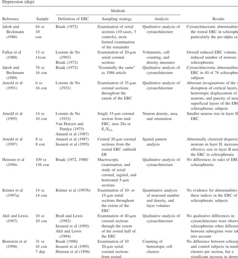
Table 1. Overview of Postmortem Studies of the Entorhinal Cortex (ERC) in Schizophrenia (sz), Control Groups (con), and Depression (dep)
Reference Sample
Methods
Results Definition of ERC Sampling strategy Analysis
Jakob and
Braak (1972) Examination of serial sections (10 cases, 3 controls), more the rostral ERC in schizophrenia, particularly the pre-alpha cell layer
Falkai et al
Examination of 20-mm serial coronal sections
Volumetric, cell counting, and density measures
Overall reduced ERC volume, and reduced number of neurons in schizophrenia
Braak (1972) “Essentially the same” as 1986 article
Qualitative analysis of cytoarchitecture
Cytoarchitectonic abnormalities in the ERC in 40 of 76 schizophrenic subjects
Examination of 35-mm coronal sections throughout the extent of the ERC
Qualitative analysis of cytoarchitecture
Aberrant invaginations of the surface, disruption of cortical layers, heterotopic displacement of neurons, and paucity of neurons in superficial layers of the ERC of schizophrenic subjects Amaral et al (1987)
Single 15-mm coronal section from mid-ERC, area 28a or Ec/ECL
Neuron density, area, and orientation
Smaller neuron size in layer II of ERC
Arnold et al (1997)
8 sz 8 con
Amaral et al (1987) Insausti et al (1995)
Central 20-mm coronal sections from the rostral ERC subfield ER
Spatial pattern analysis
Abnormally clustered dispersion of neurons in layer II, increased effective size in layer II neurons of the ERC in schizophrenia Heinsen et al
(1996)
109 sz 136 con
Braak (1972, 1980) Macroscopic examination, and study of serial coronal, sagittal, and horizontal 5-mm sections
Qualitative analysis of cytoarchitecture
No differences in sulci of ERC in schizophrenia
Krimer et al (1997a)
14 sz 14 con
Krimer et al (1997b) Examination of 10- or 15-mm serial sections throughout the extent of the ERC
Quantitative analysis of neuronal number and density, and layer volumes
No evidence for abnormalities of these indices in the ERC of schizophrenic subjects
Insausti et al (1995) Akil and Lewis
(1994)
Examination of 40-mm coronal sections through the extent of the rostral half of the ERC
Qualitative analysis of cytoarchitecture
No qualitative differences in cytoarchitecture were observed in schizophrenia when differences between subregions were taken into account Insausti et al (1995) Heinsen et al (1996)
Examination of 10
Control brains from 21 subjects without histories of neuropsy-chiatric disorders (13 males, eight females; mean age 53.8 years, range 33–72 years) were obtained during the same time period. The mean time between death and fixation of the control brains was 43 hours; that of the schizophrenic brains was 42.5 hours.
Whole brains were fixed in 10% formalin for 5–7 months. The frontal and occipital poles were separated using incisions anterior to the genu of the corpus callosum and posterior to the splenium. The central block was then embedded in paraplast and cut into 20-mm-thick whole-brain coronal sections. Every 50th section was stained for Nissl and myelin (Heidenhain-Wo¨lcke or Klu¨ver-Barrera).
Beginning with the rostralmost section from the central block, sections were reviewed moving caudally until the first section with the rostral pole of the amygdala present was identified for each brain. The ERC lies ventral to the amygdala and extends caudally adjacent to the pes hippocampi. Using Nissl-stained material, the ERC can be subdivided into six laminae containing neurons: pre-alpha, pre-beta, pre-gamma, pri-alpha, pri-beta, and pri-gamma (Braak 1972). The presence of pre-alpha cells and a cell-free lamina dissecans defines the ERC. To avoid the possible inconsistencies introduced by cytoarchitectural subdivisions we sampled the entire ERC with sections spread 1 mm apart. The rostral boundary was defined as the rostral pole of the amygdala and the caudal boundary as the last section with clear pre-alpha cell clusters. The distance between rostral and caudal boundaries was calculated, and sections assigned to anterior-half or posteri-or-half compartments of the ERC. Mean values of cluster measurements were calculated for left and right anterior and left and right posterior regions of the ERC for each subject. The anterior portion includes the core region of the ERC, as demon-strated in recent studies on the human cortex (Krimer et al 1997b). As well as dividing the ERC into compartments based on length, mean values were also calculated using the anatomic boundaries defined anteriorwise as amygdala to mamillary body and posteriorwise as mamillary body to lateral geniculate nucleus.
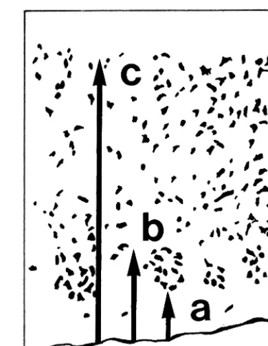
A mean of nine (range 7–15) sections were assessed per subject. Cluster measurements were made by determining the position of the superficial (parameter a) and deep (parameter b) poles of each cluster relative to the gray–white matter junction and measuring the thickness (parameter c) of the cortex (Figure 1). Test–retest reliability was determined by remeasuring all clusters from four cases. Correlation coefficients between the first and second measurements were .72 for parameter a, .81 for parameter b, and .87 for parameter c. The position of the superficial pole was normalized by cortical thickness for analy-ses. The positions of the superficial and deep poles were used to calculate cluster size, which was then normalized by cortical thickness.
Measurements were made on 1991 clusters. These were collapsed into anterior and posterior compartments resulting in two values for each brain, since preliminary analyses indicated no differences in measurements from the right and left sides. The measurements were assessed with analysis of variance using diagnosis, gender, and compartment (anterior, posterior) as main effects and age as a covariate. Diagnosis by gender and diagnosis by compartment interactions were also studied.
Results
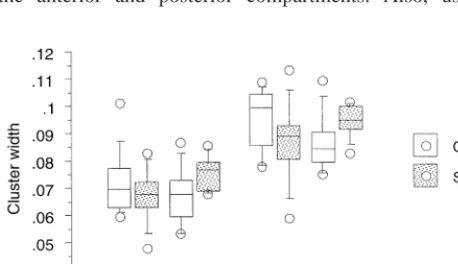
Analysis of distance of the superficial pole of the cluster from the gray-white matter junction (normalized by corti-cal thickness, [c2a]/c); Figure 2) indicated pre-alpha cell clusters were positioned closer to the gray–white matter junction in schizophrenics (diagnosis F58.27, p5.005), particularly in the anterior compartment (interaction be-tween diagnosis and compartment F 5 5.93, p 5 .02). Significant effects of gender (F5 10.66, p 5 .002) and age (F55.99, p5.02) were observed. These results were similar to calculations made when the distance to the center of the cluster was substituted for the distance to the superficial pole ([a1b]/2c). Cortical thickness alone did not show significant differences related to diagnosis, or interactions between compartment and diagnosis.
A different pattern of results was observed for the cluster size, normalized for cortical thickness ([b2 a]/c; Figure 3). No significant overall effect of diagnosis was observed, nor were age or gender effects significant. However, pre-alpha cluster size was significantly less in male schizophrenics (interaction between diagnosis and gender F 5 10.54, p 5 .002), whereas the interaction between diagnosis and compartment was not significant. To explore this finding, results are graphed in Figure 4. In the overall sample of controls, clusters positioned closer to the gray–white matter border tended to be larger. Although
clusters appeared to be larger in schizophrenic females compared with control females, this effect was associated with the position of clusters closer to the gray–white matter junction in the schizophrenic group. In contrast, in the schizophrenic males clusters were positioned some-what closer to the gray–white matter border, but were also smaller in size than controls (post hoc comparison of control vs. schizophrenic males F55.23, p5.02). Two processes, one affecting cluster position and another af-fecting cluster size, may be at work in males with schizophrenia. Effects of diagnosis and diagnosis by gender interactions remained statistically significant when the boundaries of amygdala to mamillary bodies and mamillary bodies to lateral geniculate were used to define the anterior and posterior compartments. Also, using
cortical thickness as a covariate rather than calculating ratios also resulted in the same pattern of significant findings.
Discussion
The primary finding indicates quantitative abnormalities of pre-alpha cell cluster position in schizophrenic ERCs (Figures 2 and 3). This is consistent with the findings of Jakob and Beckmann (1986) and Arnold et al (1997), demonstrating a significant difference in the distribution of pre-alpha cell clusters in schizophrenia compared with control subjects. As well as being positioned closer to the gray–white matter border than expected, clusters in males with schizophrenia were smaller in size (Figure 3).
Heterotopic neuron clusters are believed to indicate abnormalities of neurodevelopment, specifically of neuro-nal migration during the first half of gestation (Williams 1989). The process of cortical lamination is complex, and disruption at several stages could result in morphological abnormalities. Specification of the ultimate laminar posi-tion of neurons is a process that begins in the ventricular zone (Barbe 1996). Impaired neuron-to-glial interactions
Figure 2. Box plot of pre-alpha cell cluster position normalized to thickness of the cortical ribbon. In both males and females with schizophrenia, in the anterior entorhinal cortex the position of pre-alpha cell clusters was significantly closer to the gray– white matter junction than expected. Box plots illustrate percen-tiles (5th, 25th, 50th, 75th, and 95th) and individual values outside these limits. Con, control group; Sch, schizophrenia group.
Figure 3. Box plot of pre-alpha cell cluster size normalized to thickness of the cortical ribbon. For males with schizophrenia, cluster size was significantly reduced overall. Box plots illustrate percentiles (5th, 25th, 50th, 75th, and 95th) and individual values outside these limits. Con, control group; Sch, schizophrenia group.
Figure 4. Position and size relationships of pre-alpha cell clus-ters. Cluster size normalized to cortical thickness is plotted against cluster position relative to the gray–white matter junction (1.00 representing pial surface and 0.00 representing gray–white matter junction). The left member of each pair of joined values represents the mean value (6 SEM) of the posterior compart-ment of the entorhinal cortex. The right member represents the mean value of the anterior compartment. With female schizo-phrenics, pre-alpha cell clusters appear closer in position to the gray–white matter junction in schizophrenics compared with controls, especially in the anterior compartment. Note also how clusters positioned closer to the gray–white matter junction tended to be larger, in all subjects.
during the migration phase, and neuron-to-neuron interac-tions as cells progress through the subplate and cortical plate, could lead to abnormalities of final laminar position. Model systems in animals include a range of phenotypes, such as the reeler mouse (Rakic and Caviness 1995) and traumatic lesions that can induce focal neuronal ectopias (Humphreys et al 1991). In another animal model, admin-istration of methylazoxymethanol acetate to rats on 1 of 3 consecutive days of embryonal development (E9 to E11) caused morphological changes confined largely to the ERC (Talamini et al 1999). Volume reduction, change in shape, and subtle cytoarchitectonic abnormalities includ-ing disturbed lamination were described. A wide range of cortical malformations in humans are associated with neuronal migration disorders (Harding 1992). Although uncommon, associations between schizophrenia and neu-ronal migration disorders were reported (Nopoulos et al 1995). The abnormalities seen in our study are relatively subtle compared to these gross malformations.
The abnormal position of pre-alpha cells in our study could be unrelated to neuronal migration. In the context of a normal anterior–posterior gradient in cluster position in the ERC, it is possible that our findings represent mis-specification of the distribution of cytoarchitectonic sub-regions in schizophrenia, or a difference in schizophrenics in the relative size of subregions with intrinsically differ-ent pre-alpha positions. In this regard, several studies that reported no differences in cytoarchitecture of specific subregions between schizophrenics and controls may have detected quantitative differences if the overall ERC had been considered (Akil and Lewis 1997; Krimer et al 1997a). Similarly, a negative study involving counting the number of abnormally positioned clusters (Bernstein et al 1998) may have used a less sensitive technique than the measurements of cluster position and size performed in our study. Whether altered pre-alpha cell position is due to abnormalities of migration or of cytoarchitectonic parcel-ing, the hypothesis that there is a developmental distur-bance affecting the ERC in schizophrenia received support from our study. Cytoarchitectonic disturbances in the ERC are also consistent with reported disorganization of neu-rons in the hippocampus (Altshuler et al 1987; Conrad et al 1991; Kovelman and Scheibel 1984; Zaidel et al 1997), the cingulate (Benes and Bird 1987), and the frontal and lateral temporal neocortex (Akbarian et al 1993a, 1993b). While comprehensive studies are lacking, the relative absence of disorganization in the posterior ERC compared with the anterior ERC in our study suggests developmental problems in schizophrenia are not entirely diffuse.
A previous morphometric study of the ERC in schizo-phrenia indicated reduced volume of this region and a trend towards reduced neuronal density (Falkai et al 1988). Subsequently, smaller sizes of individual pre-alpha
neu-rons were described (Arnold et al 1995). These findings may have implications for understanding the differences in pre-alpha cluster size in males and females with schizo-phrenia. Although schizophrenic females had larger clus-ters than expected, this observation appeared to be related to the somewhat closer position of the clusters to the gray–white matter border in this group compared with controls. For males with schizophrenia, clusters were smaller than expected. Possible explanations for this finding include an abnormality of neurogenesis or neuro-nal loss occurring through a degenerative process. While most studies of astrocytes in schizophrenia using classic staining methods or glial fibrillary acid protein immuno-cytochemistry indicate no difference from controls (Har-rison 1995), a minority of studies have detected abnormal-ities of astrocytes (Arnold et al 1996; Stevens 1982). Specific investigation of pre-alpha cell clusters addressing this issue has not been performed. The presence of an apoptotic process in these cells also remains a possibility. In summary, our results provide further support for the model of schizophrenia as a disorder related to impaired brain development. Whether or not abnormally positioned cell clusters compromise ERC function remains unknown. The ERC receives afferent information from both limbic and association cortices. The layer II pre-alpha cell clus-ters project to the dentate gyrus while deeper layer III neurons project to the subiculum and pyramidal cell regions of the hippocampus (Van Hoesen 1997). The latter regions are reciprocally connected with deeper layers of the ERC. Disrupted lamination may result in subtle abnor-malities of connectivity. In the reeler mouse, although the major types of connections form, there is enough disrup-tion of cortical circuitry to result in behavioral deficits (Rakic and Caviness 1995). Further studies of entorhinal to hippocampal connectivity in schizophrenia could help determine whether or not this is the case in schizophrenia.
Support was provided by the Stanley Foundation (PF) and Medical Research Council of Canada (PF, WGH).
References
Akbarian S, Bunney WE, Potkin SG, Wigal SB, Hagman JO, Sandman CA, et al (1993a): Altered distribution of nicoti-namide-adenine dinucleotide phosphate-diaphorase cells in frontal lobe of schizophrenics implies disturbances of cortical development. Arch Gen Psychiatry 50:169 –177.
Akbarian S, Vin˜uela A, Kim JJ, Potkin SG, Bunney WE, Jones EG (1993b): Distorted distribution of nicotinamide-adenine dinucleotide phosphate-diaphorase neurons in temporal lobe of schizophrenics implies anomalous cortical development.
Arch Gen Psychiatry 50:178 –187.
hydrox-ylase-immunoreactive fibers in the human entorhinal cortex.
Neuroscience 60:857– 874.
Akil M, Lewis DA (1997): Cytoarchitecture of the entorhinal cortex in schizophrenia. Am J Psychiatry 154:1010 –1012. Altshuler LL, Conrad A, Kovelmn JA, Scheibel A (1987):
Hippocampal pyramidal cell orientation in schizophrenia: A controlled neurohistologic study of the Yakovlev collection.
Arch Gen Psychiatry 44:1094 –1098.
Amaral DG, Insausti R, Cowan WM (1987): The entorhinal cortex of the monkey, I: Cytoarchitectonic organization.
J Comp Neurol 264:326 –355.
Arnold SE, Franz BR, Gur RC, Gur RE, Shapiro RM, Moberg PJ, et al (1995): Smaller neuron size in schizophrenia in hippocampal subfields that mediate cortical-hippocampal in-teractions. Am J Psychiatry 152:738 –748.
Arnold SE, Franz BR, Trojanowski JQ, Moberg PJ, Gur RE (1996): Glial fibrillary acidic protein-immunoreactive astro-cytosis in elderly patients with schizophrenia and dementia.
Acta Neuropathol 91:269 –277.
Arnold SE, Hyman BT, Van Hoesen GW, Damasio AR (1991): Some cytoarchitectural abnormalities of the entorhinal cortex in schizophrenia. Arch Gen Psychiatry 48:625– 632. Arnold SE, Ruscheinsky DD, LY Han (1997): Further evidence
for abnormal cytoarchitecture of the entorhinal cortex in schizophrenia using spatial point pattern analyses. Biol
Psy-chiatry 42:639 – 647.
Barbe MF (1996): Tempting fate and commitment in the devel-oping forebrain. Neuron 16:1– 4.
Beall MJ, Lewis DA (1992): Heterogeneity of layer II neurons in human entorhinal cortex. J Comp Neurol 321:241–266. Benes FM, Bird ED (1987): An analysis of the arrangement of
neurons in the cingulate cortex of schizophrenic patients.
Arch Gen Psychiatry 44:608 – 616.
Bernstein H-G, Krell D, Baumann B, Danos P, Falkai P, Diekmann S, et al (1998): Morphometric studies of the entorhinal cortex in neuropsychiatric patients and controls: Clusters of heterotopically displaced lamina II neurons are not indicative of schizophrenia. Schizophr Res 33:125–132. Bogerts B, Falkai P, Haupts M, Greve B, Ernst S,
Tapernon-Franz U, et al (1990): Post-mortem volume measurements of limbic system and basal ganglia structures in chronic schizo-phrenics. Schizophr Res 3:295–301.
Braak H (1972): Zur Pigmentarchitektonik der Großhirnrinde des Menschen. I. Regio entorhinalis. Z Zellforsch 127:407– 438. Braak H (1980): Architectonics of the Human Telencephalic
Cortex. Berlin: Springer-Verlag.
Brodmann K (1909): Vergleichende Lokalisationslehre der
Grosshirnrinde. Leipzig, Germany: Verlag von Johann
Am-brosius Barth.
Conrad A, Abebe T, Austin R, Forsythe S, Scheibel A (1991): Hippocampal pyramidal cell disarray in schizophrenia as a bilateral phenomenon. Arch Gen Psychiatry 48:413– 417. Falkai P, Bogerts B, Rozumek M (1988): Limbic pathology in
schizophrenia: The entorhinal region—a morphometric study.
Biol Psychiatry 24:515–521.
Filimonoff IN (1947): A rational subdivision of the cerebral cortex. Arch Neurol Psychiatr 58:296 –311.
Harding BN (1992): Malformations of the nervous system. In:
Adams JH, Duchen LW, editors. Greenfield’s
Neuropa-thology, 5th ed. New York: Oxford University Press,
521– 638.
Harrison PJ (1995): On the neuropathology of schizophrenia and its dementia: Neurodevelopmental, neurodegenerative, or both? Neurodegeneration 4:1–12.
Heinsen H, Go¨ssmann E, Ru¨b U, Eisenmenger W, Bauer M, Ulmar G, et al (1996): Variability in the human entorhinal region may confound neuropsychiatric diagnoses. Acta Anat
(Basel) 157:1–12.
Humphreys P, Rosen GD, Press DM, Sherman GF, Galaburda AM (1991): Freezing lesions of the newborn rat brain: A model for cerebrocortical microgyria. J Neuropathol Exp
Neurol 50:145–160.
Insausti R, Tunon T, Sorbreviela T, Insausti AM, Gonzalo LM (1995): The human entorhinal cortex: A cytoarchitectonic analysis. J Comp Neurol 355:171–198.
Jakob H, Beckmann H (1986): Prenatal developmental distur-bances in the limbic allocortex in schizophrenics. J Neural
Transm 65:303–326.
Jakob H, Beckmann H (1989): Gross and histological criteria for developmental disorders in brains of schizophrenics. J R Soc
Med 82:466 – 469.
Kovelman J, Scheibel A (1984): A neurohistological correlate of schizophrenia. Biol Psychiatry 19:1601–1621.
Krimer LS, Herman MM, Saunders RC, Boyd JC, Hyde TM, Carter JM, et al (1997a): A qualitative and quantitative analysis of the entorhinal cortex in schizophrenia. Cereb
Cortex 7:732–739.
Krimer LS, Hyde TM, Herman MM, Saunders RC (1997b): The entorhinal cortex: An examination of cyto- and myeloarchi-tectonic organization in humans. Cereb Cortex 7:722–731. Lorente de No (1933): Studies on the structure of the cerebral
cortex. I. The area entorhinalis. J Psychol Neurol 45:381– 438.
Nopoulos PC, Flaum M, Andreasen NC, Swayze VW (1995): Gray matter heterotopias in schizophrenia. Psychiatry Res
Neuroimaging 61:11–14.
Rakic P, Caviness VS (1995): Cortical development: View from neurological mutants two decades later. Neuron 14:1101– 1104.
Rose M (1927): Die sog. Riechrinde beim Menschen und beim Affen. II. Teil des “Allocortex bei Tier und Mensch.”
J Psychol Neurol 34:261– 401.
Sgonina K (1938): Zur vergleichenden Anatomie der Entorhinal und Pra¨subikularregion. J Psychol Neurol 48:56 –163. Stevens JR (1982): Neuropathology of schizophrenia. Arch Gen
Psychiatry 39:1131–1139.
Talamini LM, Koch T, Ter Horst GJ, Korf J (1999): Methyla-zoxymethanol acetate-induced abnormalities in the entorhinal cortex of the rat parallels with morphological findings in schizophrenia. Brain Res 789:293–306.
Van Hoesen GW (1997): Ventromedial temporal lobe anatomy, with comments on Alzheimer’s disease and temporal injury.
J Neuropsychiatry Clin Neurosci 9:331–341.
rhesus monkey, I: Temporal lobe afferents. Brain Res 95:1– 24.
Vogt C, Vogt O (1919): Allgemeinere Ergebnisse unserer Hirn-forschung. J Psychol Neurol 25:279 – 462.
Williams RS (1989): Cerebral malformations arising in the first half of gestation. In: Evrard PH, Minkowski A, editors.
Developmental Neurobiology. New York: Raven Press, 11–
20.