MULTI-PATCHES IRIS BASED PERSON AUTHENTICATION SYSTEM USING
PARTICLE SWARM OPTIMIZATION AND FUZZY C-MEANS CLUSTERING
Shekar B. H.a, Sharada S. Bhatb∗
a
Mangalore University, Mangalore, Karnataka India - ([email protected]) b
Government Arts and Science College, Karwar, Karnataka, India - ([email protected])
Commission II, WG II/10
KEY WORDS:Particle swarm optimization, Fuzzy c-means, Taylor’s series expansion, weighted mean Hamming distance, Iris recog-nition system
ABSTRACT:
Locating the boundary parameters of pupil and iris and segmenting the noise free iris portion are the most challenging phases of an automated iris recognition system. In this paper, we have presented person authentication frame work which uses particle swarm optimization (PSO) to locate iris region and circular hough transform (CHT) to device the boundary parameters. To undermine the effect of the noise presented in the segmented iris region we have divided the candidate region intoNpatches and used Fuzzy c-means clustering (FCM) to classify the patches into best iris region and not so best iris region (noisy region) based on the probability density function of each patch. Weighted mean Hammimng distance is adopted to find the dissimilarity score between the two candidate irises. We have used Log-Gabor, Riesz and Taylor’s series expansion (TSE) filters and combinations of these three for iris feature extraction. To justify the feasibility of the proposed method, we experimented on the three publicly available data sets IITD, MMU v-2 and CASIA v-4 distance.
1. INTRODUCTION
Being one of the favourites of all biometric traits, iris recogni-tion system is emerged as the best human authenticarecogni-tion system. However, it is needed to develop a robust iris recognition system for authentication as well as identification. Eye images taken with visible wavelength (VW) or near infra-red (NIR) camera may in-clude images in which, the difference between the iris and ad-jacent non-iris region is not clearly distinguishable because of bad illumination, on-move or at-a-distance photography. Also, off-angled and tilted eye images made it difficult to accurately localize the iris and pupil region. After locating the position of the iris, segmenting the candidate iris region is even more com-plicated and time consuming task. Because, the iris region would normally occluded by eye lids, eye lashes, reflections of light and spectacles. Since, accuracy and speed of iris recognition system highly depend on efficient localization and segmentation process, large number of researchers are working on this issue.
Existing segmentation methods localize the pupillar and limbic boundaries first, and then apply occlusion removal techniques. Several curve fitting techniques are proposed in the literature for occlusion removal. Since both VW and NIR imaging produce degraded and noisy images, occlusion removal is challenging and time consuming task in both authentication and identification sce-narios. Moreover, located iris region may not be circular in shape. The irises with irregular shape and occlusion will result into a drastic low recognition rate. In this paper, we have proposed a novel and robust occlusion removal strategy for the degraded, noisy irises.
We have developed an iris authentication frame work which in-volves the following steps:
∗Corresponding author
1 The iris region is segmented from the input eye image us-ing particle swarm optimization (PSO) and then the pupil and limbic boundary parameters are located using circular hough transform (CHT).
2 Segmented iris region comprises of eyelids, eye lashes, re-flection of light and part of adjacent non-iris region (because of non-circular shape). To deal with this issue a novel oc-clusion removal strategy is applied. Segmented iris region is divided intoN patches using tracks and sectors. Unsuper-vised Fuzzy c-means clustering (FCM) method is applied to classify them into k clusters based on the probability den-sity function of each patch. Each patch is assigned with a weight based on the clustering output and the ground truth of the iris region.
3 Combination of Log-Gabor, Riesz and TSE filters are used to extract the features of each patch and each patch is en-coded into binary iris code using Daugman’s phase encoding technique. Weighted mean Hamming distance (WMHD) is used to find the dissimilarity scores between the two irises.
We have experimented this method on three publicly available databases, IIT Delhi, MMU v-2 and CASIA v-4 distance and ob-tained improved recognition rates.
The remainder of the paper is organised as follows: Section 2 summarises the related work, in Section 3 we have detailed the proposed iris authentication frame work, Section 4 comprises of details of experiments and discussions and Section 5 concludes the paper.
2. RELATED WORK
The state-of-art iris segmentation methods can be categorised into two types. First category locates pupil and iris boundaries
This contribution has been peer-reviewed.
based on the edge information (boundary based methods) and the second category segments the candidate iris region based on the pixel information (pixel-based methods) (Liu et al., 2016). The first boundary-based technique is the integro-differential op-erator (IDO) introduced by Daugman (Daugman, 1993), and the second one is hough transform (HT) which is first used by Wildes (Wildes, 1997). Various forms of IDO and HT (Wildes, 1997, Zuo et al., 2006, Liu et al., 2005, Jan et al., 2013, Tan et al., 2010) are proposed in the literature. Apart from these two tech-niques, other boundary based methods found in the literature are, active shape models (Abhyankar and Schuckers, 2006), binary morphology and image statistics (Kennell et al., 2006), adaptive binarisation (Basit and Javed, 2007), Adaboost cascading and elastic model (He et al., 2009), Geodesic active counter (Shah and Ross, 2009), and eyeball model (Baek et al., 2013).
Pixel based methods have used the information such as color, intensity variation and texture of the iris to extract the discrim-inative appearance feature of a pixel from the neighbourhood pixels (Liu et al., 2016). Proenca and Alexandre (Proenc¸a and Alexandre, 2006) have introduced unsupervised clustering tech-niques, Jeong et al. (Jeong et al., 2010) have used Adaboost eye detection and color segmentation, intelligent random sample con-sensus iris segmentation on four spectral images is proposed by Chou et al. (Chou et al., 2010) and clustering based coarse eye localization and integro-differential constellation are used by Tan et al. (Tan et al., 2010). Neural network classification is intro-duced by Proenca (Proenca, 2010) and Li et al. (Li et al., 2010) have used K-means clustering. Quality filters for down sampling is proposed by Du et al. (Du et al., 2011) and Gaussian Mix-ture Model have been utilised by Li and Savvides (Li and Sav-vides, 2013). Sahmoud and Abuhaiba (Sahmoud and Abuhaiba, 2013) have used K-means clustering and circular HT and Tan and Kumar (Tan and Kumar, 2014) have proposed Zernike mo-ments to extract the iris pixel information. Recently, Gangwar et al. (Gangwar et al., 2016) have proposed a boundary based course-to-fine strategy for iris localization and Liu et al. (Liu et al., 2016) have proposed iris segmentation models using convolu-tional neural network. We have proposed a segmentation strategy which exploits both, the pixel information and position with re-spect to the pixels in the neighbourhood and the geometry of the iris region.
Particle swarm optimisation (PSO) is a bioinspired theory which is first introduced by Kennedy and Eberhart (Eberhart et al., 1995) and has been applied to image segmentation problem by several researchers. Omran et al. (Omran et al., 2002) have intro-duced PSO for image classification and then used dynamic clus-tering PSO (Omran et al., 2006) for image segmentation. Chan-dar et al. have used a PSO variant adapting social and momentum components of the velocity for particle move updates (Chander et al., 2011). A simple modified PSO is proposed by Lee et al. (Lee et al., 2012) to extract both low-level features and high-level im-age semantics from the color imim-age. Tillet et al. (Tillett et al., 2005) have introduced Darwinien PSO, Ghamisi et al. (Ghamisi et al., 2014) have devised fractional-order Darwinien PSO and these techniques are evaluated on medical images (Ryalat et al., 2016). In the biometrics domain, Parez et al. have used PSO to generate the templates for face and iris localization (Perez et al., 2010) and Chen and Chu have combined probabilistic neural network and PSO to design an optimized classifier model for iris recognition (Chen and Chu, 2009). Inspired by these researches, we have used PSO for segmentation as PSO utilizes localized pixel information as well as global features of iris.
The technique of region-wise feature extraction is previously used by the researchers at the classification stage. Chen et al. (Chen et al., 2006) and Proenca and Alexandre (Proenca and Alexandre, 2007) have proposed the technique of dividing the unwrapped candidate iris intoNdivisions and obtained the mul-tiple signatures for classification. A modified version of it has been used in (Barpanda et al., 2015), and in (Bastys et al., 2011) and (Pillai et al., 2011) authors have also utilized sector divisions of iris region for noisy and uncooperative iris recognition.
3. PROPOSED IRIS AUTHENTICATION FRAME WORK
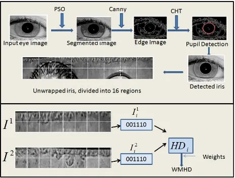
An overview of the proposed frame work is shown in Figure 1. Input eye image is preprocessed by smoothing, gamma correction and histogram equalization. We have utilised the pixel informa-tion to cluster the input eye image into iris and non-iris regions. The rationale behind using PSO clustering technique are: 1. it has been introduced as a best method for the optimization of con-tinuous nonlinear functions (Eberhart et al., 1995), 2. it has been proved better performing than K-means clustering (Omran et al., 2002, Ganta et al., 2012), 3. in case of biometrics, it is used as a best localization tool for face and iris recognition (Perez et al., 2010), and 4. coarse iris localization needs both local and global optimization and PSO does it simultaneously.
Figure 1. The proposed Iris Authentication frame work
3.1 Particle Swarm Optimisation
The PSO approach automatically determines the optimum num-ber of clusters in a given image and simultaneously clusters the data set with minimal user interference (Omran et al., 2006). It consists of a group of pixels in an image that collectively move in the neighbourhood in search of the global optimum (Ghamisi et al., 2014). Like any other genetic algorithm (GA), PSO is initialized with a population of random solutions. However, unlike GA, here potential solutions, called as particles, are as-signed with some randomized velocities (Shi et al., 2001). Each particle is treated as a point in a d-dimensional space (prob-lem space). The ith particle (ith position) is represented as
xi= (xi1, xi2, ..., xid). Each particle keeps track of its best pre-vious position, calledpbest, which is the position that gives the optimum value (best fitness) for the objective function and is rep-resented aspi= (pi1, pi2, ..., pid). Each particle also keeps track
over all best fitness value and its position are also tracked and are calledgbestvalue andgbestposition. gbestposition is repre-sented bypg = (pg1, pg2, ..., pgd). Objective function is a
pre-defined fitness function ofdvariables related to the problem to be solved. Particles are initially associated with some random offset values called as velocities, represented asvi= (vi1, vi2, ..., vid). The strategy of PSO concept is, at each step changing the ve-locity of each particle, i.e. accelerating the particle, towards its
pbestandgbestposition (Shi and Eberhart, 1998). The veloc-ity and position of the particle are changed using the following equations:
vij=w∗vij+c1∗rand()∗(pij−xij)+c2∗Rand()∗(pgj−xij) (1)
xij=xij+vij (2)
wherej∈ {1,2, . . . , d},c1andc2are positive constants, rand() and Rand() are two random functions with the range [0,1] andw
is weight factor. The current fitness value of the particle is com-pared with itspbestvalue. If the current value is better than the particle’spbestthen current position is set aspbestposition and the current value is set aspbestvalue. Current value is also com-pared with thegbestvalue and if it is better thangbestthengbest
value andgbestposition are reset to current value and current po-sition respectively. This process is continued until a predefined good fitness value is met. The parametersc1andc2are called acceleration constants and are both taken as 2.0 for almost all the applications (Shi et al., 2001). Parameterw, also called inertia weight, controls the impact of the previous values of the veloci-ties on the current velocity and hence provides a balance between local and global exploration abilities of the PSO technique (Shi and Eberhart, 1998).
Omran et al. (Omran et al., 2002) have explained how PSO is used in image classification. A swarm is a group ofK clus-ters of the input image. A particlexi is constructed asxi = (ci1, ci2, ..., cij, ..., ciK), where each cij is centroid vector of
jth
clusterCij. Whiledrepresenting the Euclidean distance, the minimum of the inter-class distances between any pair of clusters in the swarm is given by,
Dmin(xi) = min
j6=k{d(cij, cik)} (3)
Let Z be the matrix that represents the assignment of the pixels to the clusters of particlexi, i.e. an elementzijpis the pixel in the clusterCijof the particlexi. Then maximum of the average intra-class distances of the clusters of the particlexiis,
dmax(Z, xi) = max j=1,2,...,K{
X
∀zp∈Cij
d(zp, cij)/|Cij|} (4)
where|Cij|is the cardinality of the clusterCij. Objective func-tion F is defined so as to minimise the intra-class distances be-tween the pixels and their cluster means, given bydmax(Z, xi) and to maximise the inter-class distances between the clusters, given byDmin.
F(xi, Z) =w1∗dmax(Z, xi) +w2∗(zmax−Dmin) (5)
zmaxis the maximum pixel value andw1andw2are user defined constants by tuning which, we set the priorities to minimisation of intra-class (candidate iris pixels) differences and maximisation of inter-class (iris and non-iris portion of the eye) differences.
3.2 Multi-patches technique
We have used circular Hough transform to roughly identify the pupil and iris boundary parameters. At this stage, the actual pa-rameters are not identified. Because, the segmented iris is not in exact circular shape. Moreover, the candidate iris region is en-compassed with noise and occlusion such as, eye-lids, eye-lashes and photographic reflections. Some surrounding non-iris portion of the iris and some un-useful part of pupil may also be present. This will reduce the percentage of accuracy drastically. In Figure 2, the segmented iris and roughly identified circular structure of the candidate iris region are shown for the eye images taken from the three datasets. It can be seen that candidate iris region is oc-cluded by eyelids, eye lashes and some adjacent non-iris portion.
Figure 2. Row-wise : eye images from IIT Delhi, MMU v-2 and CASIA v-4 distance databases. Column-wise : Original eye
image, segmented image and roughly identified iris region.
To obtain the portion of the candidate iris region which can be used for further process we have adopted multi-patches tech-nique. The annular iris ring is divided intoNpatches usingm
tracks and nsectors. Daugman’s doubly dimensionless rubber sheet model (Daugman, 2004) is used to unwrap the iris into uni-form sized iris templates. Figure 3 shows an analytical model of annular ring which is divided into 16 patches using two sectors and eight tracks.
Figure 3. Analytical model of annular iris ring withm= 2,n= 8 andN= 16
The ground truth of the iris image is : the upper and lower region are usually occluded by the eyelids. In (Bastys et al., 2011) the
This contribution has been peer-reviewed.
sectors which contains the upper and lower iris portion are not in-volved in the feature extraction process. We have not completely omitted these regions. Because, the occluded portion is not uni-form among the eye images. It varies from image to image. Hence, we have utilised the statistical properties of the patches to classify them as best iris region and not so best iris region. The probability density function of each patch is separately com-puted. Based on these properties, patches of iris are grouped into clusters using Fuzzy c-means clustering. The motivation behind using Fuzzy clustering is that the Fuzzy matching is used by (Tsai et al., 2012) to match the two different irises. Properties of iris patches are fuzzy in nature and they possess close statistical re-lationships. FCM is a well suited tool for such data (Bezdek et al., 1984). We have observed the cluster output and the ground truth of iris and have trained the system to assign the weights to the patches.
3.3 Iris feature extraction filters
We have used Log-Gabor filter given by Masek (Masek and Kovesi, 2003), Riesz filter (Shekar and Bhat, 2015) and Taylor series expansion (TSE) filter (Shekar and Bhat, 2016) and the combinations of these filters and have compared the results. A brief introduction of these filters is given below. Readers can go through the referred papers for details.
3.3.1 Log-Gabor filter Log-Gabor is a Gabor filter which is a Gaussian on logarithmic scale. The frequency output of Log-Gabor is given by the equation,
G(f) =Exp
The filter response of Log-Gabor is encoded into binary bits us-ing Daugman’s phase encodus-ing technique (Daugman, 1993). The real and imaginary parts in the filter output are encoded based on their zero crossings. Thus, each pixel is encoded into 2 bit binary code.
3.3.2 Riesz filter Two dimension Riesz kernels are given by,
hx=
where k.k is the Euclidean norm. The components of first order monogenic Riesz signals {hxf, hyf} are obtained by convolving the input function f(x, y) with the above ker-nels. The components of second order monogenic Riesz signals {hxxf, hxyf, hyyf}are obtained by convolving the components of the first order signals with the 2D kernels given in equation (7). Each pixel in the input image is encoded into 3 bits binary code by binarising the second order monogenic signals based on the zero crossings.
3.3.3 TSE filter The partial sums of Taylor series expansion (TSE) taking the derivatives along angular axis (θ−axis) and radial axis (r−axis) are computed on multiscales.
AngularSum=
information at the zero crossings of these signals are further used to encode a pixel into binary bits.ε=θ−ηandδ=r−ξare called scale factors and are computed using the following equa-tions:
εi= 1
m(θmax−θmin)∗i, δj=
1
n(riris−rpupil)∗j (10)
whereθminand θmax are the minimum and maximum values through whichθvaries during the unwrapping process andriris andrpupilare the radii of the iris and pupil regions respectively. Each scale generates 2 binary bits. In our experiments we have taken four different scales to generate 8 bit encoding of the iris pixel.
3.4 Weighted mean Hamming distance (WMHD)
In this section the procedure to find the final dissimilarity score between the two irises is explained. LetI1
andI2
respectively. Each patch is convolved with a filter (LogGabor, Riesz or TSE) and filter output is encoded into binary bits using Daugman’s phase encoding technique. LetHDi be the dissimilarity score between the correspondent patchesI1
i and I2
i, calculated using hamming distance. Letw1i andw2i, i∈ {1,2, ..., N}, be the weights assigned to the patchesI1
i and
I2
i respectively. To compute the final set of weightswiwe have experimented the following different strategies.
S1. Arithmetic Mean :wi=
Final dissimilarity score between the irisesI1
andI2
is computed using the weighted mean distance given by,
HDI1I2=
Databases Segmentation Accuracy
IIT Delhi 98.20
MMU v-2 95.66
CASIA v-4 distance 90.50
Table 1. Segmentation accuracy obtained by the proposed method on different databases.
and each image has dual-eye iris from which patterns of left and right irises can be independently accessed. We have conducted our experiments on 1400 eyes by taking first ten left eyes and ten right eyes of first 70 subjects.
In our experiments we have used the segmentation level as 4.0 in the computation of PSO segmented image. Canny edge detec-tor (with threshold level = 2) is used to get the edge map from the PSO segmented image. The edge map thus obtained is sub-jected to Hough transform to coarsely locate the centres and radii of pupil and iris. While computing the segmentation accuracy we have manually noted down the correctly segmented irises and segmentation accuracy is computed as the ratio of the number of correctly segmented irises to the total number of eye images taken for the experiment. The percentages of segmentation accuracy obtained on IITD, MMU v-2 and CASIA v-4 distance databases are given in Table 1.
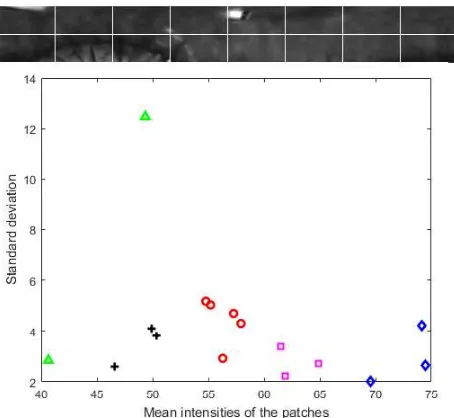
During the experiments we have selected first 20 eye images from each database. The segmented iris is divided into 16 patches us-ing 2 tracks and 8 sectors. These 16 patches are clustered into 5 groups.We have computed the probability density function of each patch and a 2D vector of mean and standard deviation is given as input to FCM. The cluster output and the ground truth of the 16 patches are compared and accordingly weights are as-signed as1,0.75,0.5,0.25,0.0representing, best iris, iris, par-tially iris, less parpar-tially iris and no iris respectively. An example of iris patches and graphical representation of the clustered output is shown in Figure 4.
Figure 4. An example of iris patches and graphical representation of the clustered output.
Further, we calculated the dissimilarity score of two irises using WMHD, using the four strategies as explained in the section 3.4
Strategy Recognition Rate
S1 92.50
S2 98.20
S3 92.50
S4 98.96
Table 2. Recognition rates using different strategies
and recognition rates obtained on IITD database taking training to test ratio as 3:2 are presented in Table 2. We have observed that strategies S2 (geometric mean) and S4 have given the good re-sults. We have experimented the proposed segmentation method and multi-patch technique on the IITD, MMU v-2 and CASIA v-4 distance databases using the feature extraction techniques ex-plained in section 3.3. Experiments are conducted using bit level fusion technology given in (Shekar and Bhat, 2015). Recognition rates obtained on the three databases applying the filters Log-Gabor, Riesz and TSE and their combinations are presented in Table 3. To compute WHMD strategy S4 is used and training to test ratio is 3:2. ROC curves of the same experiments are given in Figure 5, 6 and 7.
Method IITD MMU v-2 CASIA v-4 dist
Gabor 92.50 85.50 80.00
Riesz 95.00 86.34 82.50
Taylor 97.50 87.00 85.00
Gabor+Riesz 93.33 87.50 85.00
Gabor+Taylor 97.66 90.00 85.00
Riesz+Taylor 98.50 92.33 87.50
Gabor+Riesz+Taylor 98.96 95.67 90.00
Table 3. Recognition rates obtained by different feature extraction methods on the three databases
Figure 5. ROC curve obtained on database.
5. CONCLUSION
In this work, we have devised an iris authentication frame work comprising novel iris segmentation and occlusion elimination strategies. Extensive experiments are conducted to justify accu-racy of the proposed strategies in authentication scenario. Unlike
This contribution has been peer-reviewed.
Figure 6. ROC curve obtained on MMU v-2 database.
Figure 7. ROC curve obtained on CASIA v-4 distance database.
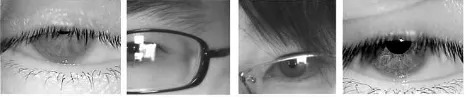
the existing methods of occlusion removal, proposed strategy is simple and easy to implement. Some of the eye images in the databases are completely degraded (refer Figure 8) and such im-ages are not counted while calculating the segmentation accuracy.
Figure 8. Exanples of degraded eye images.
REFERENCES
Abhyankar, A. and Schuckers, S., 2006. Active shape models for effective iris segmentation. In:Defense and Security Symposium, International Society for Optics and Photonics, pp. 62020H– 62020H.
Baek, S.-J., Choi, K.-A., Ma, C., Kim, Y.-H. and Ko, S.-J., 2013. Eyeball model-based iris center localization for visible image-based eye-gaze tracking systems. IEEE Transactions on Con-sumer Electronics59(2), pp. 415–421.
Barpanda, S. S., Majhi, B. and Sa, P. K., 2015. Region based feature extraction from non-cooperative iris images using triplet half-band filter bank.Optics & Laser Technology72, pp. 6–14.
Basit, A. and Javed, M., 2007. Localization of iris in gray scale images using intensity gradient. Optics and Lasers in Engineer-ing45(12), pp. 1107–1114.
Bastys, A., Kranauskas, J. and Kr¨uger, V., 2011. Iris recog-nition by fusing different representations of multi-scale taylor expansion. Computer Vision and Image Understanding115(6), pp. 804–816.
Bezdek, J. C., Ehrlich, R. and Full, W., 1984. Fcm: The fuzzy c-means clustering algorithm. Computers & Geosciences10(2-3), pp. 191–203.
Chander, A., Chatterjee, A. and Siarry, P., 2011. A new social and momentum component adaptive pso algorithm for image seg-mentation. Expert Systems with Applications38(5), pp. 4998– 5004.
Chen, C.-H. and Chu, C.-T., 2009. High performance iris recog-nition based on 1-d circular feature extraction and pso–pnn clas-sifier.Expert Systems with Applications36(7), pp. 10351–10356.
Chen, Y., Dass, S. C. and Jain, A. K., 2006. Localized iris im-age quality using 2-d wavelets. In: International conference on biometrics, Springer, pp. 373–381.
Chou, C.-T., Shih, S.-W., Chen, W.-S., Cheng, V. W. and Chen, D.-Y., 2010. Non-orthogonal view iris recognition system.IEEE Transactions on Circuits and Systems for Video Technology20(3), pp. 417–430.
Daugman, J., 2004. How iris recognition works. IEEE Transac-tions on circuits and systems for video technology14(1), pp. 21– 30.
Daugman, J. G., 1993. High confidence visual recognition of persons by a test of statistical independence.Pattern Analysis and Machine Intelligence, IEEE Transactions on15(11), pp. 1148– 1161.
Du, Y., Arslanturk, E., Zhou, Z. and Belcher, C., 2011. Video-based noncooperative iris image segmentation. IEEE Transac-tions on Systems, Man, and Cybernetics, Part B (Cybernetics)
41(1), pp. 64–74.
Eberhart, R. C., Kennedy, J. et al., 1995. A new optimizer using particle swarm theory. In:Proceedings of the sixth international symposium on micro machine and human science, Vol. 1, New York, NY, pp. 39–43.
Gangwar, A., Joshi, A., Singh, A., Alonso-Fernandez, F. and Bi-gun, J., 2016. Irisseg: A fast and robust iris segmentation frame-work for non-ideal iris images. In:Biometrics (ICB), 2016 Inter-national Conference on, IEEE, pp. 1–8.
Ganta, R. R., Zaheeruddin, S., Baddiri, N. and Rao, R. R., 2012. Particle swarm optimization clustering based level sets for image segmentation. In: 2012 Annual IEEE India Conference (INDI-CON), IEEE, pp. 1053–1056.
Ghamisi, P., Couceiro, M. S., Martins, F. M. and Benediktsson, J. A., 2014. Multilevel image segmentation based on fractional-order darwinian particle swarm optimization.IEEE Transactions on Geoscience and Remote sensing52(5), pp. 2382–2394.
Jan, F., Usman, I. and Agha, S., 2013. Reliable iris localiza-tion using hough transform, histogram-biseclocaliza-tion, and eccentric-ity.Signal Processing93(1), pp. 230–241.
Jeong, D. S., Hwang, J. W., Kang, B. J., Park, K. R., Won, C. S., Park, D.-K. and Kim, J., 2010. A new iris segmentation method for non-ideal iris images. Image and vision computing 28(2), pp. 254–260.
Kennell, L. R., Ives, R. W. and Gaunt, R. M., 2006. Binary mor-phology and local statistics applied to iris segmentation for recog-nition. In:2006 International Conference on Image Processing, IEEE, pp. 293–296.
Lee, C.-Y., Leou, J.-J. and Hsiao, H.-H., 2012. Saliency-directed color image segmentation using modified particle swarm opti-mization.Signal Processing92(1), pp. 1–18.
Li, P., Liu, X., Xiao, L. and Song, Q., 2010. Robust and accurate iris segmentation in very noisy iris images. Image and Vision Computing28(2), pp. 246–253.
Li, Y.-H. and Savvides, M., 2013. An automatic iris occlusion estimation method based on high-dimensional density estimation.
IEEE transactions on pattern analysis and machine intelligence
35(4), pp. 784–796.
Liu, N., Li, H., Zhang, M., Liu, J., Sun, Z. and Tan, T., 2016. Ac-curate iris segmentation in non-cooperative environments using fully convolutional networks. In: Biometrics (ICB), 2016 Inter-national Conference on, IEEE, pp. 1–8.
Liu, X., Bowyer, K. W. and Flynn, P. J., 2005. Experiments with an improved iris segmentation algorithm. In:Fourth IEEE Work-shop on Automatic Identification Advanced Technologies (Au-toID’05), IEEE, pp. 118–123.
Masek, L. and Kovesi, P., 2003. Matlab source code for a bio-metric identification system based on iris patterns.The School of Computer Science and Software Engineering, The University of Western Australia.
Omran, M. G., Salman, A. and Engelbrecht, A. P., 2006. Dy-namic clustering using particle swarm optimization with applica-tion in image segmentaapplica-tion. Pattern Analysis and Applications
8(4), pp. 332–344.
Omran, M., Salman, A. and Engelbrecht, A. P., 2002. Image clas-sification using particle swarm optimization. In:Proceedings of the 4th Asia-Pacific conference on simulated evolution and learn-ing, Vol. 1, Singapore, pp. 18–22.
Perez, C. A., Aravena, C. M., Vallejos, J. I., Estevez, P. A. and Held, C. M., 2010. Face and iris localization using templates de-signed by particle swarm optimization.Pattern recognition letters
31(9), pp. 857–868.
Pillai, J. K., Patel, V. M., Chellappa, R. and Ratha, N. K., 2011. Secure and robust iris recognition using random projections and sparse representations. IEEE Transactions on Pattern Analysis and Machine Intelligence33(9), pp. 1877–1893.
Proenca, H., 2010. Iris recognition: On the segmentation of de-graded images acquired in the visible wavelength. IEEE Trans-actions on Pattern Analysis and Machine Intelligence 32(8), pp. 1502–1516.
Proenc¸a, H. and Alexandre, L. A., 2006. Iris segmentation methodology for non-cooperative recognition.IEE Proceedings-Vision, Image and Signal Processing153(2), pp. 199–205.
Proenca, H. and Alexandre, L. A., 2007. Toward noncooperative iris recognition: a classification approach using multiple signa-tures.IEEE Transactions on Pattern Analysis and Machine Intel-ligence29(4), pp. 607–612.
Ryalat, M. H., Emmens, D., Hulse, M., Bell, D., Al-Rahamneh, Z., Laycock, S. and Fisher, M., 2016. Evaluation of particle swarm optimisation for medical image segmentation. In: Inter-national Conference on Systems Science, Springer, pp. 61–72.
Sahmoud, S. A. and Abuhaiba, I. S., 2013. Efficient iris segmen-tation method in unconstrained environments. Pattern Recogni-tion46(12), pp. 3174–3185.
Shah, S. and Ross, A., 2009. Iris segmentation using geodesic active contours.IEEE Transactions on Information Forensics and Security4(4), pp. 824–836.
Shekar, B. and Bhat, S. S., 2016. Iris recognition using partial sum of second order taylor series expansion. In:Proceedings of the Tenth Indian Conference on Computer Vision, Graphics and Image Processing, ACM, p. 81.
Shekar, B. H. and Bhat, S. S., 2015. Steerable riesz wavelet based approach for iris recognition. In:2015 3rd IAPR Asian Confer-ence on Pattern Recognition (ACPR), IEEE, pp. 431–436.
Shi, Y. and Eberhart, R. C., 1998. Parameter selection in particle swarm optimization. In:International Conference on Evolution-ary Programming, Springer, pp. 591–600.
Shi, Y. et al., 2001. Particle swarm optimization: developments, applications and resources. In: evolutionary computation, 2001. Proceedings of the 2001 Congress on, Vol. 1, IEEE, pp. 81–86.
Tan, C.-W. and Kumar, A., 2014. Accurate iris recognition at a distance using stabilized iris encoding and zernike moments phase features. IEEE Transactions on Image Processing23(9), pp. 3962–3974.
Tan, T., He, Z. and Sun, Z., 2010. Efficient and robust segmen-tation of noisy iris images for non-cooperative iris recognition.
Image and vision computing28(2), pp. 223–230.
Indian Institute of Technology Delhi Iris Database, n.d.
http://www.comp.polyu.edu.hk/csajaykr/IITD/ database.html.
Institute of Automation, Chinese Academy of Sciences.CASIA iris database, n.d.http://biometrics.idealtest.org/.
Malaysia Multimedia University iris database, n.d. http:// pesona.mmu.edu.
Tillett, J., Rao, T., Sahin, F. and Rao, R., 2005. Darwinian particle swarm optimization.
Tsai, C.-C., Lin, H.-Y., Taur, J. and Tao, C.-W., 2012. Iris recog-nition using possibilistic fuzzy matching on local features. IEEE Transactions on Systems, Man, and Cybernetics, Part B (Cyber-netics)42(1), pp. 150–162.
Wildes, R. P., 1997. Iris recognition: an emerging biometric tech-nology.Proceedings of the IEEE85(9), pp. 1348–1363.
Zuo, J., Kalka, N. D. and Schmid, N. A., 2006. A robust iris segmentation procedure for unconstrained subject presentation. In:2006 Biometrics Symposium: Special Session on Research at the Biometric Consortium Conference, IEEE, pp. 1–6.
This contribution has been peer-reviewed.