73
CHAPTER 4
PROTEIN STRUCTURE
W
ithin two days after the initial publication of Wilhelm Röntgen’s discovery of Xrays in 1895, a surgeon in Scotland used X rays to observe a needle as he extracted it from the palm of an unfortunate seamstress. Although this medical application resulted in the development of radiological diagnosis and treatment of disease by radiation, physical aspects
of Röntgen’s discovery also provided the means for elucidating the structure of proteins and other large molecules. The laws governing the diffraction of X rays were discovered by the two Braggs, Sir William and Sir Lawrence, who were father and son. At the Cavendish Laboratory at the University of Cambridge, where Sir Lawrence was professor, J.D. Bernal was studying the use of X-ray diffraction for the determination of the structure of large biological molecules. He had already used X rays to define the size and shape of the tobacco mosaic virus and showed it to have a regular internal structure. At the Cavendish Laboratory the group that formed around Bernal, a man of wide public and scientific interests, included the Nobel Prize winners Max Perutz and John Kendrew, who in 1937 began to use X rays to analyze two proteins fundamental to life, myoglobin and hemoglobin, both of which function in the transport of gases in the blood. Twenty-two years passed before the structures of these proteins were established; the significance of the work is that it provided the basis for an understanding of the mechanism of the action of enzymes and other proteins, an active and fruitful subject of modern investigation.
The word protein comes from the Greek proteios which means " first row ". Word coined by Jons J. Barzelius in 1938 to emphasize the importance of this group. The structure of proteins is a biomolecular structure of a protein molecule. Each protein, in particular polypeptide is a polymer which is made up of a sequence of L - α - amino (this sequence is also referred to as the residue). The deal, a chain length less than 40 residues referred to as a polypeptide, not a protein.
Protein plays an important role in almost all biological processes. A protein is a functional biological molecule that is made up of one or more polypeptides that are folded/coiled into a specific structure. Proteins are important macromolecules that serve as structural elements, transportation channels, signal receptors and transmitters, and enzymes. Proteins are linear polymer that are built up of the monomer units called amino acids. There are 20 different amino acid and they are connected by a peptide bond between the carboxyl group and the amino group in a linear chain called a polypeptide. Each protein has different side chains or the "R" groups. Proteins have many different active functional groups attached to them to help define their properties and functions.
74 Waals forces, and hydrophobic packing system. Perotein three-dimensional structure is necessary to understand the function of proteins at the molecular level.
Protein structures vary in size, from tens to thousands of residues. Proteins are classified based on their physical size as nanoparticles (1-100 nm). A protein can undergo reversible structural changes in biological function. Alternative structures of the same proteins referred to as conformation.
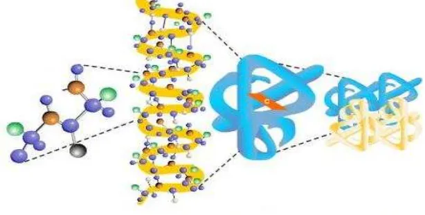
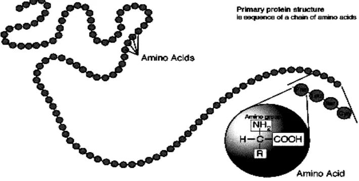
Figure 4.1. Protein structure
Plants form a protein of CO2, H2O, and nitrogen compounds. Animals that eat plants alter plant protein into animal protein. In addition to be used for the formation of the body's cells. Protein is also used as a source of energy when our body deficiency of carbohydrates and fats. Average composition of chemical elements contained in the protein are as follows: Carbon 50% , 7% hydrogen, 23% oxygen, 16% nitrogen, 0.3% sulfur, and phosphorus 0.3% .
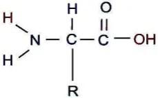
Amino acids are the basic structural units of proteins. A - amino acid consists of an amino group, carboxyl group, and the H atoms that are all specific R groups attached to the carbon. The carbon atom is called as adjacent to the carboxyl group (acid). Group R represents a side chain.
Figure 4.2. The structure of the amino acids.
75 group in the unionized form (- NH2). Have glycine carboxyl group pK of 2.3 and pK amino groups of 9.6. Thus, the midpoint of the first ionization is at pH 2.3 and for the second ionization at pH 9.6.
Figure 4.3. Ionization state of amino acids depends on the pH (en.wikipedia.org).
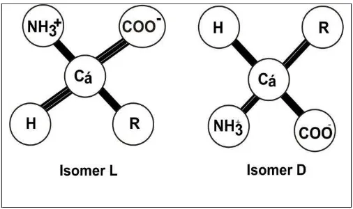
Tetrahedral arrangement of four different groups on the carbon atoms of amino acids has led to the optical activity. Two mirror image forms called isomers of L and D isomers Proteins consist of amino acids L, so the sign of the optical isomers can be ignored in the discussion and subsequent protein amino acids in question are L isomers, unless there is an explanation.
Figure. 4.4. Absolute configuration of the amino acid L -isomers and D. R describe the side chain. L and D isomers are mirror images.
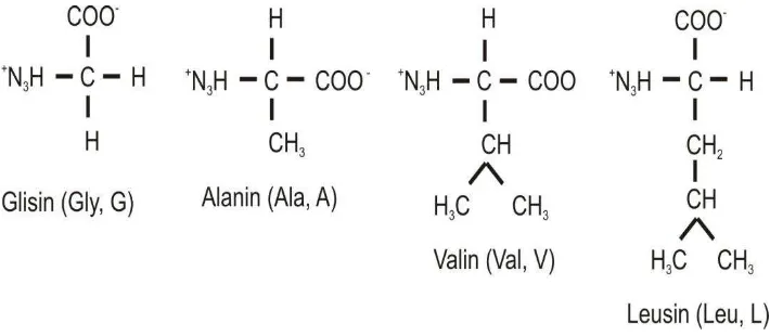
76 The simplest amino acid is glycine, which has only one hydrogen atom as a side chain (Figure 4.4). The following amino acids are alamin, with a methyl group as a side chain. Hydrocarbon side chains larger (three and four carbons) found in valine, leucine and isoleucine. Aliphatic side chains is larger hydrophobic, water-repellent and tend to form groups. As will be discussed later, the three -dimensional structure of a protein that is soluble in water would be stabilized by the hydrophobic side chains are flocking to avoid contact with the water. Differences in size and shape of the hydrocarbon side chain allows the protein to form a structure that is concise and compact holes.
Figure 4.5. Amino acids with aliphatic side chains.
A. Protein Structure
Proteins fold into secondary, tertiary, and quaternary structures based on intra-molecular bonding between functional groups or interintra-molecular bonding (quaternary only) and can obtain on a variety of three-dimensional shapes depending on the amino acid sequence. All proteins have primary, secondary and tertiary structures but quaternary structures only arise when a protein is made up of two or more polypeptide chains. The folding of proteins is also driven and reinforced by the formation of many bonds between different parts of the chain. The formation of these bonds depends on the amino acid sequence. The study of their structures is important because proteins are essential for every activity in the human body as well as they are the key components of biological materials. Primary structure is when amino acids are linked together by peptide bonds to form polypeptide chains. Secondary structure is when the polypeptide chains fold into regular structures like the beta sheets, alpha helix, turns, or loops. A functional protein is much more than just a polypeptide, it is one or more polypeptides that have been precisely folded into a molecule with a very specific, unique shape which is critical to its function.
77 structure and tertiary structure is less clear. In addition, the presence of well known quaternary structures and structures that will be discussed at a glance supersekunder in this section.
1. Primary structure
In 1953, Frederick Sanger determines the amino acid sequence of insulin, a protein hormone. This is an important event for the first time show unequivocally that the protein having the amino acid sequence of a particular right. Amino acid sequence is then known as the primary structure. The primary structure of a protein is the level of protein structure which refers to the specific sequence of amino acids. When two amino acids are in such a position that the carboxyl groups of each amino acid are adjacent to each other, they can be combined by undergoing a dehydration reaction which results in the formation of a peptide bond.
Amino acids in a polypeptide (protein) are linked by peptide bonds that begin with the N-terminal with a free amino group and ends at C-terminal with a free carboxyl group. rts . The peptide bond is planar and cannot rotate freely due to a partial double bond character. While there is a restricted rotation about peptide bond, there are two free rotations on (N-C) bond and (C-C) bond, which are called torsion angles, or more specifically the phi and psi angles. The freedoms of rotation of these two bonds are also limited due to steric hindrance. Genes carry the information to make polypeptides with a defined amino acid sequence. An average polypeptide is about 300 amino acids in length, and some genes encode polypeptides that are a few thousand amino acids long.
It's important to know the primary structure of the protein because the primary structure encodes motifs that are of functional importance in their biological function; structure and function are correlated at all levels of biological organization. It is also shown that insulin is composed of only L amino acids that are interconnected via a peptide bond between amino - and carboxyl group - achievement stimulate other researchers to study the amino acid sequences of various proteins. Currently known complete amino acid sequence of more than 10,000 proteins. A striking fact states that each protein has a unique amino acid sequence with the sequence of very precise.
78 Figure 4.6. Peptide bond formation.
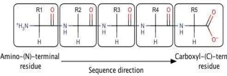
Many amino acids bonded by peptide bonds to form polypeptide chains are not branched (Figure 2.7). One unit of amino acid residues in a polypeptide chain called. Because the direction of the polypeptide chain has constituent units have different ends, namely amino - and carboxyl - groups. Under the agreement, the amino end of the polypeptide chain is placed at the beginning; meaningful sequence of amino acids in a polypeptide chain are written with prefixed by aminoterminal residue. In a tripeptide Ala - Gly - Trp (AGW), alanine is a residue aminoterminal and Tryptophan are carboxyl - terminal residues. It should be noted that the Trp - Gly - Ala (WGA) is a tripeptide that is different.
Figure 4.7. Amino acid residues contained in the box, chain starting at the amino end.
79
Figure 4.8. Polypeptide chain is formed from main chain repeated on a regular basis (spine) and the side chains of certain (R1, R2, R3 are colored yellow).
A number of proteins have disulfide bonds. Antarrantai disulfide bonds in the chain and is formed by oxidation of cysteine residues. Are generated cysteine disulfide (Figure 4.8). Intra- cell proteins generally do not have disulfide bonds, whereas extracellular proteins often have several. Non cross - sulfur bond derived from lysine side chains are found in several proteins. For example, the collagen fibers in the connective tissue is strengthened in this way, just as fibrin in blood collection.
80 Figure 4.9b Model sulfide bonds in the primary structure.
2. Secondary structure
The amino acid sequence of a polypeptide, together with the laws of chemistry and physics, cause a polypeptide to fold into a more compact structure. Amino acids can rotate around bonds within a protein. This is the reason proteins are flexible and can fold into a variety of shapes. Folding can be irregular or certain regions can have a repeating folding pattern. The coils and folds that result from the hydrogen bonds between the repeating segments of the polypeptide backbone are called secondary structures. Although the individual hydrogen bonds are weak, they are able to support a specific shape for that part of the protein due to the fact that they are repeated many times over a long part of the chain. Secondary structures of a protein are proposed by Pauling and Corey. Its structures are formed by amino acids that are located within short distances of each other. Because of the planar nature of the peptide bonds, only certain types of secondary structure exist. The three important secondary structures are
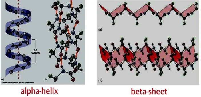
α-helix, β-sheets, and β-turns. Also, the beta sheets can be parallel, antiparallel, or mixed. Antiparallel beta sheets are more stable because the hydrogen bonds are at a nighty degree angles. The a-helix is a coiled structure stabilized by intrachain hydrogen bonds.
Pauling and Corey polypeptide conformation study various possibilities to create molecular models. They are very obey observations bond angles and distances on amino acids and small peptides. In 1951, they revealed two polypeptide structure called helix and pleated sheets. This structure is related to the setting position of the amino acid residues of space in a linear sequence.
Characteristics of the Secondary Structures (http://en.wikibooks.org/wiki/Structural_ Biochemistry/ Proteins):
81 most common helical structure is a right-handed helix with its hydrogen bonds parallel to its axis. The hydrogen bonds are formed between carbonyl oxygen and amine hydrogen groups of four amino acid residues away. Each amino acid advances the helix, along its axis, by 1.5 Å. Each turn of the helix is composed of 3.6 amino acids; therefore the pitch of the helix is 5.4 Å. There is an average of ten amino acid residues per helix with its side chains orientated outside of the helix. Different amino acids have different propensities for forming x-helix, however proline is a helix breaker because proline does not have a free amino group. Amino acids that prefer to adopt helical conformations in proteins include methionine, alanine, leucine, glutamate and lysine (malek).
Figure 4.10.
α
Helix (en.wikibooks.org)2. β-sheet: ß-sheets are stabilized by hydrogen bonding between peptide strands. In a
82
are more extended than an α-helix, and the distance between adjacent amino acids
is 3.5 Å. Hydrogen bonding in β-strand can occur as parallel, anti- parallel, or a
mixture. Amino acid residues in β- parallel configuration runs in the same orientation. Pleated sheets makes up the core of many globular proteins and also are dominant in some fibrous proteins such as a spiders web. The large aromatics such as: tryptophan, tyrosine and phenylalanine, and beta-branched amino acids
like: isoleucine, valine, and threonine prefer to adopt β-strand conformations.This orientation is energetically less favorable because of its slanted, non-vertical hydrogen bonds. Trytophan, tyrosine, and phenylalanine are hydrophobics while the other amino acids are hydrophilics.
Figure 4.11. Another type of secondary structure, a beta sheet (en.wikibooks.org)
3. β-turns: Poly peptide chains can change direction by making reverse turns and
loops. Loop regions that connect two anti-parallel β-strands are known as reverse
turns or β-turns. These loop regions have irregular lengths and shapes and are usually found on the surface of the protein. The turn is stabilized by hydrogen bond between the backbone of carbonyl oxygen and amine hydrogen. The CO group of the residue, in many reverse turns, which is bonded to the NH group of residue i + 3 . The interaction stabilizes abrupt changes in direction of the polypeptide chain. Unlike the alpha-helices and ß-strands, loops do not have regular periodic structures. However, they are usually rigid and well defined. Since they loops lie on the surface of the proteins, they are able to participate in interactions between proteins and other molecules. Ramachandran plot is a plot that shows the available torsion angles of where proteins can be found. However, in the plot, if there are many dots that locate all over the place, it means that there exists a loop.
83 sequence. Means all the CO group and the main chain NH groups form hydrogen bonds. Each subsequent acid residues with residues along the helical axis of Figure 2.10. Helix has a range of 1.5 to 100° rotation, so that there are 3.6 amino acid residues per helical twist.
In helical amino acids within three and four will be located in a linear sequence in the opposite helix so not interconnected. The distance between the two helical twist is multiplication translational distance (1.5) and the number of residues in each round of the same 3.6 to 5.4. Helical twist direction as the screw can be turn right (clockwise) and turn left (counter clockwise) rotating helical proteins are right. Helical content of the protein varies widely from almost nothing to 100% . For example, the enzyme chymotrypsin contains no helix. In contrast, 75% protein myoglobin and hemoglobin helical. The length of single-stranded helix is usually less than 45. But two or more helices can spiral into each other to form a stable structure, the length can reach 1000 (100 nm or 0.1 m) or more. Helical twisting each myosin and tropomyosin found in muscle, fibrin clots in blood and in hair keratin. The helical shape of the protein has a role in the formation of mechanically rigid fiber bundle such spikes. Cytoskeleton (inner buffer) a cell containing many filaments which are the two strands of the helix spiral into each other.
Helical structure was deduced by Pauling and Corey six years before the structure is evident in myoglobin by using X-ray examination The description of the helical structure is an important event in the history of molecular biology because it shows that the conformation of the polypeptide chain can be predicted if the known properties of its components carefully and precisely.
Figure 4.11. The main structure of amino acids.
84 Figure 4.13. The structure of the helix spiral.
Pauling and Corey find another mode of periodic structures called pleated sheet (so-called because it is the structure that they found while the second helix as the first structure). Pleated sheets of 0 different from a helical rod-shaped. Polypeptide chain pleated sheet called a strand, straight shape is not stretched taut like a coiled helix. Axis distance between adjacent amino acids is 3.5 A, while the helix is 1.5 A. Another difference is that the pleated sheets stabilized by hydrogen bonds between NH and CO groups on different polypeptide chains, whereas the helix there are hydrogen bonds between NH and CO groups on the same chain.
Figure 4.14. R. pleated sheet
85
3. Tertiary structure
Tertiary structure depicting spatial arrangement of amino acid residues far apart in the linear sequence and the pattern of disulfide bonds. The difference between the secondary and tertiary structure is less clear (see Figure 4.15). Collagen shows a special type of a helix and is the most abundant protein found in mammals. Collagen is the main component of the fiber in the skin, bones, tendons, cartilage and teeth. This extracellular protein containing three helical polypeptide chains, each of which is along the nearly 1000 residues. The sequence of amino acids in collagen are very irregular: every third residue is nearly always glycine. Compared with other proteins in the collagen content of proline is also high. Furthermore, collagen -containing 4 - hydroxyproline are rarely found in other proteins. Sequence glycine - proline - hydroxyproline (Gly - Pro - Hyp) is often encountered.
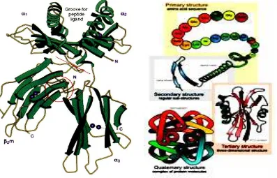
Figure 4.15. Comparison between the structure of primary, secondary and tertiary.
Collagen is a rod -shaped molecule, with a length of approximately 3000 with a diameter of only 15. Helical pattern of the combined three polypeptide chains, is totally different from the one strand helix hydrogen bonds can not be found. However, each strand helix of collagen is stabilized by power repel pyrrolidine ring of proline and hydroxyproline residues. In this helical shape that is more open than the twisted helical tense, pyrrolidone rings farther apart. The third strand to form superhelical beating each other polypeptides.
86 on different chains act as a hydrogen acceptor. Hydroxyl group of hydroxyproline residues also play a role in the formation of hydrogen bonds.
With glycine is understandable why put yourself at every position in the range of one thousand three residues that form helical collagen. The interior of the three- strand helix is very solid. It turns out that glycine is the only residue that fits on the inside. Because there are three helical residues in each round, then every third residue in each strand should be of glycine. Amino acid residues adjacent to the glycine located on the outside of the strand and the space is enough for proline and hydroxyproline residues were great.
Proteins are made up of more than one polypeptide chain has an additional level of structural organization. Each polypeptide chain is called sub- units. Describe the quaternary structure of the protein subunit arrangement in space. For example, hemoglobin, consists of two chains and two chains of hemoglobin subunit composition of the tetramer is instrumental in binding antartempat communication O2, C O 2, and H + are apart. Viruses are very limited utilize genetic information to form a sheath composed of sub - units of the same sub- units repeated in a symmetrical arrangement.
Figure 4.16. Myoglobin space model with the same orientation.
B. Protein Structure Determination Method
1. X-ray crystallography
87 Figure 4.17. Crystallization myoglobin (www.scienceinschool.org).
Slow salting produce irregular crystals, instead of amorphous precipitates. Some proteins crystallize easily, while others require a greater effort. Crystallization is an art, because it requires perseverance, patience and a cool hand. The number of large and complex proteins that have been crystallized on the rise. For example, the polio virus by 8500 - which is the unity of the 240 kd protein subunits surrounding a core of RNA, has been known to be crystallized and its structure by X-ray methods.
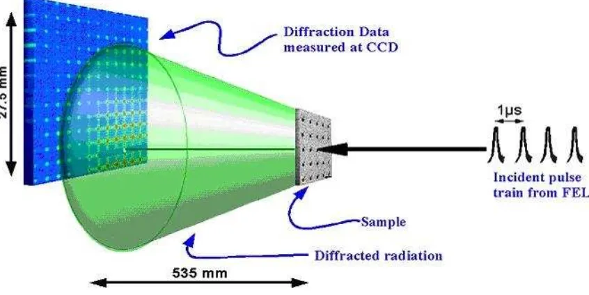
The three components that play a role in the analysis of X-ray crystallography is the X-ray source, a protein crystal and detector (Figure 2.18). Beam with a wavelength of 1.54 A obtained by accelerating electrons to copper. A beam of X -rays directed at a protein crystal. Most of the X-rays will directly penetrate the crystal and the rest will be scattered into various directions. Files are scattered (or experiencing diffraction) can be detected with X-ray film. Blackish color of the film is directly proportional to the intensity of the X-ray detector decentralized or with solid state electronics. The basic principle of X-ray crystallography:
1) dipencar X-rays by electrons. The amplitude of the wave is decentralized by atom is directly proportional to the number of electrons. Carbon atoms will scatter X-rays six times more powerful than a hydrogen atom.
88 Figure 4.18. Basic experimental X-ray crystallography, crystal and detector
(http://photon-science.desy.de).
Protein crystals are inserted in the capillary and placed in the right position on the X-ray beam and the film. With careful crystal motion will be generated in the form of X-ray photography composition dots called regular reflection. The intensity of each point on the X-ray photography can be measured and is the basic data for the analysis of X-ray crystallography. The next stage was to reconstruct the picture of the protein based on intensity. In the light microscope or an electron microscope, the scattered beam is focused by the lens that instantly gives an overview. But the lens to focus the X-rays do not exist. Picture can be obtained by using a mathematical calculation called the Fourier transform. Each point depicts the electron density waves, which correspond to the magnitude of the square root of the intensity of the point. Each wave also has phases, namely tops and bottoms of the waves. Phase of each wave determines whether the waves are coming from another point of amplified or deleted. This phase can be determined by the diffraction pattern produced by a standard tagging heavy metals such as uranium or mercury in certain places in the protein.
Now it can be interpreted electron density map, which gives the electron density at points regularly spread in the crystal. Three- dimensional picture of the electron density is shown as a parallel sections and stacked. Each piece is a transparent plastic sheet with the electron density distribution is shown by contour lines, together with the contour lines on a map to illustrate the height of the geological survey. The following stage is the interpretation of electron density maps. Critical factor is the resolution of X-ray analysis were determined by the amount of scattered intensity used in the Fourier synthesis. The recent results of X-ray analysis is determined by the degree of crystalline perfection. For proteins, the resolution limit is usually about 2.
89 recognize and bind other molecules, how it functions as an enzyme, how proteins fold and how it goes. This remarkable result will continue to grow rapidly and will give a great influence on the field of biophysics.
2. Spectroscopy NMR (Nuclear Magnetic Resonance)
Nuclear magnetic resonance spectroscopy (NMR) is a widely used and powerful method that takes advantage of the magnetic properties of certain nuclei. The basic principle behind NMR is that some nuclei exist in specific nuclear spin states when exposed to an external magnetic field. NMR observes transitions between these spin states that are specific to the particular nuclei in question, as well as that nuclei's chemical environment. However, this only applies to nuclei whose spin, I, is not equal to 0, so nuclei where I = 0 are ‘invisible’ to NMR spectroscopy. These properties have led to NMR being used to identify molecular structures, monitor reactions, study metabolism in cells, and is used in medicine, biochemistry, physics, industry, and almost every imaginable branch of science.
X-ray crystallography is equipped with NMR spectroscopy, which is able to reveal the atomic structure of a molecule in solution. Nuclei of certain atoms such as hydrogen (1H) magnetic intrinsically (see Table 4.1). Round protons are positively charged, the same as other charged particles that produce a rotating magnetic moment. Magnetic moment is contained in one of two orientations (called and) when affected by external magnetic field (Figure 4.19). The energy difference between the two orientations is proportional to the magnetic field strength is given.
Table 4.1. The most important point in biological NMR signal.
90 Figure 4. 19. Basic NMR spectroscopy.
Status has a slightly lower energy so slightly more dense (by a factor of 1.00001) because according to the magnetic field. The transition from an isolated low level (α) to
β level occurs when the nucleus absorbs electromagnetic radiation with a frequency that is appropriate.
0 0
γ H
v =
2
π
Ho is the magnetic field strength is fixed and is a constant (called the magnetogyric ratio) for a particular core. For example, in the 1H resonance frequency of 100 kilogauss magnetic field (10 tesla) is 426 megahertz (MHz), which lies in the area of radio frequency spectrum. The relationship between the energy absorbed by the frequency will show a peak at 426 MHz.
91 ppm. Most of the proton chemical shifts in protein molecules located between 1 and 9 ppm. NMR spectral absorption peak called lin (lines). Particular proton is usually more than one cause lin nonekuivalen influenced by adjacent protons; This effect is called spin - spin coupling. Hydrogen atoms separated by three or less covalent bonds be linked in this way.
Brief on the sample magnetization induced by radio -frequency pulses will disappear with time, the sample will experience relaxation and return to a balanced state. This relaxation process can explain the structure and dynamics of macromolecules because it is very sensitive to the geometry and motion. Another thing that gives a lot of information is NOE (Nuclear Overhauser Effect), an interaction between the core is inversely proportional to the distance between the nucleus rank of six. Magnetization is transferred from the excited to the core nucleus that is not excited when they are separated less than approximately 5 (Figure 4.20). Overhauser spectroscopy spectrum of the two - dimensional core and improved (NOESY = nuclear Overhauser Enhancement Spectroscop) graph showing pairs of adjacent protons. Diagonal line NOESY spectrum corresponding to the spectrum of one - dimensional chemical shift. Peaks outside the diagonal line gives new information: identifies pairs of protons at a distance of less than 5 (Figure 4.20). Overlapping peaks in the NOESY spectrum can usually be separated by using NMR spectra of proteins are characterized by 15N and 13C. Irradiation of these cores will be separated NOE peak along the axis, which is an approach called multidimensional NMR spectroscopy. Three-dimensional structure of proteins can be determined from a number of these relationships.
92 Only the NMR spectroscopic techniques and X-ray crystallography can express three-dimensional atomic structure of proteins and biomolecules detailed else. X-ray method gives a good overview of the resolution, but requires a crystal. NMR method, on the contrary, effective for proteins in solution and requires a very concentrated solution (1 mM - or 15 mg / ml for the 15 - kd protein). Biggest size currently in use for NMR method is 30 kd, for larger proteins do not give accurate results. However, much can be done within the boundaries of these domains because proteins are usually smaller than 30 kd. Additionally NMR spectroscopy rays can also explain the dynamics. NMR techniques and X-rays are complementary in the study of the structure.
To improve your understanding of the material above, do the exercises below! 1) Explain the basic principles kristaligrafi 3 X-ray!
2) Describe the structure of the amino acids in the form of amino acids was isolated and dipolar ionic form!
3) Briefly describe the difference in the architecture (structure) of proteins and function!
4) How to obtain protein crystals in X-ray crystallography techniques?
5) Explain briefly the physical principles associated with the NMR technique!
If you have difficulty in answering the questions above, to help you read the following explanation:
1) The basic principle of X-ray crystallography:
a) X-rays by electrons dipencar. The amplitude of the wave is decentralized by atom is directly proportional to the number of electrons. Carbon atoms will scatter X-rays six times more powerful than a hydrogen atom.
b) the scattered waves recombine. Each atom in the molecule plays a role in X-ray diffraction of waves. In the film or detector decentralized waves reinforce each other when in the same phase and will cancel out when not in the same phase.
c) How has scattered waves recombine depends only on the arrangement of atoms.
EXERCISE
93 2)
3) The primary structure is the sequence of amino acids. Secondary structure associated with the setting position space adjacent amino acid residues in the linear sequence. This steric arrangement gives the periodic structure. Helix - and strand showed secondary structure. Tertiary structure depicting spatial arrangement of amino acid residues far apart in the linear sequence and pattern sulfide bonds.
4) protein crystals can be obtained by adding ammonium sulfate or other salts into concentrated protein solution to reduce solubility. For example, myoglobin will crystallize in a solution of ammonium sulfate 3 M.
5) using the NMR resonance mechanism of magnetic field caused by the movement of electrical charges in the protein molecules.
Protein plays an important role in almost all biological processes. Proteins are essential components or major components of animal or human cells. Therefore it is forming cells of our body, the protein contained in the food serves as a major agent in the formation and growth of the body. Average composition of chemical elements contained in the protein are as follows: Carbon 50% , 7% hydrogen, 23% oxygen, 16% nitrogen, 0.3% sulfur, and phosphorus 0.3% .
Amino acids are the basic structural units of proteins. A - amino acid consists of an amino group, carboxyl group, and the H atoms that are all specific R groups attached to the carbon. The carbon atom is called as adjacent to the carboxyl group (acid).
Proteins fold into secondary, tertiary, and quaternary structures based on intra-molecular bonding between functional groups or intermolecular bonding (quaternary only) and can obtain on a variety of three-dimensional shapes depending on the amino acid sequence. All proteins have primary, secondary and tertiary structures but quaternary structures only arise when a protein is made up of two or more polypeptide chains. The folding of proteins is also driven and reinforced by the formation of many bonds between different parts of the chain. The formation of these bonds depends on the amino acid sequence..
An understanding of the structure and function of proteins greatly assisted by X-ray crystallography, which is a technique that can declare a three -dimensional positions of atoms in the protein molecule to the right. X-ray crystallography is equipped with NMR spectroscopy, which is able to reveal the atomic structure of a
