Kinase pathways in dominant and subordinate
ovarian follicles during the first wave of
follicular development in sheep
A.C.O. Evans
a,∗, F. Martin
baDepartment of Animal Science and Production, Conway Institute for Biomolecular and Biomedical Research, University College Dublin, Belfield, Dublin 4, Ireland
bDepartment of Pharmacology, Conway Institute for Biomolecular and Biomedical Research, University College Dublin, Belfield, Dublin 4, Ireland
Received 15 June 2000; received in revised form 1 September 2000; accepted 25 September 2000
Abstract
The mechanism by which one or more dominant ovarian follicles continue development while other subordinate follicles regress is not known. The mitogen activated protein kinases (MAPKs) are a group of kinases that are activated by hormonal factors and form a cascade of processes that regulate cell growth, division and differentiation. The aim of the present experiment was to characterise the presence of the MAPKs, Erk 1/Erk 2 and Akt in healthy dominant follicles and regressing subordinate follicles. Following in vivo monitoring of ovarian follicle development, three ewes were ovariectomised and the follicular fluid and follicle wall (theca and granulosa cells) saved from the dominant and largest subordinate follicle. The dissected diameter and follicular fluid oestradiol concentration of the dominant follicle was larger (P <0.01) than the largest subordinate follicle (6.5±0.0 mm and 41.3±4.9 ng/ml versus 4.7±0.3 mm and 0.6±0.4 ng/ml). Western blot analyses showed that there was more Akt (202.7±6.4 versus 59.6±32.7 units;P <0.05) and Erk 1/Erk 2 (104.5±10.6 versus 0.3±0.2 units;P <0.01) present in follicle wall samples from the dominant compared to the largest subordinate follicles. Phosphorylated forms of Akt and Erk 1/Erk 2 were detected in samples from dominant but not subordinate follicles. We suggest that signal transduction pathways involving Akt and Erk 1/Erk 2 may play an important role in determining the outcome of ovarian follicle growth and development in sheep. © 2000 Elsevier Science B.V. All rights reserved.
Keywords: Sheep-ovary; Follicle; Intracellular pathways
∗Corresponding author. Tel.:+353-1-706-7771; fax:+353-1-706-1103.
E-mail address: [email protected] (A.C.O. Evans).
1. Introduction
In cattle, sheep and humans, the dominant ovulatory follicle(s) is selected from a cohort of growing follicles 5–7 days prior to ovulation (Leyendecker et al., 1990; Roche et al., 1998; Evans et al., 2000). This follicle grows to a larger diameter than other follicles and appears to suppress the development of smaller (subordinate) follicles. Unlike the menstrual cycle of humans, cattle and sheep have two or more successive waves of follicular development during oestrous cycles (Webb et al., 1999; Evans et al., 2000). Healthy dominant follicles produce large quantities of oestradiol compared to atretic subordinate follicles that produce relatively more progesterone (Evans and Fortune, 1997; Evans et al., 2000).
The demise of cells within follicles occurs by apoptosis (Jolly et al., 1997; Van Wezel et al., 1999). All the follicles of a cohort develop in the same endocrine environment, yet how the dominant follicle survives while the neighbouring subordinate follicles die by apop-tosis is not known. It seems plausible that individual follicles respond to their endocrine environment by regulating their own intracellular environments thereby controlling degra-dation or continued growth. Follicles produce intrafollicular growth factors that contribute to the success of folliculogenesis (Webb et al., 1999) and the later stages of follicle de-velopment are reliant upon the gonadotrophic hormones LH and FSH (Webb et al., 1992; Campbell et al., 1995). The actions of both the gonadotrophins and growth factors are mediated via cell surface receptors that rely on signal transduction mechanisms to relay information to the cell nucleus (Hill and Treisman, 1995). The nature of these pathways is complex and their interaction with the events that control apoptosis in ovarian cells is not clear.
The mitogen-activated protein kinases (MAPKs) are a group of serine/threonine kinases that are activated by a variety of extracellular stimuli and mediate signal transduction from the cell surface to the nucleus (Blumer and Johnson, 1994). Upon activation, MAPKs translocate to the nucleus and phosphorylate transcription factors (Cobb and Goldsmith, 1995). The MAPK cascade is involved in processes regulating cell growth, division and differentiation. The most widely studied cascades are the extracellular signal-regulated kinases-1 (Erk 1; p44 MAPK) and Erk 2 (p42 MAPK) one of whose functions when acti-vated is to phosphorylate a range of transcription factors including signal transducers and activators of transcription (Stat) in the nucleus (Pircher et al., 1999). Most recently Erk has been shown to control the synthesis of nucleotides, the first step in the production of DNA and RNA (Graves et al., 2000). Another common signal transduction pathway involves the activation of protein kinase B (also known as Akt) by PI3 kinase (Coffer et al., 1998). Akt is a general mediator of cell survival and has been shown to suppress the apoptotic death of a number of cell types induced by a variety of stimuli (Downward, 1998). Akt regulates the activity of transcription factors and modulates the activity of members of the Bcl2 family of proteins (Brunet et al., 1999; Kops et al., 1999) thus preventing the proapoptotic actions of a family of proteins referred to as caspases (Datta et al., 1997; Cardone et al., 1998; Kulik and Weber, 1998) that serve as regulators and effectors of apoptosis (see Mehmet, 2000). The activation of both Erks and Akt involve phosphorylation of an inactive form of the protein by specific upstream kinases (Khokhlatchev et al., 1998).
has been shown to induce the activation of Erks in porcine (Cameron et al., 1996) and rat (Das et al., 1996) granulosa cells in vitro. Insulin and/or IGF-I have been shown to activate Akt in fibroblasts, neuronal cells (Campana et al., 1999), epithelial cells (Alessi and Downes, 1998), Chinese hamster ovary cells (Takata et al., 1999) and porcine granulosa cells (Westfall et al., 1999) in vitro. However, the presence of Erk 1/Erk 2 and Akt pathways in ovarian follicles in vivo and their relative roles in dominant and subordinate follicles is not known.
The aim of the present experiment was to characterise the presence of Akt and Erk 1/Erk 2 in healthy dominant follicles and regressing subordinate follicles. This was achieved by Western blot analyses of proteins from follicles collected during the first follicular wave of follicular development on day 5 of the oestrous cycle in sheep.
2. Materials and methods
2.1. Animals, ultrasonography and tissue collection
During November and December, three Suffolk-cross cyclic maiden ewe lambs (10 months old) were kept at pasture (53◦18′N) with free access to water. A raddled vasec-tomised ram was kept with the ewes to detect oestrus. They were fed 0.5 kg of a 19% protein ration each per day when they were brought in for ultrasonographic examinations. All procedures were licensed by the Department of Health and Children, Ireland, in accor-dance with the cruelty to animals act, 1876 and European Community Directive 86/609/EC. Prior to the start of the experiment oestrous cycles were synchronised using a progestogen releasing intravaginal device (Veramix; Upjohn Ltd., Cranley, UK) left in place for 13 days. Starting on day 0 (day of heat) and continuing for 20–23 days (until day 5 of the subsequent oestrous cycle), the ovaries of each animal were examined daily by transrectal ultrasonog-raphy using a rigid 7.5 mHz linear-array transducer (Concept 500; Dynamic imaging Ltd., Livingston, Scotland). Each day the position and diameter of individual follicles≥2 mm in diameter and corpora lutea were recorded on a diagram as previously described (Evans et al., 2000). From the diagrams, the growth profiles of the dominant (largest) and largest subordinate (second largest) follicles of the first wave of follicle development in each cycle were determined. The day of emergence of identified follicles was retrospectively identified as the day on which the follicle was 2 or 3 mm in diameter. The days of detection was the number of days between which the follicle was first and last identified at 2 or 3 mm in diameter.
2.2. Radioimmunoassay
Estradiol concentrations were measured in follicular fluid samples as previously described (Prendiville et al., 1995) using a Biodata Estradiol MAIA kit (S.P.A radioimmunoassay kit, Code 12264, Biochem Immunosystems, Italy). The follicular fluid was diluted in assay buffer and no extraction of the sample was needed. The sensitivity was 0.031 pg per tube and the intra-assay coefficients of variation were 11.1 and 8.4% for samples containing 0.14 and 0.68 pg per tube, respectively. Progesterone concentrations were measured in follicular fluid samples in a single assay (Evans et al., 2000). The sensi-tivity of the assay was 3 pg per tube and intra-assay coefficients of variation were 7.4 and 7.0% for reference samples containing 7.0 and 26.0 pg per tube, respecti-vely.
2.3. Immunoblotting analyses
Protein was extracted from the frozen follicle-wall samples by homogenisation in 200ml
of cold extraction buffer (10 mM HEPES, pH 7.9, 1 mM EDTA, 0.5 mM DTT, 10% glycerol, 400 mM KCl, 5 mM benzamidine, 5mg/ml each of pepstatin and aprotinin). The supernatant
was retained after centrifugation at 14 000 g for 5 min at 4◦C. Protein concentrations were determined using a spectrophotometric assay (BIORAD protein assay).
Follicle-wall proteins were separated using SDS–polyacrylamide gel electrophoresis (20mg total protein per lane) and were then electrophoretically transferred onto a
nitrocellu-lose membrane. Equal loading was confirmed visually after staining with Ponceau S solution (Sigma–Aldrich, Ireland). Membranes were blocked for 1–3 h in TBS-T (25 mM Tris–HCl, pH 7.6, 150 mM NaCl, 0.1% Tween-20) containing 5% non-fat dried milk and then overnight (14–16 h) with the appropriate primary antibody diluted in 5% BSA in TBS-T at 4◦C. The an-tibodies (all rabbit anti-mouse and supplied by New England BioLabs) for Akt and Erk 1/Erk 2 were used at a dilution of 1:2000, the antibodies for phospho-Akt and phospho-Erk 1/Erk 2 were used at a dilution of 1:1000, the antibody against Stat-1 was used at a dilution of 1:4000. Membranes were then rinsed twice in TBS-T for 10 min and incubated with the secondary antibody raised against rabbit IgG conjugated to horseradish peroxidase (Dako, Cambridge, UK) at room temperature for 60 min. After rinsing five times for 7 min in TBS-T the bands were visualised using enhanced chemiluminescence (Supersignal® West Dura, Pierce) and autoradiography.
The intensity of protein bands were quantified using Scion Image software (http://www. scioncorp.com). Autoradiograms were digitised, the area of each band of interest was se-lected, the mean pixel intensity was determined (0 =white, no intensity; 256 =black, maximum intensity) and background intensities (measures on adjacent areas of each au-toradiogram) were subtracted from individual values.
2.4. Statistical analyses
Mean values were compared using t-test and all values are given as the mean ±
3. Results
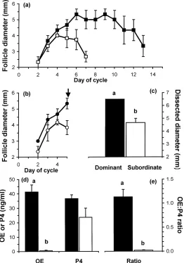
As expected, the dominant follicle of the first follicle wave of the first complete oestrous cycle was detected for a longer period (11.3±2.2 days;P <0.05) than the largest subor-dinate follicle (5.3±0.3 days; Fig. 1a). After ovariectomy on day 5 of the second oestrous cycle, the dominant follicle had a larger (P < 0.01) dissected diameter than the largest
Fig. 2. Immunoblotting signal intensity (top panel) and Western blot analyses of Akt and Erk 1/Erk 2 in follicle wall samples from dominant (Dom, solid bars) and subordinate (Sub, open bars) follicles on day 5 of the oestrous cycle in three cyclic ewe lambs. Columns with no common superscript are different (xy,P <0.05; ab,P <0.01).
subordinate follicle (Fig. 1c). Oestradiol and the ratio of oestradiol to progesterone concen-trations in follicular fluid were greater (P <0.01) in the dominant compared to the largest subordinate follicles (Fig. 1d and e) indicating the good and poor health of the dominant and subordinate follicles, respectively.
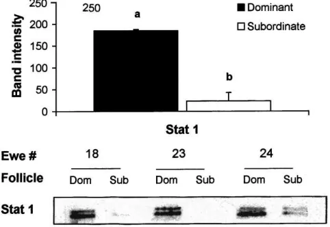
There was more Akt (P < 0.05) and Erk 1/Erk 2 (P < 0.01) present in follicle wall samples from the dominant compared to the largest subordinate follicles (Fig. 2) and the phosphorylated forms of Akt and Erk 1/Erk 2 were detected in samples from dominant follicles (Fig. 3). Phosphorylated forms of Akt and Erk 1/Erk 2 were not detected in samples from subordinate follicles (data not shown). More Stat 1 was detected in follicle wall samples from the dominant compared to the largest subordinate follicles (P <0.01; Fig. 4).
Fig. 4. Immunoblotting signal intensity (top panel) and Western blot analyses of Stat 1 in follicle wall samples of dominant (Dom) and subordinate (Sub) follicles on day 5 of the oestrous cycle in three cyclic ewe lambs. Columns with no common superscript are different (ab,P <0.01).
4. Discussion
The presence of Akt and Erk 1/Erk 2 in dominant or subordinate follicles of a follicular wave has not been described previously. In dominant follicles, the signalling pathways that include Akt and Erk 1/Erk 2 are active as shown by the presence of the native protein (Fig. 2) and the activated phosphorylated forms (Fig. 3). In addition, the high concentration of oestradiol in follicular fluid (Fig. 1) and the abundant presence of Stat 1 in follicle wall samples (Fig. 4) indicates that on day 5 of the cycle the dominant follicles were healthy (Jolly et al., 1997) and transcriptionally active. In contrast, the smaller subordinate follicles had a very low ratio of oestradiol to progesterone in follicular fluid (Fig. 1), indicating an advanced atretic state, and much reduced levels of Akt, Erk 1/Erk 2 and Stat 1 compared to the dominant follicle (Figs. 2 and 4). We suggest that the mechanisms that regulate the continued development of dominant and regression of subordinate follicles are mediated by signal transduction pathways that utilise the Akt and Erk 1/Erk 2 pathways. It remains to be established if these pathways play a role in the process of dominant follicle selection earlier in the wave.
Insulin and IGFs have been shown to activate Akt in a wide variety of cells in vitro, including porcine granulosa cells (Westfall et al., 1999). The regulation of IGF availabil-ity within follicles is regulated by IGF binding proteins (IGFBPs) and it has been shown that IGFBP concentrations are lower in healthy dominant follicles compared to regress-ing subordinate follicles in cattle (Echternkamp et al., 1994; Armstrong et al., 1998) and sheep (Besnard et al., 1996). Hence, differences between follicle types for levels of Akt (Fig. 2) may be an eventual consequence of differing levels of stimulation by growth factors.
While the weight of literature would suggest that FSH acts via Erk 1/Erk 2 and that insulin/IGFs act through Akt (Price and Silva, 1999), it is likely that there is crossover and that other pathways are involved. IGF-I activates Erk 1/Erk 2 in rat adipocytes (Porras et al., 1998) and Chinese hamster ovary cells (Sellers, 1999) in vitro and FSH stimulates the activation of p38 MAPK in rat granulosa cells in vitro (Maizels et al., 1998). Consequently, elucidation of the exact intracellular pathways by which FSH and IGFs control follicle growth and development remain to be established.
The characteristics of dominant compared to subordinate follicles are that they continue to grow and to produce large quantities of oestradiol (Roche et al., 1998). It is widely assumed that the insulin/IGF system controls growth and development and that FSH controls aromatase production. While this may largely be the case there is some crossover as FSH can stimulate the development of granulosa cells (Robker and Richards, 1998) and insulin and IGF-I can stimulate oestradiol production by ovine granulosa cells in vitro (Campbell et al., 1996). The interaction of these systems is further complicated as each system seems to augment the actions of the other by regulating binding protein production (Armstrong et al., 1998) and receptor expression (Zhou et al., 1997). To completely understand the role of these hormones in controlling development and oestradiol production we must fully understand the intracellular signalling pathways within cells. To date, research has concentrated on the relationships among follicle development and endocrine and local factors. However, how these factors regulate intracellular events within the ovary and the biochemical links between hormonal changes and apoptosis and oestradiol production need to be understood. The present data demonstrate the presence of AKT and Erk 1/Erk 2 pathways in dominant and subordinate follicles and provide a basis for further studies on the hormonal regulation of follicle development.
5. Conclusions
continued development of dominant follicles in sheep and that these proteins may play a role during follicle selection; however, this has yet to be determined.
Acknowledgements
We thank P. Duffy and M.P. Boland for assistance with the animals; N. Hynes for carry-ing out the radioimmunoassays, and D. Finlay and V. Healy for advice and assistance on immunoblotting. This work was partially funded by a UCD President’s Research Award to AE.
References
Alessi, D.R., Downes, C.P., 1998. The role of PI 3-kinase in insulin action. Biochim. Biophys. Acta 1436, 151–164. Armstrong, D.G., Baxter, G., Gutierrez, C.G., Hogg, C.O., Glazyrin, A.L., Campbell, B.K., Bramley, T.A., Webb, R., 1998. Insulin-like growth factor binding protein-2 and -4 messenger ribonucleic acid expression in bovine ovarian follicles: effect of gonadotropins and developmental status. Endocrinology 139, 2146–2154. Besnard, N., Pisselet, C., Monniaux, D., Locatelli, A., Benne, F., Gasser, F., Hatey, F., Monget, P., 1996. Expression
of messenger ribonucleic acids of insulin-like growth factor binding proteins-2, -4 and -5 in the ovine ovary: localization and changes during growth and atresia of antral follicles. Biol. Reprod. 55, 1356–1367. Blumer, K.J., Johnson, G.L., 1994. Diversity in function and regulation of MAP kinase pathways. Trends Biochem.
Sci. 19, 236–240.
Brunet, A., Bonni, A., Zigmond, M.J., Lin, M.Z., Juo, P., Hu, L.S., Anderson, M.J., Arden, K.C., Blenis, J., Greenberg, M.E., 1999. Akt promotes cell survival by phosphorylating and inhibiting a Forkhead transcription factor. Cell 96, 857–868.
Cameron, M.R., Foster, J.S., Bukovsky, A., Wimalasena, J., 1996. Activation of mitogen-activated protein kinases by gonadotropins and cyclic adenosine 5′-monophosphates in porcine granulosa cells. Biol. Reprod. 55, 111–
119.
Campana, W.M., Darin, S.J., O’Brien, J.S., 1999. Phosphatidylinositol 3-kinase and akt protein kinase mediate IGF-I- and prosaptide-induced survival in schwann cells. J. Neurosci. Res. 57, 332–341.
Campbell, B.K., Scaramuzzi, R.J., Webb, R., 1995. Control of antral follicle development and selection in sheep and cattle. J. Reprod. Fertil. Suppl. 49, 335–350.
Campbell, B.K., Scaramuzzi, R.J., Webb, R., 1996. Induction and maintenance of oestradiol and immunoreactive inhibin production with FSH by ovine granulosa cells cultured in serum-free media. J. Reprod. Fertil. 106, 7–16.
Cardone, M.H., Roy, N., Stennicke, H.R., Salvesen, G.S., Franke, T.F., Stanbridge, E., Frisch, S., Reed, J.C., 1998. Regulation of cell death protease caspase-9 by phosphorylation. Science 282, 1318–1321.
Carson, R.S., Findlay, J.K., Burger, H.G., Trounson, A.O., 1979. Gonadotropin receptors of the ovine ovarian follicle during follicular growth and atresia. Biol. Reprod. 21, 75–87.
Cobb, M.H., Goldsmith, E.J., 1995. How MAP kinases are regulated. J. Biol. Chem. 270, 14843–14846. Coffer, P.J., Jin, J., Woodgett, J.R., 1998. Protein kinase B (c-Akt): a multifunctional mediator of
phosphatidylinositol 3-kinase activation. Biochem. J. 335, 1–13.
Das, S., Maizels, E.T., DeManno, D., St. Clair, E., Adam, S.A., Hunzicker-Dunn, M., 1996. A stimulatory role of cyclic adenosine 3′,5′-monophosphate in follicle-stimulating hormone-activated mitogen-activated protein
kinase signaling pathway in rat ovarian granulosa cells. Endocrinology 137, 967–974.
Datta, S.R., Dudek, H., Tao, X., Masters, S., Fu, H., Gotoh, Y., Greenberg, M.E., 1997. Akt phosphorylation of BAD couples survival signals to the cell-intrinsic death machinery. Cell 91, 231–241.
Echternkamp, S.E., Howard, H.J., Roberts, A.J., Grizzle, J., Wise, T., 1994. Relationships among concentrations of steroids, insulin-like growth factor-I, and insulin-like growth factor binding proteins in ovarian follicular fluid of beef cattle. Biol. Reprod. 51, 971–981.
Evans, A.C.O., Duffy, P., Hynes, N., Boland, M.P., 2000. Waves of follicle development during the estrous cycle in sheep. Theriogenology 53, 699–715.
Evans, A.C.O., Fortune, J.E., 1997. Selection of the dominant follicle in cattle occurs in the absence of differences in the expression of messenger ribonucleic acid for gonadotropin receptors. Endocrinology 138, 2963–2971. Graves, L.M., Guy, H.I., Kozlowski, P., Huang, M., Lazarowski, E., Pope, R.M., Collins, M.A., Dahlstrand, E.N.,
Earp III, H.S., Evans, D.R., 2000. Regulation of carbamoyl phosphate synthetase by MAP kinase. Nature 403, 328–332.
Hansson, V., Skalhegg, B.S., Tasken, K., 1999. Cyclic-AMP-dependent protein kinase (PKA) in testicular cells. Cell specific expression, differential regulation and targeting of subunits of PKA. J. Steroid Biochem. Mol. Biol. 69, 367–378.
Henderson, K.M., Kieboom, L.E., McNatty, K.P., Lun, S., Heath, D., 1985. Gonadotropin-stimulated cyclic AMP production by granulosa cells from Booroola and Romney ewes with and without a fecundity gene. J. Reprod. Fertil. 75, 111–120.
Hill, C.S., Treisman, R., 1995. Transcriptional regulation by extracellular signals: mechanisms and specificity. Cell 80, 199–211.
Jolly, P.D., Tisdall, D.J., De’ath, G., Heath, D.A., Lun, S., Hudson, N.L., McNatty, K.P., 1997. Granulosa cell apoptosis, aromatase activity, cyclic adenosine 3′,5′-monophosphate response to gonadotropins, and follicular
fluid steroid levels during spontaneous and induced follicular atresia in ewes. Biol. Reprod. 56, 830–836. Jolly, P.D., Tisdall, D.J., Heath, D.A., Lun, S., McNatty, K.P., 1994. Apoptosis in bovine granulosa cells in
relation to steroid synthesis, cyclic adenosine 3′,5′-monophosphate response to follicle-stimulating hormone
and luteinizing hormone, and follicular atresia. Biol. Reprod. 51, 934–944.
Khokhlatchev, A.V., Canagarajah, B., Wilsbacher, J., Robinson, M., Atkinson, M., Goldsmith, E., Cobb, M.H., 1998. Phosphorylation of the MAP kinase ERK2 promotes its homodimerization and nuclear translocation. Cell 93, 605–615.
Kops, G.J., de Ruiter, N.D., De Vries-Smits, A.M., Powell, D.R., Bos, J.L., Burgering, B.M.T., 1999. Direct control of the Forkhead transcription factor AFX by protein kinase B. Nature 398, 630–634.
Kulik, G., Weber, M.J., 1998. Akt-dependent and -independent survival signaling pathways utilized by insulin-like growth factor I. Mol. Cell. Biol. 18, 6711–6718.
Leyendecker, G., Waibeltreber, S., Wildt, L., 1990. The central control of follicular maturation and ovulation in the human. Oxf. Rev. Reprod. Biol. 12, 93–146.
Maizels, E.T., Cottom, J., Jones, J.C., Hunzicker-Dunn, M., 1998. Follicle stimulating hormone (FSH) activates the p38 mitogen-activated protein kinase pathway, inducing small heat shock protein phosphorylation and cell rounding in immature rat ovarian granulosa cells. Endocrinology 139, 3353–3356.
Mehmet, H., 2000. Caspases find a new place to hide. Nature 403, 29–30.
Pircher, T.J., Petersen, H., Gustafsson, J.A., Haldosen, L.A., 1999. Extracellular signal-regulated kinase (ERK) interacts with signal transducer and activator of transcription (STAT) 5a. Mol. Endocrinol. 13, 555–565. Porras, A., Alvarez, A.M., Valladares, A., Benito, M., 1998. p42/p44 mitogen-activated protein kinases activation
is required for the insulin-like growth factor-I/insulin induced proliferation, but inhibits differentiation, in rat fetal brown adipocytes. Mol. Endocrinol. 12, 825–834.
Prendiville, D.J., Enright, W.J., Crowe, M.A., Finnerty, M., Hynes, N., Roche, J.F., 1995. Immunization of heifers against gonadotropin-releasing hormone: antibody titers, ovarian function, body growth, and carcass characteristics. J. Anim. Sci. 73, 2382–2389.
Price, C.A., Silva J.M., 1999. Intracellular regulation of P450 aromatase by FSH & insulin in bovine granulosa cells. J. Reprod. Fertil. Abstr. Ser. 23 (Abstract #5).
Richards, J.S., 1995. Ovarian cell differentiation: a cascade of multiple hormones, cellular signals, and regulated genes. Recent Prog. Horm. Res. 50, 223–254.
Robker, R.L., Richards, J.S., 1998. Hormone-induced proliferation and differentiation of granulosa cells: a coordinated balance of the cell cycle regulators cyclin D2 and p27Kip1. Mol. Endocrinol. 12, 924–940. Roche, J.F., Mihm M., Diskin M.G., Ireland J.J., 1998. A review of regulation of follicle growth in cattle. J. Anim.
Sellers, L.A., 1999. Prolonged activation of extracellular signal-regulated kinase by a protein kinase C-dependent and N17Ras-insensitive mechanism mediates the proliferative response of G(i/o)-coupled somatostatin sst(4) receptors. J. Biol. Chem. 274, 24280–24288.
Takata, M., Ogawa, W., Kitamura, T., Hino, Y., Kuroda, S., Kotani, K., Klip, A., Gingras, A.C., Sonenberg, N., Kasuga, M., 1999. Requirement for Akt (Protein kinase B) in insulin-induced activation of glycogen synthase and phosphorylation of 4E-BP1 (PHAS-1). J. Biol. Chem. 274, 20611–20618.
Van Wezel, I.L., Dharmarajan, A.M., Lavranos, T.C., Rodgers, R.J., 1999. Evidence for alternative pathways of granulosa cell death in healthy and slightly atretic bovine antral follicles. Endocrinology 140, 2602–2612. Webb, R., Godsen, R.G., Telfer, E.E., Moor, R.M., 1999. Factors affecting folliculogenesis in ruminants. Anim.
Sci. 68, 257–284.
Webb, R., Gong, J.G., Law, A.S., Rusbridge, S.M., 1992. Control of ovarian function in cattle. J. Reprod. Fertil. Suppl. 45, 141–156.
Westfall, S.D., Obholz K.L., Davis J.S., 1999. Follicle-stimulating hormone (FSH) enhances insulin-like growth factor (IGF-I) survival signalling in porcine granulosa cells. Biol. Reprod. 60 (Suppl. 1) (Abstract 173). Zhou, J., Kumar, T.R., Matzuk, M.M., Bondy, C., 1997. Insulin-like growth factor I regulates gonadotropin