ANTICANCER ACTIVITY OF MANGOSTEEN PERICARP DRY
EXTRACT AGAINST MCF-7 BREAST CANCER CELL LINE
THROUGH ESTROGEN RECEPTOR -
α
Agustina Setiawati1, Florentinus Octa Dika Riswanto1, Sri Hartati Yuliani1, Enade Perdana Istyastono2,
Breast cancer has very complex morphological and molecular characteristic. Estrogen receptor is one of biomarker in breast cancer progression, more than 60% breast cancer overexpress estrogen receptor α (ERα). Xanthone in Garcinia mangostana was investigated whether to have anticancer activity on colorectal, prostate, lung, blood and breast cancer. This research was focused on molecular mechanism of anticancer activity of mangosteen pericarp extract (MPE) on ER-α. This study used MCF-7 cells as a model of ER-α overexpressed breast cancer cells. Cytotoxic study towards MCF-7 cells was designed by using MTT test, further apoptotic induction assay was determined by double staining method using acridine orange and ethidium bromide. Extract molecular mechanism against breast cancer was assayed by immunocytochemistry. The MTT data was analyze using probit analysis to get IC50 then apoptosis and immunocytochemistry data were analysis qualitative analysis. MPE had strong cytotoxic activity on MCF-7 cells with IC50 of 45μg/mL and morphological changes passed through apoptosis induction. The expression of ER-α in MPE treated cells was same as untreated cells. MPE did not suppress ER-α in both nucleus and cytoplasm. Anticancer activity of MPE misht be mediated by other gene involved in ER-α signaling pathway in breast cancer cells.
Key words: Breast cancer, Garcinia mangostana peel extract, estrogen
receptor-α
INTRODUCTION
The most commonly diagnosed types of cancer among women in 2012 was breast cancer and breast cancer is expected to account for 29% (226,870) of all new cancer cases cancer in Indonesia (Idamardi, 2007).
Breast cancer has very complex morphological and molecular characteristic. One important marker of breast cancer for
stop the progression of breast cancer.
Mangosteen (Garcinia mangostana Linn)
pericarp contains various phytochemicals, primary xanthones. Xanthone isolated from
mangosteen pericarps were: α-mangostin, β
-mangostin, γ-mangostin, mangostinone and 2-isoprenyl-1,7 dihydroxy-3-methoxy xanthone inhibited human leukimia HL60 cell line.
Among them, α-mangostin suppresed proliferation and induced apotosis of HL60
cells at 10μM (Matsumoto et al., 2003).
Xanthone extract (81% α-mangostin and γ
mangostin) suppressed HT116 colon cancer
cell (Aisha et al., 2012; Nabandith et al., 2004;
Nakagawa et al., 2007). Xanthone: α-mangostin,
β-mangostin, γ-mangostin had anticancer
activity on DLD-1 (Akao et al., 2008). Alpha
mangostin had antiproliferative activity against SKBR3 human breast adenocarcinoma cell line
(Moongkarndi et al., 2004) and MDA-MB231
(Kurose et al., 2012). This compound were also
metastasis in xenograft model in Balb/c mice
(Shibata et al., 2011). Garcinia mangostana Linn
pericarp extract has potential property to be chemopreventive agent against breast cancer.
This study investigated Garcinia
mangostana pericarp extract (MPE) activity
against breast cancer. Furthermore, it focused
on ERα as molecular target to suppress
proliferation of MCF-7 cell, in vivo model of ER-α over expressed breast cancer cell.
MATERIAL AND METHODS
MPE was purchased from PT.
Borobudur had 28.10% α-mangosteen. Fructus
cortex of Garcinia mangostana Linn was extracted
use 70% ethanol with ratio botanical extract 10:1 and used maltodextrine as its excipient. MCF-7 breast cancer cell line was cultured in Parasitology laboratory, Faculty of Medicine, Gadjah Mada University. Apoptosis staining using ethidium bromide and acridine orange from SIGMA. This research used US BIO
estrogen receptor α primary antibody.
MTT Cytotoxic Assay
MCF-7 cells breast cancer cell line were subcultured until confluent. Approximately
5x10³ cells were seeded into 96-well microplate
and incubated at 37°C and 5% CO2 for 24h.
Medium was fride treatment control, were prepared from Tamofen® tablet. MPE and tamoxifen were dissolved in DMSO as stock solutions. Various concentration of MPE and tamoxifen in medium were added into 96-well
plate 100μL each and incubated in the same as
previous condition for 24h. Each concentration was assayed in triplicates (n=3). The medium culture was removed and rinsed by PBS 10%,
one hundreds micro Liter I 100μL contain
5mg/mL MTT was added into each well and incubated feather for 4h. Event, medium
containing MTT was removed and 100μL SDS
was added each well to dissolve the optical density of the resulting solution. The 96-well microplate was incubated for 24h and avoid from light contact. Formazan crystals were detected by ELISA reader using 595nm wavelength.
Apoptosis Assay
MCF-7 were seeded 5x10⁴/100μL into
coverslip in 24-well plate, then adapted in 37°C
and 5% CO2 for 24h. Further, the medium was
removed and rinsed by PBS. Seventeen and
34µg/mL of MPE in Dulbecco’s Modified
Eayle’s Medium were added into the well plate and incubated for 24h. These concentrations
were determined fariure calculation IC50, as
estimated concentration closed to IC50. The
medium was removed from MCF-7 cells and rinsed by PBS. The coverslip was removed from well plate and transferred to object glass, and acridine orange-ethidium bromide (AE) was dropped at the coverslip. This slide was soon observed under flouresence microscope.
Immunocytostaining of ER-α
MCF-7 cells in DMEM was seeded 105/100μL on 6-well plate and incubated under
5% CO2 for 24h. MPE in concentration of 17
incubated at least for an hour and washed three time using PBS. Secondary antibody of biotinylated goat anti-polyvalent was added and incubated at room temperature for 10min and washed four times in PBS. DAB as chromogen the mistune was dropped and incubated for and 3-8min, washed using aquadest. Finally, hematoxylin solution was added and incubated for 3-4 minutes. This slides were dried and observed under light microscope. Expression
of ER-α was showed by brown cell colour.
Analysis
The cytotoxic data of MPE and
tamoxifen were analyzed by excel. IC50 was
then calculated using probit analysis (Finney and Stevens, Any new references?
RESULT AND DISCUSSION
This study investigated another cytotoxic
properties of Garcinia mangostana pericarp
against breast cancer cell line. MCF-7 was used
as in vivo model of over expressed ER-α cells.
MPE and tamoxifen were dissolved into excess
DMSO as inert solvent in cell culture. Cavalli et
al. (2011) and Licciardi et al. (2010) showed
cytotoxic activity against MCF-7 cells. This research used 0.02% v/v DMSO for MPE and oxifen. MTT was absorbed into living cells and converted into purple coloured-formazan complex by succinate dehydrogenase in
mitochondria (Doyle and Grifith, 2000). MPE
exhibit cytotoxic parabolic profile (Figure 1),
probit analysis yielded MPE IC50 of45µg/mL.
The urage of tamoxifen as positive
control is a estrogen receptor α partial agonis,
was regarded as first line drug in ER-α over
expressed breast cancer treatment (Ao et al.,
2011; Fowler et al., 2004). Tamoxifen, was also
tested against MCF-7 cells by the same procedure. Tamoxifen had parabolic cytotoxic
profile and had IC50 47µM or equal to 12,65.10
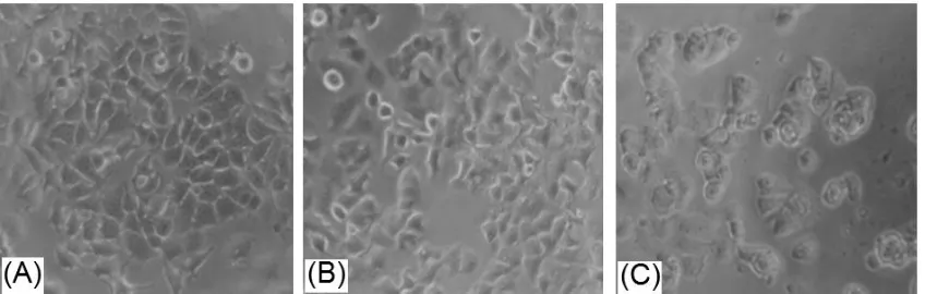
-7µg/mL by probit analysis. Tamoxifen had
strong cytotoxic activity making MCF-7 cells broken into debris but MPE (Figure 2). This
could be explained that tamoxifen is pure chemical compound having spesific target on the cells. MPE also drived morphological changes in MCF-7 cells even it did not make the cells broken into debris. MPE contained various natural compounds so it did not have
spesific target compared to THAT
OFtamoxifen.
Assay then must be designed in other study to confirm molecular mechanism of MCF-7 cells. This study used double staining method using acridine orange and ethidium bromide. Viable cells were green and apoptotic cells were red. MPE showed apoptosis
induction towards MCF-7 cells were
dose-dependent.
This research used apoptotic cells 17 and 34µg/mL MPE to investigate apoptotic staining. MCF-7 cells were orange and those Figure 1. Effect of MPE on MCF-7 viability after 24 hour incubation
membrane integrity was disturbed (Figure 3). MPE has lower cytotoxic and apoptosis induction activity compare to that of tamoxifen, it might be due to complexity of MPE natural compound. These novel findings underline the benefit of in vitro study to
elucidate molecular mechanisms of
chemopreventive agent.
The molecular mechanism of apoptosis
induction of MPE on ER-α was confirmed
using immunocytochemistry method.
Tamoxifen, a Selective Estrogen Receptor
Modulators (SERMs), suppress ER-α
expression. It down regulated ER-α by
changing conformation of ER-α thus AF-2
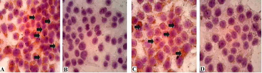
domain is hide so AF-2 dependent co-activator hardly bind to the receptor (Brufsky, 2011). Untreated cells showed high expression of
ER-α both in nucleus and cytoplasm. ER-α is located in nucleus and cytoplasm near cell
membrane in the absense and presence of
estrogen (Cheskis et al., 2007). MCF-7 cells
treated with 34µg/mL made cells being fragmented into debris there fore this study MPE with concentratum of 17µg/ml, was used
is detect ER-α expression. Even MPE had
strong cytotoxic and apoptosis induction
activity, it hardly suppressed ER-α expression.
Is inhibited ER-α signaling pathway and might
have other protein targets on MCF-7. There
were 83 genes involved in ER-α signaling
pathway in positive ER-α cell lines (Chisamore
et al, 2009). Therefore, other genes involved in
ER- α signaling pathway must be investigated.
ACKNOWLEDGEMENT
This work was funding by Internal Research Grant, Center for Research and Public Services Sanata Dharma University, Yogyakarta
Figure 3. Apoptotic cells observation using double staining method under flourencence
microscope using 400x magnification.(A)Untreated cells, (B)Cells treated 17µg/mL MPE (Cpoint
C: MPE concentration should be "cells treated 34ug/mL MPE" (as described on the text) not 'cells
treated 13ug/mL'; (D) Cells treated 12,65.10-7µg/mL tamoxifen living cells; apoptotic cells.
Figure 4. The observation of ER-α expression using immunocytochemistry method under light
microscope using 400x magnification.(A)Untreated cells, (B) Untreated cells without antibody (C)
REFERENCES
Ao A., Morrison BJ., Wang H., Lopez A.,
Reynolds BA., Lu J., 2011, Response of
Estrogen Receptor-Positive Breat Cancer Tumorspheres to Antiestrogen Treatments, 6(4): e18810.
Aisha AF., Abu-Salah KM., Ismail Z., Majid AMSA. 2012. In vivo and in vivo
anti-colon cancer effect of Garcinia
mangostana xanthones extract. BMC
Complementary and Alternative Medicine, 12: 104- 113
Akao Y., Nakagawa Y., Linuma M., Nozawa Y. 2008. Anti-Cancer Effects of Xanthones
from pericarps of Mangosteen, Int. J.
Mol. Sci. 9, 355 – 70.
Brufsky. 2011. Undestanding the estrogen receptor signaling pathway: focus on current endocrine agent for breast cancer
in postmenopausal women., Community
Oncology, 8(8): 343- 352.
Cavalli A., Bisazza R., Bussano, 2011, Poly(amidoamine)-cholesterol conjugate
nanoparticles obtained by
electrospraying as novel tamoxifen
delivery system, Journal of Drug Delivery,
Article ID 587604, 9 pages.
Center for Disease Control and Prevention
(CDC). 2009. Breast Cancer, available
online at http://www.cdc.gov/cancer/ breast/, accesed on March 3, 2014. Cheskis BJ., Greger JG., NagpalS., Freedman
LP. 2007. Signaling by estrogens. J.Cell
Physiol. 213: 610-617.
Chisamore MJ., Wilkisonm HA., Flores O.,
Chen JD. 2009. Estrogen-related receptor-α
antagonist inhibits both estrogen receptor-positive and estrogen receptor-negative breast tumor growth in mouse xenograft, 8: 672- 681.
Doyle A. and Griffiths JB. 2000. Cell and Tissue
Culture for Medical Research, John Willey & Sons LTD, England.
Finney DJ., Stevens WL. 1948. A Table for Calculation of Working Probits and
Weights in Probit Analysis, Biometrika,
35(1-2): 191- 201.
Flowler AM., Solodin N., Mara T., Preisler M., Zhang P., Lee AV, Alarid ET. 2004.
Increases in estrogen receptor-α
concentration in breast cancer cells
promote serine
118/104/106-independent AF-1 transactivation and
growth in the absence of estrogen,
FASEB Journal, 18: 81-93
Hanstein B., Djahansouzi S., Dall P., Beckmann
MW., Bender HG. 2004. Insight into
molecular biology of the estrogen receptor define novel therapeutic targets for breast cancer, 150: 243- 255.
Giacinti L., Claudio PP., Lopez M., Giordano A. 2006. Epigenetic Information and Estrogen Receptor Alpha Exoression in
Breast Cancer. The Oncologist. 11(1): 1-8
Idamardi, 2007. Kanker Payudara, Yayasan
Kanker Payudara Indonesia, Jakarta. Kurose H., Shibata MA., Iinuma M., Otsuki Y.
2012. Alteration in Cell Cycle and Induction of Apoptotic Cell Death in
Breast Cancer Cells Treated with α -Mangostin Extracted in Mangosteen
Pericarp, J Biomee and Biotec. 1-9
Licciardi G., Cavallaro M., Di Stefano G., Pitarresi, C., Fiorica G., Giammona.
2010. New self-assembling
polyaspartylhydrazide copolymer
micelles for anticancer drug delivery, Int
Journal Pharm, 396(1-2):219–228.
Matsumoto K., Akao Y., Kobayashi E., Ohguchi K., Ito T., Tanaka T., Linuma M., Nozawa Y. 2003. Induction of
Apoptosis by Xanthones from
Mangosteen in Human Leukimia Cell mangostana (mangosteen) on SKBR3
human breast cancer cell line.
J.Ethnopharmacol..90(1): 161-6.
Nabandith V., Suzui M., Kaneshiro T., Kinjo T., Matsumoto K., Akao Y., Linuma M., Yoshimi N., Inhibitory Effect of Crude Alpha-Mangostin. 2004. a Xanthone derivative, on two different categories of colon preneoplastic lesion induced by
1,2-dimethylhidrazine in rat. Asian Pac J
Cancer Prev. 5(4): 433-8.
Nakagawa Y. Linuma M., Nozawa Y., Akao Y.
increased miRNA-143 expression in human colorectal cancer DLD-1 cells.
Bioorg.Med. Chem. 15: 5620- 5628.
Siegel R., Naishadham D., Jemal A. Cancer
Statistics. 2012. CA Cancer J. Clin. 2012.
62:10 – 29.
Shibata MA., Linuma M., Morimoto J., Kurose H., Akamatsu A., Okuno Y., Akao Y.,
Otsuki. 2011. Α-mangostin extracted from the pericarp of the mangosteen (Garcinia mangostana Linn) reduces
tumor growth and lymph node
metastasis in an immunocompetent xenograft model of metastatic mammary
cancer carrying a p53 mutation, BMC
Medicine, (9): 69.
Ueda J., Tezuka Y., Baskota AH., Tran QL., Tran QK., Hariyama Y., Saiki I., Kadota S., 2002. Antiproliferative Activity of
Vietnamese Medicinal Plants. Biol.Pharm.