Role of the macrophage galactose lectin in the uptake of
desialylated LDL
Anna L. Bartlett
a,1, Thomas Grewal
b,1, Elena De Angelis
c, Simon Myers
d,
Keith K. Stanley
a,*
aCentre for Immunology,Uni6ersity of NSW and St Vincent’s Hospital,Darlinghurst,NSW2010,Sydney, Australia bUni6ersity Hospital Eppendorf,1.Medical Clinic,Martinistr.52,D-20246Hamburg,Germany
cUni6ersity of Cambridge Department of Medicine and Cambridge Institute for Medical Research,Addenbrooke’s Hospital,Hills Road,
Cambridge CB2 2XY, UK
dHeart Research Institute,145Missenden Road,Camperdown,NSW2050Sydney,Australia
Received 7 July 1999; received in revised form 13 December 1999; accepted 14 January 2000
Abstract
Desialylated low density lipoprotein (LDL) is rapidly taken up and accumulated by both peripheral blood monocytes and cells isolated from human arterial intima consisting predominantly of smooth muscle cells. It is shown that thioglycollate (TG)-elicited mouse macrophages and mouse peritoneal macrophages stimulated with lipopolysaccharide (LPS) show increased expression of a membrane-bound, galactose-specific lectin that could be responsible for this uptake. In LPS-stimulated macrophages accumula-tion of desialylated LDL is increased ca. 2.6-fold. Accumulaaccumula-tion of acetylated LDL in the same cells is reduced, suggesting that the galactose-specific lectin might be responsible for the uptake of desialylated LDL. Transfection of cells with the mouse macrophage Gal/GalNAc-specific lectin (MMGL) increased their capacity to take up asialofetuin (ASF) and, to a smaller extent, desialylated LDL. The uptake of desialylated LDL was small, most likely due to the high kd of MMGL for biantennary
oligosaccharides as found on LDL, and low concentration of LDL achieved in tissue culture experiments. The data suggest that the expression of galactose-specific lectins can be elevated under inflammatory conditions, and that these receptors could contribute to foam cell formation under conditions of high desialylated LDL concentration, as might be found in arterial intima. © 2000 Elsevier Science Ireland Ltd. All rights reserved.
Keywords:Desialylated LDL; Lectin; Lipopolysaccharide; MMGL; Scavenger receptor
www.elsevier.com/locate/atherosclerosis
1. Introduction
Fatty streak formation in the arteries of patients suffering from atherosclerosis has often been attributed
to the expression of scavenger receptors in macrophage foam cells leading to the unregulated uptake of modified lipoproteins from the blood [1,2]. Acetylation or oxidation of low density lipoprotein (LDL) in vitro effectively converts LDL into a ligand for the scavenger receptor, but it is not known if oxidation is the primary cause of LDL modification in vivo.
An alternative modification of LDL that might lead to atherosclerosis is desialylation [3]. Apolipoprotein (apo) B-100 isolated from native LDL of normolipi-demics was found to be only partially sialylated with approximately half of the N-linked lactosamine oligosaccharides having one exposed Gal residue [4]. Patients with coronary heart disease (CHD) have been shown to have lower levels of sialic acid on LDL apo B [5] which could therefore contain biantennary
oligosac-Abbre6iations: AcLDL, acetylated low density lipoprotein; ASF, asialofetuin; ASGPR, asialoglycoprotein receptor; FACE, fluores-cence assisted carbohydrate electrophoresis; HBSS, Hanks’ buffered salt solution; LDL, low density lipoprotein; LDLR, LDL receptor; LPDS, lipoprotein deficient serum; LPS, lipopolysaccharide; MMGL, mouse macrophage Gal/GalNAc-specific lectin; NT-LDL, neu-raminidase-treated LDL; PAGE, polyacrylamide gel electrophoresis; PBS, phosphate buffered saline; PI, periodate; PMA, phorbol 12-myristate 13-acetate; TCA, trichloroacetic acid; TG, thioglycollate. * Corresponding author. Tel.: +612-93612833; fax: + 612-93612391.
E-mail address:[email protected] (K.K. Stanley). 1Both authors contributed equally to this publication.
charides terminating in Gal residues — making them a high affinity ligand for lectin receptors [6]. LDL lipids are also found to be desialylated in CHD patients [5], leading to a lower overall sialic acid content of LDL particles in patient groups in several studies [7,8]. Previ-ous publications have suggested that uptake of desialy-lated LDL by macrophages could be via scavenger receptors [9], although our data, based on competition with asialofetuin (ASF), argued for uptake by a Gal-specific lectin receptor [10]. The uptake of desialylated LDL has now been examined in macrophages stimu-lated with different agents. Lipopolysaccharide (LPS)-stimulated macrophages, which have down-regulated scavenger receptors, were found to take up desialylated LDL more avidly than resting macrophages Furthermore, we show that LPS-stimulated macrophages express increased amounts of a galactose/ galactosamine-specific lectin (Gal/GalNAc-specific lectin). These data argue for a lectin-mediated uptake of desialylated LDL that is independent of the scav-enger receptor. In cells transfected with the mouse macrophage Gal/GalNAc specific lectin (MMGL) an increase in the uptake of ASF, and to a smaller extent, desialylated LDL, could be demonstrated. Thus it is possible that the human orthologue of MMGL might be responsible for the uptake of desialylated LDL by macrophages in atheromatous plaque.
2. Experimental procedures
2.1. Materials and methods
Neuraminidase attached to agarose beads, ASF and bovine serum albumin (BSA) were obtained from Sigma, bicinchoninic acid assay kit and sulfo-NHS-bi-otin were from Pierce. Antibody against apo B-100 (monoclonal), goat-anti-mouse Ig-rhodamine and re-striction enzymes were purchased from Boehringer Mannheim. Anti-FLAG M2 monoclonal antibody was from KODAK. Secondary antibodies, linked to horseradish peroxidase (HRP), Rainbow molecular weight markers (full range), streptavidin – fluorescein, PD-10 columns (Sephadex G-25 M) and ECL kit and film were from Amersham Pharmacia Biotech. Invitro-gen TA cloning kit (pCR 2.1 vector) was obtained from Bresatec, SA. For DNA extractions QIAquick Gel Extraction kit from QIAGEN was used. Lipofec-tAMINE reagent (GibcoBRL) was purchased from Life Technologies, Geneticin (G418) and trichloroacetic acid (TCA) were from Calbiochem. Oligonucleotides for polymerase chain reaction (PCR) and DNA sequencing were obtained from Bresatec. The Glyco fluorescence assisted carbohydrate electrophoresis (FACE) kit was obtained from Gradipore.
2.2. Preparation of LDL and modified proteins
Blood was drawn from healthy, fasting normolipi-demic individuals having plasma cholesterol levels be-tween 4.2 and 5.09 mmol/l. The blood was added immediately to a concentrated protease inhibitor solu-tion containing, at final dilusolu-tion, 90 U aprotinin/ml, 20 mg soybean trypsin inhibitor/ml and 3 mM EDTA. The blood cells were removed by centrifugation at 400×g for 30 min. LDL was then prepared by the rapid procedure of Sattler et al. [11], which involves a single KBr gradient centrifugation in a Beckman TL-100 cen-trifuge. The LDL was ready in about 3 h after collect-ing the blood. Small molecules were removed from the LDL prior to use by gel filtration on PD-10 columns and elution with nitrogen-purged phosphate buffered saline (PBS).
LDL was desialylated in vitro with neuraminidase attached to agarose beads as described by Camejo et al. [12]. More than 85% of the apo B-associated sialic acid was removed in this process. For binding, uptake and degradation by MMGL-transfected cells, LDL was de-sialylated for 4 h only to prevent any LDL precipita-tion. Analysis of apo B-100 oligosaccharides after neuraminidase treatment by FACE was described pre-viously [13].
LDL was acetylated as described previously [10,14] using 6 ml acetic anhydride/mg LDL protein. [125I]Iodination of neuraminidase-treated LDL (NT-LDL) and ASF was carried out with Na125I (2 – 5 mCi/mg protein) by the iodine monochloride method [15]. Iodinated proteins (specific activity 0.1 – 1.1×10⁶ and 1.6 – 2.1×10⁶ cpm/pmol protein for NT-LDL and ASF, respectively) were separated from unreacted iodine using PD-10 column followed by dialysis for 24 h against PBS.
Biotinylation of proteins was carried out in 50 mM NaHCO3buffer using sulfo-NHS-biotin in 6-fold molar excess for 1 h at room temperature. The unreacted biotin was removed with PD-10 column followed by dialysis for 24 h against PBS.
2.3. Preparation and use of macrophages
Resident peritoneal macrophages were isolated from QS mice as described [16]. For binding studies 0.5 – 1× 10⁶ mouse peritoneal macrophages were incubated at 4°C for 4 h with 10 mg/ml [125I]NT-LDL in DMEM containing 1% BSA. The medium was aspirated and the unbound material was removed with five washes in cold PBS. Cells were then scraped off in 200ml of cold PBS and the radioactivity determined. Protein content was determined by the bicinchoninic acid assay [17].
Degradation of [125
I]NT-LDL was determined by incubation of 1×10⁶
of 5 – 10mg protein/ml. A total of 100 ml of media was removed and 3 M TCA was added to a final concentra-tion of 10%. The sample was vortexed vigorously and incubated for 10 min on ice. After addition of 250ml of 0.7 M AgNO3to remove free iodine [18] the sample was centrifuged at 2000 rpm and the amount of radioactiv-ity in the remaining supernatant was determined.
2.4. Biosensor assay
Approximately 27 ng/mm2
of ASF was coupled to the surface of a carboxy-methyl dextran biosensor cu-vette using 1-ethyl-3-(3-dimethylaminopropyl) carbodi-imide and N-hydroxysuccinimide according to the manufacturers instructions (Fisons). After quenching and washing in PBS containing 0.05% Tween 20 the binding of solubilised membrane protein was deter-mined in a Fisons IAsys biosensor at 25°C. A total membrane fraction was prepared from the control, elicited or stimulated macrophages by homogenisation of approximately 1×10⁸ cells [19] and centrifugation for 1 h at 100 000×g in a Beckman TL-100 ultracen-trifuge. Each membrane pellet was then resuspended in 100ml of 1% NP-40 containing 20 mM HEPES pH 6.8, 100 mM KCl, 0.1 mM Pefabloc (Boehringer Mannheim), 1 mM benzamidine and 5mg/ml aprotinin at 4°C (NP-40 buffer). For each assay the cuvette was first equilibrated in 1% NP-40 buffer until a steady baseline was achieved. A total of 50 ml of solubilised membranes were then added (approx. equivalent to 107 cells). After 4 min the membranes were washed out of the cuvette with 1% NP-40 buffer and the new baseline determined.
2.5. Lipid loading of macrophages
For lipid-loading experiments native (LDL), acety-lated (AcLDL), or desialyacety-lated-LDL (NT-LDL) was added to the cells at a final concentration of 50mg/ml in Hanks’ buffered salt solution (HBSS) containing 10% lipoprotein deficient serum (LPDS). The cells were then incubated for 24 h at 37°C. Control cells were prepared under the same conditions without addition of LDL. At the end of this time cholesterol and cholesterol esters were extracted with hexane/methanol as described [20]. HPLC analysis was by a reversed-phase C-18 column (Supelco, 25×0.46 cm, 5 mm particle size). Cholesterol and cholesterol esters were detected at 210 nm after elution with acetonitrile:isopropanol (30:70 v/v) at a flow rate of 1 ml/min.
2.6. Western blot
A peptide corresponding in sequence to the car-boxyterminal 9 amino acid of MMGL (CEMK-LAKES) [21] was coupled to diphtheria toxin and used
to immunise rabbits by multiple intradermal injections. Serum was prepared from blood taken 10 days after the second monthly injection of 300 mg of peptide.
Membranes from various macrophage preparations, or total cell protein was solubilised in SDS gel sample buffer and a portion (approx. 30 and 100 mg, respec-tively) loaded on a 10 or 12.5% polyacrylamide gel. Transfer onto nitrocellulose and Western blotting was as previously described [22] using 1:1000 antiserum followed by anti-rabbit Ig-HRP (1:10 000) and en-hanced chemiluminescence (ECL) detection (Amer-sham). For the detection of MMGL-FLAG, the KODAK M2 monoclonal anti-FLAG antibody (1:2000), followed by anti-mouse Ig-HRP (1:10 000) and ECL detection were used.
2.7. Cloning MMGL
The cDNA encoding MMGL was obtained from RAW 264.7, a mouse macrophages-like cell line, by PCR using the following oligonucleotides: 5%MMGL:
5%-GCG TGT CGA CAT GAT ATA CGA AAA CCT
CCA GAA C-3% and 3%FLAG MMGL: 5%-GCG AAG
CTT CTA CTA CTT GTC ATC GTC GTC CTT GTA GTC GCT CTC CTT GGC CAG CTT C-3%.
These oligonucleotides amplified the complete open reading frame of MMGL [21] and introduced a se-quence encoding the FLAG epitope at the 3% end and flanking Sal I/Hind III restriction enzyme sites. The PCR product was first cloned into pCR 2.1 vector (Invitrogen) using the overhanging A residues intro-duced by Taq polymerase. Sequencing revealed a single mutation: 853 TC that resulted in a change in the amino acid sequence: ValAla. This mutation occurs in the carbohydrate recognition domain (CRD), but this amino acid is not preserved in other gal-specific lectin receptors [23]. It is unlikely therefore that this conservative replacement would have any functional significance. The DNA fragment encoding MMGL (945 bp) was cut out from the PCR 2.1 vector with restric-tion enzymes (SalI/Hind III), and cloned into pAXne-oRX vector.
2.8. Transfecting ECV 304 cells and
immunofluorescence analyses
expressing cells, then checked by immunolabelling for the cell surface presence of the MMGL-FLAG receptor and active internalisation. The MMGL expression in the transfected cells did not decrease even after ten passages, or freezing and thawing.
For all immunofluorescence analyses, cells were cul-tured directly on glass coverslips and grown for 2 – 3 days. For anti-FLAG antibody binding, cells were incu-bated with the antibody for 1 h at 4°C then fixed with methanol; for internalisation experiments, after incuba-tion at 4°C for 1 h, cells were placed in fresh media and warmed up to 37°C for 1 h prior to fixing in methanol. To confirm the lectin’s ability to bind and internalise ASF, cells were incubated with biotinylated ASF and anti-FLAG antibody for 1 h at 37°C, in the presence of leupeptin or chloroquine to prevent lysosomal degradation. Incubation with the secondary antibodies, goat-anti-mouse Ig-rhodamine and/or streptavidin – fluorescein, was carried out for 30 min at 37°C.
In some cases cells were fixed in 3% paraformalde-hyde90.2% Triton X-100, then incubated with anti-FLAG antibody followed by goat-anti-mouse Ig-rhodamine to look at the steady state distribution of the lectin receptor.
2.9. Binding, uptake and degradation of125I-labelled
proteins
ASF and NT-LDL were iodinated using the ICl method [15]. Transfected and untransfected cells were passaged into 6-well plates and grown for 3 days in media containing 10% foetal calf serum (FCS) before the experiments; in some cases cells were pre-in-cubated with LDL (0.1 mg/ml) 24 h prior to the experiment to downregulate the LDL receptor (LDLR).
For surface binding after equilibrating at 4°C, the cells were incubated with [125I]ASF or [125I]NT-LDL9 competitors (either 100×excess of ASF or 30 – 40 ×ex-cess of LDL) in 1 ml serum-free media for 2 h at 4°C. Total cell-associated counts were measured in cells in-cubated with [125I]ASF or [125I]NT-LDL9competitors for 2 h at 37°C. Medium was then removed by aspira-tion, and the wells were rinsed 5× with 2 ml PBS. The cells were dissolved in 1 ml 0.2 M NaOH and the bound radioactivity was determined with a COBRA auto-gamma counter.
The rate of degradation was measured as the increase in TCA-soluble radioactivity accumulating in medium after 18 h incubating with [125I]ASF or [125I]NT-LDL9 competitors at 37°C under a 5% CO2. To 0.9 ml of medium 100mg of BSA and 0.1 ml of 100% TCA were added and after at least 10 min on ice, the precipitates were centrifuged at 10 000 rpm for 10 min, then the amount of radioactivity was measured in the superna-tant fluid.
Protein determination in all experiments was done on cells rinsed 5× with 2 ml PBS and dissolved in 1 ml 0.2 M NaOH using bicinchoninic acid assay and BSA standard curve. In the case where in each individual experiment the protein concentrations between wells were insignificantly different for given cell line, average of 18 – 36 (all in triplicates) protein concentrations were used in calculations of cpm/mg cell protein.
3. Results
3.1. Membrane-bound lectins are expressed in
TG-elicited and LPS-stimulated macrophages
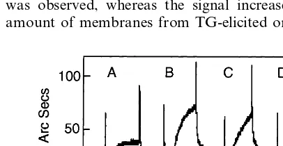
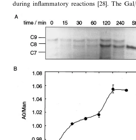
Expression of membrane bound Gal-specific lectins in mouse macrophages stimulated with different agents was tested by measuring the binding of solubilised macrophage membrane proteins to ASF in a biosensor cuvette. A total membrane fraction was prepared from equal numbers of control, elicited or stimulated macrophages and dissolved in 200 ml of 1% NP-40 buffer. When 50ml of these preparations were added to the biosensor cuvette, significant binding could only be detected when the membranes were derived from TG-elicited macrophages or from macrophages stimulated with LPS. This was evident from the steeply sloped binding curve, and by the inability to wash off the bound material in PBS – Tween buffer (Fig. 1). In con-trast, when solubilised membranes from control or periodate (PI)-elicited macrophages were used, the binding isotherm rapidly plateaued, and the signal re-turned to baseline on washing the cuvette. Even after addition of 150 ml of control membranes no binding was observed, whereas the signal increased when the amount of membranes from TG-elicited or
LPS-stimu-Fig. 1. Induction of lectins in macrophage membranes. Mouse peri-toneal macrophages (A) untreated, (B) treated with 1 mg/ml
lated macrophages added to the cuvette was increased (data not shown). Similar results were obtained in two to three different experiments using different biosensor cuvettes.
3.2. Induction of Gal/GalNAc-specific lectin in
TG-elicited and LPS-stimulated macrophages
Membrane fractions from control, TG-elicited, PI-elicited and LPS-activated macrophages were im-munoblotted with a polyclonal antibody raised against the carboxy terminal nine amino acids of the MMGL [21]. This antibody detected several bands in whole cell lysates from control RAW 264.7 cells which express the protein, but only one band at ca. 39 kDa in purified plasma membrane protein fraction. A band, migrating with the same mobility, was detected in the membranes of TG-elicited macrophages and LPS-stimulated macrophages but not in untreated peritoneal macrophages or PI-elicited macrophages (data not shown), consistent with the lack of Gal-specific binding in these cells (Fig. 1).
3.3. Lectins expressed in TG-elicited macrophages are
not surface located
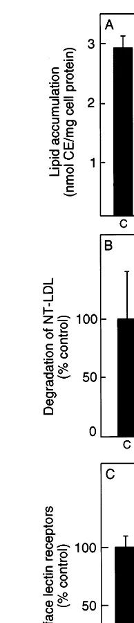
Previous studies have shown that desialylated LDL degradation by macrophages is competed by ASF sug-gesting that the macrophage Gal-specific lectin may be responsible for the uptake of desialylated LDL [10]. Since TG-elicited macrophages exhibited elevated levels of membrane associated lectins in the biosensor assay we tested the ability of these cells to accumulate desia-lylated LDL by measuring the accumulation of choles-terol esters by reversed phase HPLC (Fig. 2A). Surprisingly it was found that TG-elicited macrophages demonstrated less accumulation than control cells. To determine if this was caused by a decreased rate of uptake of the NT-LDL cells were incubated with 125
I-labelled NT-LDL and the release into the medium of TCA soluble radioactivity was measured (Fig. 2B). The TG-elicited macrophages were considerably less able to degrade 125I-labelled NT-LDL, suggesting that the in-duced lectin receptors might be intracellularly located rather than on the cell surface. This hypothesis was confirmed by measuring the amount of binding of 125I-labelled ASF to the cell surface (Fig. 2C). This
experiment indicates that fewer Gal-specific lectins were present on the surface of the TG-elicited macrophages despite the increased lectin associated with total mem-brane extracts of these cells.
3.4. LPS gi6es increased uptake of NT-LDL with
decreased uptake of AcLDL
Thioglycollate (TG) can contain endotoxin therefore
Fig. 2. Decreased cell surface lectin expression in thioglycollate (TG)-elicited macrophages. (A) Cholesterol esters associated with mouse peritoneal and TG-elicited macrophages after 24 h incubation with 50mg/ml of neuraminidase-treated low density lipoprotein
(NT-LDL). (B) Trichloroacetic acid (TCA) soluble radioactivity released by 1×10⁶ mouse peritoneal macrophages or 1×10⁶ TG-elicited macrophages after incubation with 10mg/ml [125I]NT-LDL for 4 h at
37°C. (C) Binding of [125I]asialofetuin (ASF) for 4 h at 4°C to 1×10⁶ mouse peritoneal macrophages or TG-elicited macrophages. C= un-treated mouse peritoneal macrophage control, TG=thioglycollate elicited macrophages.
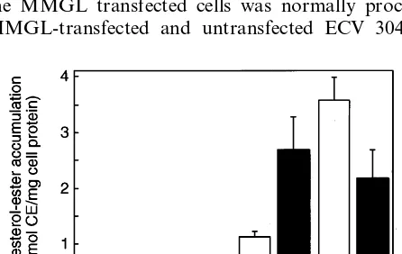
con-sistent with the increase in surface expression of lectin receptors detected by the biosensor assay (Fig. 1). In two separate experiments this stimulation of uptake was 2.6- and 6-fold. In contrast, AcLDL accumulation was decreased after LPS-stimulation as previously re-ported [24,25], presumably due to lowered scavenger receptor expression, and native LDL uptake was not changed by LPS-stimulation (Fig. 3).
3.5. Transfection of ECV304 cells with MMGL
Three lines of evidence suggest that MMGL might be involved in the uptake of desialylated LDL leading to atherosclerosis: (a) macrophages after inflammatory stimulation express a lectin which has been tentatively identified as MMGL; (b) these macrophages accumu-late desialyaccumu-lated, but not native LDL; and (c) MMGL is specifically induced in rat cardiac allografts with atherosclerosis [26]. To test this hypothesis, a cDNA fragment encoding the open reading frame of mouse MMGL from a macrophage-like cell line, RAW 264.7 was cloned and used to transfect ECV 304 cells, an immortalised cell line derived from human umbilical vein endothelial cells (HUVEC). The 3% oligonucleotide used to amplify the MMGL cDNA encoded the FLAG epitope, to provide a convenient tag for immunolocali-sation. As a result, the C-terminus of the receptor contained 8 extra amino acid residues. The tag was located in the extracellular domain, MMGL being a type II transmembrane glycoprotein. For transfection experiments ECV 304 cells were used, as these cells have low endogenous MMGL-like activity as measured by binding of [125
I]ASF (see below).
To verify that the FLAG-tagged protein expressed in the MMGL transfected cells was normally processed, MMGL-transfected and untransfected ECV 304 cells
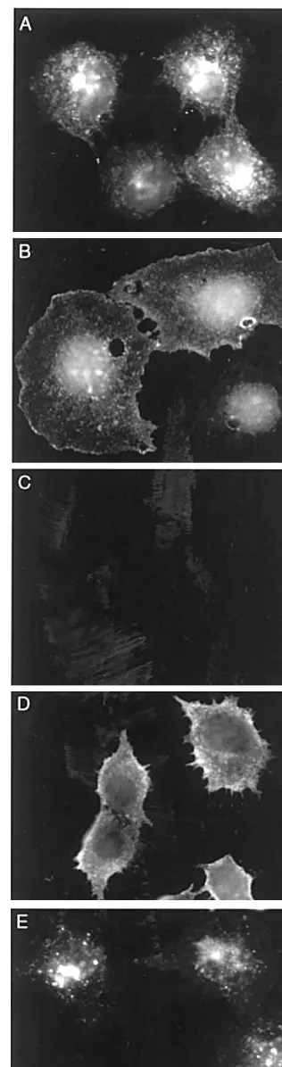
were solubilised in Laemmli sample buffer, separated the proteins by SDS-polyacrylamide gel electrophoresis (PAGE) (12.5%) and electrotransferred onto nitrocellu-lose. The recombinant MMGL-FLAG was identified using the monoclonal anti-FLAG antibody followed by anti-mouse Ig-HRP and compared to the MMGL band detected in RAW cells using an anti-peptide antiserum followed by anti-rabbit Ig-HRP. The size of the bands corresponding to the MMGL-FLAG and MMGL were approximately 39 kDa (data not shown). The control ECV cells did not show any bands with either antibody. The level of expression of MMGL in the cloned, transfected cell-lines was assessed by indirect immu-nofluorescence using a monoclonal FLAG anti-body. In Fig. 4A a strong surface and intracellular punctate staining was observed in cells fixed in formal-dehyde and permeabilised in 0.2% Triton X-100. When formaldehyde fixation in the absence of Triton was used, only the surface staining remained (Fig. 4B) confi-rming that the punctate staining arose from intracellu-lar vesicles. In order to verify that the surface receptor was capable of being internalised cells were incubated with anti-FLAG monoclonal at 4°C, washed, and then either fixed with methanol (Fig. 4D) or placed in fresh media and incubated at 37°C for 1 h, followed by fixation with methanol (Fig. 4E). Untransfected cells showed no fluorescence at comparable exposures (Fig. 4C). It can be seen that surface MMGL receptor tagged with monoclonal anti-FLAG antibody is capable of being internalised in the transfected cells.
3.6. Binding, association and degradation of [125I]ASF
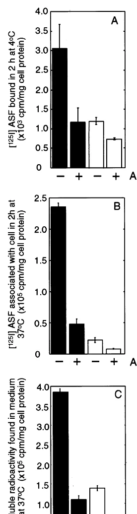
To determine if the MMGL receptor was functional in the transfected cells, the surface binding, uptake into endosomal compartments, and release of low molecular weight degraded [125I]ASF were measured. For surface binding, the cells were incubated with [125I]ASF for 2 h at 4°C (Fig. 5A). The transfected cells (solid bars, Fig. 5A) showed between 2.5-fold (P=0.04) (Fig. 5A, bars 1 and 3) and 3-fold (P=0.01) increase in surface binding in two separate experiments compared to un-transfected cells. Adding 100-fold excess unlabelled ASF decreased binding of [125I]ASF by 60% (P=0.06) (Fig. 5A, bars 1 and 2) and 30% (P=0.0002) in these experiments, respectively, reducing binding to levels similar to those seen in untransfected cells (Fig. 5A, bars 3 and 4).
The total cell associated counts were also measured after incubating cells with [125I]ASF for 2 h at 37°C, as an indication of the ability of cells to internalise ligand into endocytic compartments (Fig. 5B). After 2 h incu-bation with ligand, endosomal pathways are saturated but little lysosomal degradation has occurred. In trans-fected cells (solid bars, Fig. 5B), total cell associated radioactivity increased nearly 10-fold (PB0.0001) com-Fig. 3. Stimulated uptake of neuraminidase-treated low density
lipo-protein (NT-LDL) after lipopolysaccharide (LPS)-treatment. Choles-terol esters were measured in mouse peritoneal macrophages before (open bars) and after stimulation with 1mg/ml LPS (solid bars) using
Fig. 4. (Continued)
Degradation of [125I]ASF in transfected and untrans-fected cells was estimated by measuring the release of TCA soluble radioactivity into the medium after incu-bating cells with [125I]ASF for 18 h at 37°C (Fig. 5C). In medium incubated with transfected cells, the level of degradation products was increased 3-fold (PB0.0001) (Fig. 5C, bars 1 and 3) and the increase was abolished by competition with 100-fold excess of non-radioactive ASF (Fig. 5C, bar 2). Similar results were obtained in four separate experiments.
3.7. Binding and uptake of ASF by indirect
immunofluorescence
In Fig. 5 it can also be seen that competition with an excess of non-radioactive ASF caused a significant de-crease in the binding, uptake and degradation of [125I]ASF by untransfected ECV304 cells, suggesting that also some endogenous receptors for ASF are present on ECV cells. ECV304 cells were chosen for the transfection experiments because they have low levels of endogenous Gal lectin activity compared to other cells (e.g. CHO cells, data not shown). To confirm that the increased binding and internalisation of ASF in the transfected cells compared to the untransfected was due to the expression of functional MMGL, cells grown on coverslips were incubated with biotinylated ASF and anti-FLAG antibody for 1 h at 37°C in the presence of chloroquine to inhibit lysosomal degradation. The cells were then fixed with methanol, and labelled with goat-anti-mouse Ig-rhodamine and streptavidin – fluorescein, so the ligand and the antibody could be monitored in the same cells using different excitation and emission wavelengths. In Fig. 6 it can be seen that the cells taking up large quantities of biotinylated ASF (Fig. 6A), are the same cells that show high levels of staining for the lectin receptor (Fig. 6B).
Fig. 4. Immunofluorescence of positive transfected cells showing surface and/or internal staining. Cells were fixed in (A) 3% paraformaldehyde90.2% Triton X-100 and (B) in 3% paraformalde-hyde without Triton X-100, then incubated with anti-FLAG antibody followed by goat-anti-mouse Ig-rhodamine showing the intracellular and surface steady state distribution of the lectin, respectively; (C) labelling of control (untransfected) ECV 304 cells with goat-anti-mouse Ig-rhodamine after anti-FLAG antibody binding at 4°C for 1 h followed by methanol fixation; (D) labelling of positive transfected cells with goat-anti-mouse Ig-rhodamine after anti-FLAG antibody binding at 4°C for 1 h followed by methanol fixation; and (E) anti-FLAG antibody uptake at 4°C for 1 h followed by incubation at 37°C for 1 h in fresh medium, methanol fixation and labelling with goat-anti-mouse Ig-rhodamine as described in Section 2.
3.8. Binding, association and degradation of [125I]NT
-LDL
All LDL particles isolated from plasma are recovered in a partially sialylated state [13] however very few particles contain apo B-100 with biantennary oligosac-charides in their di-desialylated form [4]. The Gal-spe-cific lectins have 1000×lower kd for di-desialylated biantennary oligosaccharides (i.e. with two exposed Gal residues) than for mono-desialylated oligosaccharides (with only one exposed Gal) [6]. Therefore, to convert native LDL into a higher affinity ligand for MMGL, freshly isolated LDL was treated with neuraminidase bound to agarose beads for 4 h. This time was sufficient to remove most of the apo B-bound sialic acid and to generate di-desialylated oligosaccharides. Fig. 7 shows FACE analysis of neutral oligosaccharides isolated from LDL protein after treatment with neuraminidase for various times. The di-desialylated oligosaccharide was identified by its mobility compared to a glucose ladder and a standard A0 (Fig. 7A, last lane), generated by mild acid hydrolysis of mono- and di-sialylated oligosaccharides from apo B. The degree of desialyla-tion of LDL at each time point was estimated from the ratio of the intensities of the di-desialylated oligosac-charide band to one of the high mannose chains, Man8GlcNAc2 (Fig. 7B). It has been found that longer treatment with neuraminidase does not remove any additional sialic acid and is detrimental, as it causes LDL to aggregate and precipitate.
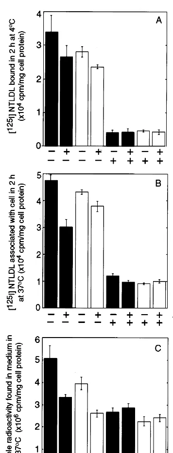
Purified NT-LDL was iodinated and incubated with the transfected and untransfected cells to determine the role of MMGL in the binding, uptake and degradation of desialylated LDL. Prior to the experiments, cells were grown for 24 h in medium containing 10% FCS supplemented with 0.1 mg/ml human LDL to ensure that the LDLR was fully downregulated.
Binding of [125I]NT-LDL at 4°C to MMGL-trans-fected cells (solid bars, Fig. 8) was increased 21% (Fig. 8A, bars 1 and 3) and 65% compared to untransfected cells in two separate experiments. Addition of 100-fold excess unlabelled ASF caused the binding of [125 I]NT-LDL in the transfected cells to decrease to levels similar to untransfected cells (Fig. 8A, bars 1 and 2), suggest-ing that the lectin receptor is responsible for some binding of [125I]NT-LDL. However, the majority of the binding was due to residual LDLRs as excess LDL reduced binding in all cells by 50 – 80%. The reason for the unexpectedly high level of LDLR binding might be its higher affinity at 4°C compared to 37°C [27].
Internalisation of NT-LDL into the endocytic path-way was measured as before by the association of ligand with cells after 2 h incubation at 37°C. In MMGL-transfected ECV cells a small increase in cell associated [125
I]NT-LDL was observed compared to untransfected cells (Fig. 8B, bars 1 and 3). Adding a Fig. 5. Binding, association and degradation of [125I]asialofetuin
Fig. 6. Immunofluorescence of transfected cells after incubation with biotinylated asialofetuin (ASF) and anti-FLAG antibody for 1 h at 37°C in the presence of chloroquine, followed by methanol fixation and incubation with goat-anti-mouse Ig-rhodamine and streptavidin – fluorescein. (A) Fluorescein channel showing accumulation of biotinylated ASF in lysosomes; (B) rhodamine channel showing cells with different levels of expression of mouse macrophage Gal/GalNAc-specific lectin (MMGL)-FLAG.
100-fold excess of unlabelled ASF, reduced cell associ-ated counts in transfected cells by 36 (Fig. 8B, bars 1 and 2) and 80% in two separate experiments indicating a role for MMGL rather than the LDLR in this internalisation. Addition of excess of unlabelled LDL substantially reduced the cell associated radioactivity (Fig. 8B, bars 1 and 5) indicating that over half of the cell associated radioactivity was due to uptake by LDLRs.
Degradation of [125I]NT-LDL in MMGL transfected cells, measured by TCA soluble release of radioactivity into the medium over 18 h, was increased by 30% (Fig. 8C, bars 1 and 3) and 15% in two separate experiments, compared to the untransfected cells (Fig. 8C, bars 1 and 3). This increase was abolished by competition with 100-fold excess of non-radioactive ASF, reducing it to levels similar to those observed using untransfected cells (Fig. 8C, bars 1 and 2). Addition of unlabelled LDL still caused a reduction in detected counts (30 – 50% in Fig. 8C, bars 5 – 8). The smaller effect of LDL in these longer-term experiments is presumably due to consump-tion of the LDL during the experiment.
Although a trend was observed in Fig. 8 suggesting a role for Gal-specific lectins in the internalisation of desialylated LDL, the P-values for increased binding, uptake and degradation of [125
I]NT-LDL in transfected compared to untransfected cells were not significant (P=0.07 toP=0.29).
4. Discussion
It has recently been shown that uptake of desialylated LDL by mouse peritoneal macrophages could be com-peted by ASF, suggesting the involvement of a
galactose-specific receptor [9]. Several lectins have been described in activated macrophages where they are thought to take part in the recruitment of macrophages during inflammatory reactions [28]. The
Fig. 8. Binding, association and degradation of [125 I]neuraminidase-treated LDL (NT-LDL) by mouse macrophage Gal/GalNAc-specific lectin (MMGL)-transfected (solid bars) and untransfected ECV304 cells (open bars). The cells were incubated with [125I]NT-LDL (107 cpm/well)9100-fold excess of unlabelled asialofetuin (ASF) and/or 30 – 40-fold excess of unlabelled LDL in 1 ml serum-free media for: (A) 2 h at 4°C; and (B) 2 h at 37°C; for surface binding and cell association, respectively. Medium was removed and the cells were rinsed 5 times with 2 ml phosphate buffered saline (PBS), dissolved in 1 ml 0.2 M NaOH and the radioactivity was determined with a gamma counter. (C) Degradation of [125I]NT-LDL was determined by incubating cells with [125I]NT-LDL9100-fold excess of unlabelled ASF and/or 30 – 40-fold excess of unlabelled LDL in 1 ml serum-free media for 18 h in a CO2 incubator at 37°C. trichloroacetic acid (TCA) soluble radioactivity was measured in the medium.
and by TG-elicitation of macrophages, but is not ex-pressed significantly in other tissues or in bone-marrow derived macrophages stimulated with a variety of cy-tokines [26].
It has been shown that functional Gal-binding recep-tors are induced in TG-elicited macrophages and by LPS-activation of mouse peritoneal macrophages. An anti-peptide antibody raised in rabbits was able to detect a 39 kDa protein after both TG-elicitation and LPS-activation, suggesting that the mouse macrophage Gal/GalNAc-specific lectin was indeed present, al-though the action of other lectins as well can not be ruled out. PI elicitation, which did not result in elevated lectin expression by the biosensor assay, did not give a cross-reacting band of this mobility.
In LPS-activated macrophages, but not in TG-elic-ited macrophages, we have demonstrated increased de-sialylated LDL uptake, presumably because the lectins induced in TG-elicited macrophages are not accessible at the cell surface. This would not be surprising in view of the large amount of phagocytosis occurring in TG-elicited cells and the similar reports of latent receptors by other groups [30,31]. The receptor responsible for internalisation of desialylated LDL was not the scav-enger receptor, since LPS-stimulation of macrophages caused an increase in cholesterol ester accumulation from desialylated LDL whilst inhibiting the uptake of acetylated LDL in the same cells. It has been previously shown that the scavenger receptor is down regulated under these conditions [24,25]. In addition, previous reports that desialylated LDL might be taken up by scavenger receptors [9] are unlikely to be true, as scav-enger receptors normally take up ligands with increased negative charge [32] whereas desialylated LDL has lost acidic sialic acid residues.
In a previous study [33] it was shown that high concentrations of LPS (\10 mg/ml) could cause an increased uptake of LDL, suggesting an increase in the expression of the LDLR. At a concentration of 1 mg/ml, however, no increase of LDL uptake was ob-served in agreement with our data (Fig. 2). Funk et al. [34] have reported an increase in LDL uptake at LPS concentrations as low as 1mg/ml, but the LDL was not characterised for degree of desialylation, and direct measurements of receptor concentration were not re-ported. In this study it was possible to measure an upregulation of the Gal/GalNAc-specific lectin after treatment with 1 mg/ml LPS concomitant with an in-crease in cholesterol-ester accumulation after incuba-tion of mouse peritoneal macrophages with NT-LDL. The lack of increase in LDL uptake makes it most probable that the increased uptake of desialylated LDL was via the lectin receptor.
Transfection of ECV cells with DNA encoding for MMGL resulted in a cell line expressing high levels of surface Gal/GalNAc-specific lectin. This lectin was specific lectin is a type II membrane spanning protein
shown to act as a receptor capable of binding cell surface ligand and delivering it to lysosomes by immu-nofluorescence detection of biotinylated ASF uptake, and by the increased binding, uptake and degradation of [125I]ASF in MMGL transfected cells compared to untransfected control. By using desialylated LDL as ligand a small, but non-significant metabolism of desia-lylated LDL by MMGL receptors that is independent of the LDLR has also been demonstrated.
Although the LDLR was downregulated by growing cells in high-serum media, it was estimated from the amount of LDL bound at 4°C that approximately 4 – 7000 LDLRs remain on the cell surface. A similar calculation using the 4°C binding of ASF suggests that approximately 100 000 MMGL are present per cell. Although these figures are both underestimates (since one has not extrapolated to infinite ligand concentra-tion using a Scatchard analysis) they give a measure of the relative numbers of these two receptors on the transfected cells. Despite the excess of MMGL recep-tors the LDLR is still able to compete for a substantial portion of desialylated LDL binding due to its low dissociation constant (2×10⁻⁹ M [27]). Binding of glycoproteins to the Gal-specific lectins depends strongly on the structure of the carbohydrate chains: mono-, bi-, tri- and tetraantennary Gal-terminal oligosaccharides bind with increasing affinities, with dissociation constants of 10−3, 10−6, 5
×10−9 and 10−9 M, respectively [6]. The
kd of MMGL for the bi-antennary di-desialylated oligosaccharides present in NT-LDL, therefore, would be ca. 10−6 M, over 500 times less than that of LDL for the LDLR.
In the experiments only 0.03 mM NT-LDL was added, of which approximately 85% (0.025 mM) con-tained di-desialylated biantennary oligosaccharides, thus the concentration of ligand for the MMGL would be much less than itskd. LDL in serum is present at a 64-fold higher concentration (ca. 1.6 mM [35]) and in atherosclerotic lesions, desialylated LDL may be spe-cifically retained through interactions with proteogly-cans and other matrix components [12]. Under these conditions the uptake by the Gal-specific lectin will be increased since it has a highkd, while that of the LDLR will remain constant. It has been shown that MMGL is drastically and specifically upregulated in rat cardiac allografts with atherosclerosis, suggesting that this re-ceptor might be a marker for chronic rejection [24]. The hypothesis was that this receptor might also account for lipid accumulation if it could bind to circulating desia-lylated LDL. The data demonstrate that relatively little desialylated LDL is taken up by a Gal-specific lectin at concentrations achievable in tissue culture experiments due to the low affinity of these receptors. It remains a possibility, however, that a human orthologue of MMGL might contribute to atherosclerosis in humans in the microenvironment of the arterial intima.
Acknowledgements
Thomas Grewal was a recipient of a post-doctoral scholarship from the Deutsche Forschungsgemeinschaft (DFG). Simon Myers is a recipient of an Australian Postgraduate Award and Australian Atherosclerosis Society Trust Fund scholarship. Anna L. Bartlett is supported by a grant from NH&MRC of Australia. This project was partially funded by a New South Wales Health Research and Development infrastructure grant.
References
[1] Goldstein JL, Ho YK, Basu SK, Brown MS. Binding site on macrophages that mediates uptake and degradation of acetylated low density lipoprotein, producing massive cholesterol deposi-tion. Proc Natl Acad Sci USA 1979;76:333 – 7.
[2] Fogelman AM, Van-Lenten BJ, Warden C, Haberland ME, Edwards P. Macrophage lipoprotein receptors. J Cell Sci Suppl 1988;9:135 – 49.
[3] Orekhov AN, Tertov VV, Mukhin DN. Desialylated low density lipoprotein — naturally occurring modified lipoprotein with atherogenic potency. Atherosclerosis 1991;86:153 – 61.
[4] Taniguchi T, Ishikawa Y, Tsunemitsu M, Fukuzaki H. The structures of the asparagine-linked sugar chains of human apolipoprotein B-100. Arch Biochem Biophys 1989;273:197 – 205. [5] Tertov VV, Orekhov AN, Sobenin IA, Morrisett JD, Gotto AM, Jr, Guevara JG, Jr. Carbohydrate composition of protein and lipid components in sialic acid-rich and -poor low density lipo-proteins from subjects with and without coronary artery disease. J Lipid Res 1993;34:365 – 75.
[6] Lee YC, Townsend RR, Hardy MR, Longren J, Arnarp J, Haraldsson M, Lonn H. Binding of synthetic oligosaccharides to the hepatic Gal/GalNAc lectin. Dependence on fine structural features. J Biol Chem 1983;258:199 – 202.
[7] Tertov VV, Sobenin IA, Gabbasov ZA, Popov EG, Jaakkola O, Solakivi T, Nikkari T, Smirnov VN, Orekhov AN. Multiple-modified desialylated low density lipoproteins that cause intra-cellular lipid accumulation. Isolation, fractionation and characterization. Lab Invest 1992;67:665 – 75.
[8] Ruelland A, Gallou G, Legras B, Paillard F, Cloarec L. LDL sialic acid content in patients with coronary artery disease. Clin Chim Acta 1993;221:127 – 33.
[9] Orekhov AN, Tertov VV, Sobenin IA, Smirnov VN, Via DP, Guevara J, Jr, Gotto AM, Jr, Morrisett JD. Sialic acid content of human low density lipoproteins affects their interaction with cell receptors and intracellular lipid accumulation. J Lipid Res 1992;33:805 – 17.
[10] Grewal T, Bartlett A, Burgess J, Packer N, Stanley KK. Desialy-lated LDL uptake in human and mouse macrophages can be mediated by a lectin receptor. Atherosclerosis 1996;121:151 – 63. [11] Sattler W, Mohr D, Stocker R. Rapid isolation of lipoproteins and assessment of their peroxidation by high-performance liquid chromatography postcolumn chemiluminescence. Methods Enzy-mol 1994;223:469 – 89.
[12] Camejo G, Lopez A, Lopez F, Quinones J. Interaction of low density lipoproteins with arterial proteoglycans. The role of charge and sialic acid content. Atherosclerosis 1985;55:93 – 105. [13] Bartlett AL, Stanley KK. All low density lipoprotein particles
[14] Basu SK, Goldstein JL, Anderson GW, Brown MS. Degradation of cationized low density lipoprotein and regulation of choles-terol metabolism in homozygous familial hypercholescholes-terolemia fibroblasts. Proc Natl Acad Sci USA 1976;73:3178 – 82. [15] Bilheimer DWS, Eisenberg S, Levy RI. The metabolism of very
low density lipoprotein proteins. I. Preliminary in vitro and in vivo observations. Biochim Biophys Acta 1972;260:212 – 21. [16] Jessup W, Mander EL, Dean RT. The intracellular storage and
turnover of apolipoprotein B of oxidized LDL in macrophages. Biochim Biophys Acta 1992;1126:167 – 77.
[17] Smith PK, Krohn RI, Hermanson GT, Mallia AK, Gartner FH, Provenzano MD, Fujimoto EK, Goeke NM, Olson BJ, Klenk DC. Measurement of protein using bicinchoninic acid. Anal Biochem 1985;150:76 – 85.
[18] Kim M-J, Dawes J, Jessup W. Transendothelial transport of modified low-density lipoproteins. Atherosclerosis 1994;108:5 – 17.
[19] Mander EL, Dean RT, Stanley KK, Jessup W. Apolipoprotein B of oxidized LDL accumulates in the lysosomes of macrophages. Biochim Biophys Acta 1995;1212:80 – 92.
[20] Kritharides L, Jessup W, Gifford J, Dean RT. A method for defining the stages of low-density lipoprotein oxidation by the separation of cholesterol- and cholesteryl ester-oxidation prod-ucts using HPLC. Anal Biochem 1993;213:79 – 89.
[21] Sato M, Kawakami K, Osawa T, Toyoshima S. Molecular cloning and expression of cDNA encoding a galactose/ N-acetyl-galactosamine-specific lectin on mouse tumoricidal macrophages. J Biochem 1992;111:331 – 6.
[22] Burnette WN. ‘Western blotting’: electrophoretic transfer of proteins from sodium dodecyl sulfate- — polyacrylamide gels to unmodified nitrocellulose and radiographic detection with anti-body and radioiodinated protein A. Anal Biochem 1981;112:195 – 203.
[23] Drickamer K, Hamon JF, Binns G, Leung JO. Primary structure of the rat liver asialoglycoprotein receptor. Structural evidence for multiple polypeptide species. J Biol Chem 1984;259:770 – 8. [24] Van Lenten BJ, Fogelman AM, Seager J, Ribi E, Haberland
ME, Edwards PA. Bacterial endotoxin selectively prevents the expression of scavenger-receptor activity on human monocyte-macrophages. J Immunol 1985;134:3718 – 21.
[25] Van Lenten BJ, Fogelman AM. Lipopolysaccharide-induced in-hibition of scavenger receptor expression in human monocyte-macrophages is mediated through tumor necrosis factor-alpha. J Immunol 1992;148:112 – 6.
[26] Russell ME, Utans U, Wallace AF, Liang P, Areci RJ, Karnovsky MJ, Wyner LR, Yamashita Y, Tarn C. Identification and upregulation of galactose/N-acetylgalactosamine macrophage lectin in rat cardiac allografts with arteriosclerosis. J Clin Invest 1994;94:722 – 30.
[27] Goldstein JL, Brown MS. The low-density lipoprotein pathway and its relation to atherosclerosis. Ann Rev Biochem 1977;46:897 – 930.
[28] Nagia-Makker P, Ochieng J, Christman JK, Raz A. Regulation of the expression of galactoside-binding lectin during human monocytic differentiation. Cancer Res 1993;53:5033 – 7. [29] Ii M, Kurata H, Itoh N, Yamashina I, Kawasaki T. Molecular
cloning and sequence analysis of cDNA encoding the macrophage lectin specific for galactose and N-acetylgalac-tosamine. J Biol Chem 1990;265:11295 – 8.
[30] Lau W, Devery JM, Geczy CL. A chemotactic S100 peptide enhances scavenger receptor and Mac-1 expression and cholesteryl ester accumulation in murine peritoneal macrophages in vivo. J Clin Invest 1995;95:1957 – 65.
[31] Skiba PJ, Keesler GA, Tabas I. Interferon-gamma down-regu-lates the lipoprotein(a)/apoprotein(a) receptor activity on macrophage foam cells. Evidence for disruption of ligand-in-duced receptor recycling by interferon-gamma. J Biol Chem 1994;269:23059 – 67.
[32] Haberland ME, Fogelman AM, Edwards PA. Specificity of receptor-mediated recognition of malondialdehyde-modified low density lipoproteins. Proc Natl Acad Sci USA 1982;79:1712 – 6. [33] Lopes-Virella MF, Klein RL, Stevenson HC. Low density
lipo-protein metabolism in human macrophages stimulated with mi-crobial or microbial-related products. Arteriosclerosis 1987;7:176 – 84.
[34] Funk JL, Feingold KR, Moser AH, Grunfeld C. Lipopolysac-charide stimulation of RAW 264.7 macrophages induces lipid accumulation and foam cell formation. Atherosclerosis 1993;98:67 – 82.
[35] Gotto AM, Pownall HJ, Havel RJ. Introduction to the plasma lipoproteins. In: Segrest JP, Albers JJ, editors. Methods in Enzymology, vol. 128. London: Academic Press, 1986.