Comparison of Helicobacter pylori Detection Using
Immunohistochemistry and Giemsa and Its Association
with Morphological Changes in Active Chronic Gastritis
Wildawati Nurdin*, Ening Krisnuhoni**, Kusmardi**
*Faculty of Medicine, Universitas Indonesia/Dr. Cipto Mangunkusumo General National Hospital, Jakarta **Division of Gastroentero-hepatology, Department of Patology Anatomy, Faculty of Medicine,
Universitas Indonesia/Dr. Cipto Mangunkusumo General National Hospital, Jakarta
Corresponding author:
Ening Krisnuhoni. Division of Gastroentero-hepatology, Department of Patology Anatomy, Dr. Cipto Mangunkusumo General National Hospital. Jl. Diponegoro No. 71 Jakarta Indonesia. Phone: +62-21-31930373; Facsimile: +62-21-3912477. E-mail: [email protected]
ABSTRACT
Background:*DVWULWLVLVDQLQÀDPPDWLRQRIWKHJDVWULFPXFRVDDVDUHVSRQVHWRLQIHFWLRQRULUULWDWLRQRI
the gaster. The most common aetiology of chronic gastritis is Helicobacter pylori (H. pylori) infection. Presence RI+S\ORULLVDVVRFLDWHGZLWKWKHRFFXUUHQFHRILQÀDPPDWLRQDWURSK\DQGLQWHVWLQDOPHWDSODVLD,QWHUPV of morphology, H. pylori is known in 2 forms, which are rod-shaped and coccoid-shaped. Coccoid-shaped EDFWHULDDUHGLI¿FXOWWRGHWHFWXVLQJ*LHPVDVWDLQLQJ7KHUHIRUHLPPXQRKLVWRFKHPLVWU\VWDLQLQJRI+S\ORUL and evaluation of the sensitivity of coccoid-shaped of H. pylori are needed.
Method:Cross-sectional study on 90 biopsy tissues of chronic gastritis patients in year 2015 and 2014, which included 30 Giemsa cases with positive H. pylori, 30 cases of active chronic gastritis with negative +S\ORULEXWFRFFRLGVKDSHGZDVIRXQGDQGQRQDFWLYHFKURQLFJDVWULWLVZHUHVXEVHTXHQWO\VWDLQHGZLWK immunohistochemistry staining of H. pylori.
Results: ([SUHVVLRQRIFRFFRLGVKDSHG+S\ORULLQDFWLYHFKURQLFJDVWULWLVZDVVLJQL¿FDQWO\GLIIHUHQWS
LQLPPXQRKLVWRFKHPLVWU\VWDLQLQJ7KHUHZDVDVLJQL¿FDQWGLIIHUHQFHEHWZHHQDFWLYHFKURQLFJDVWULWLV ZLWKSRVLWLYH+S\ORULDQGQHJDWLYH+S\ORULLQLPPXQRKLVWRFKHPLVWU\VWDLQLQJZLWKGHJUHHRILQÀDPPDWLRQ 6HQVLWLYLW\DQGVSHFL¿FLW\WHVWEHWZHHQ*LHPVDDQGLPPXQRKLVWRFKHPLVWU\VWDLQLQJVKRZHGVHQVLWLYLW\RI DQGVSHFL¿FLW\RI
Conclusion: Immunohistochemistry staining in active chronic gastritis was more sensitive compared to Giemsa staining in detecting H. pylori, particularly the coccoid-shaped bacteria.
Keywords: active chronic gastritis, H. pylori immunohistochemistry
ABSTRACT
Method: Studi potong lintang terhadap 90 jaringan biopsi pasien gastritis kronik pada tahun 2015 dan 2014 yang meliputi 30 kasus Giemsa dengan H. pylori positif, 30 kasus gastritis kronik aktif dengan H. pylori negatif tapi ditemukan bentuk coccoid, dan 30 kasus gastritis kronik non-aktif, kemudian dilakukan pewarnaan imunohistokimia H. pylori.
Results:(NVSUHVL+S\ORULEHQWXNFRFFRLGSDGDNURQLNDNWLIPHPLOLNLSHUEHGDDQ\DQJEHUPDNQDS
pada pulasan imunohistokimia. Terdapat perbedaan yang bermakna antara gastritis kronik aktif H. pylori positif GDQ+S\ORULQHJDWLISDGDSXODVDQLPXQRKLVWRNLPLDGHQJDQGHUDMDWLQÀDPDVL8MLVHQVLWLYLWDVGDQVSHVL¿VLWDV DQWDUDSHPHULNVDDQ*LHPVDGDQSXODVDQLPXQRKLVWRNLPLDKDVLOVHQVLWLYLWDVGDQVSHVL¿VLWDVQ\D
Conclusion: Pewarnaan imunohistokimia pada gastritis kronik aktif lebih sensitif dibandingkan dengan pewarnaan giemsa untuk mendeteksi H. pylori terutama jenis coccoid .
Keywords: gastritis kronik aktif, Giemsa, imunohistokimia H. pylori
INTRODUCTION
Gastritis is one of the most common digestive tract problems. Worldwide, the incidence of gastritis is 1.8 - 2.1 million, while in South East Asia, 583,635 per year. The incidence of gastritis in Indonesia is quite high, which is 247,396 cases from 238.452.952 population.1 *DVWULWLVLVDQLQÀDPPDWRU\FRQGLWLRQRIWKHJDVWULF PXFRVDDVDQLQÀDPPDWRU\UHVSRQVHWRZDUGVLQIHFWLRQ or irritation of the gaster.2 Generally, the cause of chronic gastritis is the infection of Helicobacter pylori (H. pylori), a gram-negative bacteria, presence of autoimmune disease and reaction towards chemical and drugs.3
Globally, the prevalence of H. pylori varies. Infection is more common to be found in the developing countries compared to developed countries. Infection may affect all ages, starting from childhood to adulthood.4H. pylori infection in developing countries can reach up to 25-30%, where 5-27% are found in early childhood and 50-60% are found in adults aged more than 60 years old.5 Based on the report from several studies, it was known that the prevalence of H. pylori infection in Indonesia varied. H. pylori infection in Dr. Mohammad Husein Palembang Hospital reached 46.7% in 2009, 24.3% in Tugurejo Semarang Hospital in year 2004-2010, 20.1% in Surakarta in year 1997.6,7,8 Meanwhile, in Jakarta based on serologic examination in 150 primary school children, the obtained prevalence was 27%.9
Along with the increased prevalence of H. pylori
infection, various methods have been developed to detect it, either using invasive or non-invasive methods. Some known non-invasive methods include urea breath test, nitrogen excretion test, blood immunoglobulin (Ig) G and IgA serologic examination, and faecal H. pylori antigen test. On the other hand, invasive methods include microbial culture test, urease examination in
biopsy tissue, histopathology, and polymerase chain reaction (PCR) of the biopsy tissue. Appropriate H. pylori diagnostic test is chosen based on the sensitivity DQGVSHFL¿FLW\RIWKHPHWKRGVEHLQJXVHGFRVWDQG equipment availability.10 Invasive detection of H.
pylori is performed through gastric biopsy. Gastric ELRSV\WKDWIXO¿OOHG6\GQH\V\VWHPFULWHULDLVELRSV\ from the antrum and corpus.11 H. pylori detection in gastric biopsy can be performed through Giemsa staining, immunohistochemistry staining, McMullen modification method, and silver staining method. Rotimi et al studied 63 gastric samples to detect H. pylori. Sensitivity of all the four methods being used were immunohistochemistry using H. pylori antibody (98.3%), McMullen modification (90%), Giemsa (86.7%), and silver staining(85%), respectively.12
Based on its morphology, H. pylori bacteria are known in two forms, rod-shaped and coccoid-shaped. Rod-shaped H. pylori has tendency to become coccoid-shaped in several environmental conditions, such as: oxygen exposure, base pH, starvation, long-term treatment, and inadequate proton pump inhibitor (PPI) or antibiotic administration.13,14Coccoid-shaped bacteria is hard to detect using Giemsa staining due to the difficulty in differentiating coccoid-shaped bacteria from artefact or other bacteria. Therefore, immunohistochemistry staining of H. pylori is needed. A different opinion from several researchers stated that the coccoid-shaped is a transformation form leading to degenerative state or death, while some others considered it as active and viable form.13,14
Coccoid-shaped H. pylori may also be caused by increased oxygen pressure and antibiotic administration. Coccoid-shaped H. pylori is form that cannot be cultured but is still alive and can be induced back to the virulent form (spiral). Coccoid-shaped H. pylori
some are responsible in the recurrence of infection after antimicrobial treatment; however, the pathogenesis of coccoid-shaped H. pylori is still unclear and has not been much studied.13,14 In the study performed by She et al, there were 3 strains of coccoid-shaped H. pylori
which changed from spiral-shaped due to exposure to metronidazole.14 In this study, we would like to know the correspondence between immunohistochemistry and Giemsa staining to detect H. pylori in chronic gastritis and to observe the morphological or histological difference of chronic gastritis with rod-shaped and coccoid-shaped H. pylori. Immunohistochemistry staining method becomes a consideration in increasing the sensitivity in the detection of H. pylori as it relies RQPRUHVSHFL¿FDQWLJHQDQGDQWLERG\ELQGLQJ
METHOD
This study used cross-section design, performed in Department of Anatomical Pathology Faculty of Medicine University of Indonesia/Dr. Cipto Mangunkusumo Hospital (FMUI/CMH) in November 2015 to January 2016. Accessible population of this study was active chronic gastritis cases which were diagnosed in Anatomical Pathology Department FMUI in year 2014-2015 with topographic code C15, C16, and morphologic code H544 in accordance with ,QWHUQDWLRQDO&ODVVL¿FDWLRQRI'LVHDVH,&' standard. Samples of active chronic gastritis with positive H. pylori in Giemsa staining and active chronic gastritis with negative H. pylori in Giemsa staining, but had coccoid-shaped were obtained through consecutive method. Samples of non-active chronic gastritis were obtained through simple random sampling. Estimation of the sample size counted with formula (paired categorical) was 51 cases.
Search and exploration of cases were performed in Anatomical Pathology Department FMUI/CMH in January 2015 to September 2015, and if results were not adequate, samples were further taken from the previous years. Anatomical pathology examination form and slides were collected; subsequently, re-evaluation towards active chronic gastritis H & E slides and Giemsa positivity were conducted. (YDOXDWLRQRILQÀDPPDWLRQDWURSK\DQGPHWDSODVLD were performed using visual analog scale. Later, XQVWDLQHGVOLGHVIURPSDUDI¿QEORFNZKLFKIXO¿OOHG the criteria were made and H. pylori (BC 7) antibody which was incubated for 1-2 hours with 1:50 dilution was examined.
Assessment of the results of H. pylori (BC 7 ) i m m u n o h i s t o c h e m i s t r y s t a i n i n g w a s performed by researcher using light microscope. Immunohistochemistry staining evaluation was based on the presence of H. pylori staining in gastric mucosa. Staining results evaluation was performed by researchers together. Statistical analysis was performed using Chi-square test and if criteria had not EHHQIXO¿OOHG)LVKHU¶VH[DFWWHVWZRXOGEHXVHGDVDQ alternative. These statistical tests were performed using SPSS 20 software.
RESULTS
Active chronic gastritis samples with Giemsa positive H. pylori and active chronic gastritis with Giemsa negative H. pylori but contained coccoid-shaped were obtained through consecutive technique. Non-active chronic gastritis samples were collected through simple random sampling in one year period, which was January to December 2015, in each studied group, and if results were not adequate, samples were further taken from the previous years. This study evaluated 3 categories, which consisted of 30 cases with positive H. pylori with Giemsa, 30 cases of active chronic gastritis, and 30 cases of non-active chronic gastritis. Patients’ age data distribution showed non-normal distribution, which was median of age 51.50 years old, the youngest age was 7 years old and the eldest was 86 years old, with the age range of 79 years old and mean age of 49.08 years old.
Table 1. Characteristic of samples
Table 2. Results of Gastric Biopsy Assessment
Absence of atrophy 6 (6.7) Mild atrophy 57 (63.3) Moderate atrophy 22 Severe atrophy 5 (5.6) Intestinal metaplasia
Absent
Present 6 (6.7)
Evaluation was performed to 60 cases of active chronic gastritis which were divided into 2 categories; ¿UVWFDVHVRIDFWLYHFKURQLFJDVWULWLVZLWKQHJDWLYH
H. pylori in Giemsa staining but had coccoid-shaped, in the immunohistochemistry staining positive H. pylori
was found in 16 cases (53.3%), but had coccoid-shaped
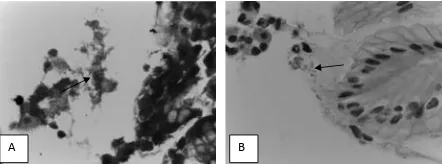
H. pylori morphology (Figure 1 and 2). While, 30 cases which initially showed positive H. pylori with Giemsa staining, the immunohistochemistry staining also revealed positive results (Figure 3 and 4).
In this study, we also performed staining to 30 biopsy samples of non-active chronic gastritis with results of 1 sample which Giemsa staining was positive
H. pylori, but the immunohistochemistry staining turned out to be negative H. pylori (Table 5).
Table 5. Staining Results in non-active chronic gastritis
Giemsa Immunohistochemistry
+ - +
-Non-active 0 30
A B
Figure 1. Evaluation of H. Pylori: (A) Giemsa staining, negative
H. Pylori; (B) Immunohistochemistry staining, positive coccoid-VKDSHG[PDJQL¿FDWLRQ
A B
Figure 2. Evaluation of H. Pylori: (A) Giemsa staining, negative
H. Pylori; (B) Immunohistochemistry staining, positive coccoid-VKDSHG[PDJQL¿FDWLRQ
A B
Figure 3. Evaluation of H. Pylori: (A) Positive H. pylori in Giemsa staining; (B) Positive H. pylori in immunohistochemistry VWDLQLQJ[PDJQL¿FDWLRQ
B A
Figure 4. Evaluation of H. Pylori: (A) Positive H. pylori in Giemsa staining; (B) Positive H. pylori in immunohistochemistry VWDLQLQJ[PDJQL¿FDWLRQ
Table 3. Evaluation Results of Active Chronic Gastritis Based on
,QÀDPPDWLRQ$WURSK\DQG0HWDSODVLDDQGH. pylori in Giemsa
and Immunohistochemistry Staining
Giemsa
Immunohisto-Table 4. Evaluation Results of Active Chronic Gastritis Based on
,QÀDPPDWLRQ$WURSK\DQG0HWDSODVLDDQG6KDSHRIH. pylori
DISCUSSION
Gastritis cases in this study were found in patients aged 7 years old to 86 years old with the peak incidence found in 51-60 years old age group with the mean age of 49 years old. This is in concordance with the literature which state that the incidence of chronic gastritis may happen in all age group from childhood to adulthood.4 The results of this study was not so different from the study conducted by Dhakwa et al which revealed that the average age of gastritis patients were 41.5 years old. This was also in agreement with the study performed by Kalebi et al which concluded that the mean age was 43 years old with variation of 18-86 years old.14,15 The incidence of gastritis is higher in female (57.8%) as compared to male. This was in accordance with the study done by Al Ammar et al which reported 58.19% female, and different from the study conducted by Capelle et al that found 55% chronic gastritis incidence were male.27,16
*DVWULWLVFDVHVZLWKPLOGLQÀDPPDWLRQZHUHIRXQG LQFDVHVPRGHUDWHLQÀDPPDWLRQLQFDVHV DQGVHYHUHLQÀDPPDWLRQLQFDVHV There was no atrophy in 6 cases (6.7%), mild atrophy in 57 (63.3%), moderate atrophy in 22 cases (24.4%), and severe atrophy in 5 cases (5.6%). Metaplasia was found only in 6 cases (6.7%). Hashemi et al found gastric histopathological appearance of normal mucosa in 8.7%, inactive chronic gastritis in 37.7%, active chronic gastritis in 47.1%, atrophy changes in 25%, and intestinal metaplasia in 8.9%. Zhang et al reported gastric histology appearance of patients with chronic gastritis (non-ulcer dyspepsia) H. pylori infection was IRXQGLQLQÀDPPDWLRQLQDWURSK\RIWKH mucosa in 36.8%, and intestinal metaplasia in 37.0%.17,18 Active chronic gastritis cases with negative H. pylori Giemsa staining, but with positive H. pylori
in immunohistochemistry staining were found in 53.3% cases with coccoid-shaped morphology. Study performed by Tajalli et al towards 54 samples found that the positivity of H. pylori with immunohistochemistry method was as many as 43 cases (79.63%), while the positivity with Giemsa method was as many as 24 cases (44.44%) and 18 (33.33%) with H & E staining. The results of this study revealed that classical method was not sensitive enough to identify H. pylori particularly the coccoid-shaped.43 Key et al detected 37% H. pylori with H & E staining, 55% with Giemsa staining, 62% with Warthin starry, 66% with immunohistochemistry and 45% were detected using PCR. Immunohistochemistry staining was positive in all cases where H. pylori was detected using other methods.19
Study performed by Orhan et al revealed that with immunohistochemistry method, low-density coccoid-shaped H. pylori could be observed easily. Positive H. pylori with immunohistochemistry staining were found in 3 from 10 cases of negative urea breath test (UBT). This study concluded that LPPXQRKLVWRFKHPLVWU\ VWDLQLQJ ZDV PRUH VSHFL¿F compared to Giemsa and UBT in detecting H. pylori
infection.20 Immunohistochemistry examination statistical test was performed towards the coccoid-shaped H. pylori REWDLQHGWKDWWKHUHZDVVLJQL¿FDQW
difference between the two (Appendix 1).
Results of statistical test showed that there was VLJQL¿FDQWGLIIHUHQFHSEHWZHHQH. pylori in active and non-active chronic gastritis with Giemsa staining. Statistical test was also performed towards
H. pylori in active and non-active chronic gastritis towards immunohistochemistry staining; fisher exact test was performed and found that there was VLJQL¿FDQWGLIIHUHQFHSEHWZHHQH. pylori
in active and non-active chronic gastritis towards immunohistochemistry staining (Appendix 2-3). Statistical test towards H. pylori in active chronic gastritis with immunohistochemistry staining with LQÀDPPDWLRQZDVSHUIRUPHGDQGIRXQGWKDWWKHUHZDV VLJQL¿FDQWGLIIHUHQFHSEHWZHHQDFWLYHFKURQLF gastritis with immunohistochemistry staining and LQÀDPPDWLRQ$SSHQGL[7KLVZDVLQDFFRUGDQFH with the literature that stated that the density of mononuclear cells and activation of polymorphonuclear cells in general were proportional with the density of
H. pylori.5,19,20,21,22 Study conducted by Yakoob et al on 176 cases of chronic gastritis concluded that there was VLJQL¿FDQWDVVRFLDWLRQS EHWZHHQLQIHFWLRQ and activity of H. pylori. Aggregated lymphoid was VLJQL¿FDQWO\DVVRFLDWHGZLWKDFWLYHFKURQLFJDVWULWLV23
Different result was found in atrophy and intestinal PHWDSODVLDZKHUHWKHUHZDVQRVLJQL¿FDQWGLIIHUHQFH (p < 0.05) between positive and negative H. pylori
7KHUHZDVQRVLJQL¿FDQWGLIIHUHQFHEHWZHHQWKH degree of inflammation in active chronic gastritis with rod-shaped and coccoid-shaped H. pylori in immunohistochemistry staining (p > 0.05)(Appendix 7); similar result was also found in atrophy and intestinal PHWDSODVLDZKHUHWKHUHZDVQRVLJQL¿FDQWGLIIHUHQFH between the degree of atrophy and the presence of intestinal metaplasia in chronic active gastritis with rod-shaped and coccoid-shaped H. pylori in immunohistochemistry staining (Appendix 8-9). Soylu et al studied H. pylori using immunohistochemistry method found that from positive H. pylori samples there was diffuse staining pattern, vaguely in 90.9%, with smooth granules on the surface in 90.9%, granule-like dot pattern in 54.5%, and spiral-shaped in 9.1%.24
(YDOXDWLRQRQWKHVHQVLWLYLW\DQGVSHFL¿FLW\EHWZHHQ Giemsa and immunohistochemistry staining had been performed; it was found that the sensitivity was 65% DQGWKHVSHFL¿FLW\ZDV7KLVZDVLQFRQFRUGDQFH with the study conducted by Monteiro et al who in their study of culture obtained sensitivity value of 93.8%, VSHFL¿FLW\WKHSRVLWLYHYDOXHRIH. pylori culture FRQ¿UPHGWKHSUHVHQFHRILQIHFWLRQEXWQHJDWLYHFXOWXUH did not exclude the suspicion of H. pylori.25 Study performed by Dogar et al compared haematoxylin eosin staining with immunohistochemistry staining in WKHLGHQWL¿FDWLRQRIH. pylori found 27.2% H. pylori
were detected using H & E staining, and 31.4% H. pylori were detected using immunohistochemistry staining. H & E sensitivity test was performed upon immunohistochemistry showed sensitivity value of DQG VSHFL¿FLW\ YDOXH RI 7KLV VWXG\ concluded histopathological examination from gastric ELRSV\ZDVVWLOODQDFFXUDWHDQGHI¿FLHQWPHWKRGWR diagnose H. pylori, immunohistochemistry staining might increase diagnostic results.26
Urease examination of biopsy tissue (campylobacter like organism test/CLO test), Monteiro et al obtained sensitivity value of 83%, specificity 96.4%.25 Histopathology method, Monteiro et al found VHQVLWLYLW\YDOXHRIVSHFL¿FLW\:KLOH with polymerase chain reaction (PCR) of the biopsy WLVVXHWKH\IRXQGVHQVLWLYLW\VSHFL¿FLW\25 Rotimi et al (year 2000) evaluated 63 gastric samples to detect H. pylori. Observation results using combination RI ¿YH WHVWV UDSLG ELRSV\ XUHDVH WHVW XUHD EUHDWK test, culture, serology, and histology) found that from interobserver consensus the best method from all the four methods were H. pylori antibody (98.3%), 0F0XOOHQPRGL¿FDWLRQ*LHPVDDQG silver staining(85%), respectively.12
T h i s s t u d y a l s o e v a l u a t e d G i e m s a a n d immunohistochemistry staining in non-active chronic gastritis from 30 samples with the result that 1 case with positive H. pylori in Giemsa staining but the immunohistochemistry staining showed negative H. pylori results; this is possible that the positive result in Giemsa staining was a false positive result.
CONCLUSION
Morphological appearance of active chronic gastritis in positive H. pylori and negative H. pylori
in immunohistochemistry staining has significant difference with the degree of inflammation. Morphological appearances of active chronic gastritis in the mucosa that are infected with coccoid-shaped and rod-shaped H. pylori were not significantly different; probably this coccoid-shaped is the active form of H. pylori. Immunohistochemistry staining to detect H. pylori is more sensitive compared to the Giemsa staining, particularly in the coccoid-shaped. False positive H. pylori was found in Giemsa staining in non-active chronic gastritis case. The results of this study may be extended to study further about coccoid-shaped H. pylori, both in terms of coccoid bacteria activity or even the morphological changes it caused. Immunohistochemistry examination is recommended to be used in diagnosing H. pylori in active chronic gastritis, particularly the coccoid-shaped.
REFERENCES
1. Tarigan P. Tukak gaster. In: Sudoyo AW, Setiyohadi B, Alwi I, eds. Buku Ajar Ilmu Penyakit Dalam. 4th ed. Jakarta: Interna
Publ 2007.p.513-4.
2. Desai HG. Chronic gastritis-clinical relevance. Med Update 2005;80:381-5.
3. Yuwono V, Krisnuhoni E, Handjari DR. Saluran cerna dan pancreas. In: Nasar IM, Himawan S, Marwoto W, eds. Buku Ajar Patologi II (Khusus). 1st ed. Jakarta: Sagung Seto 2010.p141-3.
4. Rugge M, Genta RM. Staging and grading of chronic gastritis. Hum Pathol 2005;36:228-33.
5. Bhattachaya B. Non-neoplastic Disorders of the stomach. In: Montgomery E, Donahue CA, eds. Gastrointestinal and liver pathology. 2nd ed. Foundations in Diagnostic Pathology:
Saunders Elsevier 2012.p.65-89.
6. Triana N, Unita M, Fantoni J, Theodorus, Yuwono. Pemeriksaan Helicobacter pylori dengan PCR dari lambung penderita gastritis kronis. Majalah Patologi 2010;19:6-12. 7. Albertus J, Rani AA, Simadibrata M, Abdullah M, Syam AF.
Helicobacter pyloriLQIHFWLRQLQVXSHU¿FLDOJDVWULWLVHURVLYH
gastritis and gastric ulcer. Indones J Gastroenterol Hepatol Dig Endos 2012;13:74-8.
9. Hegar B, Magdalena, Firmansyah A. Infeksi Helicobacter pylori pada murid sekolah dasar di Jakarta. Majalah Kedokteran Indonesia 2000;48:209-12.
10. Patel SK, Pratap CB, Jain AK, Gulati AK, Nath G. Diagnosis of helicobacter pylori: what should be the gold standar? World J Gastroenterol 2014;20:12847-59.
11. Rugge M, Pennelli G, Pilozzi E, Fassan M, Ingravallo G, Russo VM, et al. Gastritis : the histology report. Digest Liver Dis 2011;43:373-84.
12. Rotimi O, Cairns A, Gray S, Moayyedi S, Dixon MF.
+LVWRORJLFDOLGHQWL¿FDWLRQRIHelicobacter pylori: comparison of staining methods. J Clin Pathol 2000;53:756-9.
13. Mizoguchi H, Fujioka T, Kishi K, Nishizono A, Kodama R, Nasu M. Diversity in protein synthesis and viability of Helicobacter pylori coccoid froms in response to various stimuli. Infect Immun 1988;66:5555-60.
14. Dhakhwa R, Acharya IL, Shresta HG. Histophatologic study of chronic antral gastritis. J Nepal Health Res Counc 2012;10:57-60.
15. Kalebi A, Rana F, Mwanda W, Lule G, Hale M.
+LVWRSDWKRORJLFDO SUR¿OH RI JDVWULWLV LQ DGXOW SDWLHQV VHHQ
at a referral hospital in Kenya.World J Gastroenterol 2007;13:4117-21.
16. Al-Ammar NS, Hamadi SS, Al Saimary I. Demographical study of Helicobacter pylori associated gastritis. Advances in Bioresearch 2011;2:47-61.
17. Hashemi MR, Rahnavardi M, Bikdeli B, Zahadeni MD. H. pylori infection among 1000 Southern Iranian dyspeptic patients. World J Gastroentero 2006;12:5479-82.
18. Zhang C, Yamada N, Wu Y, Wen M, Matsuhisa T, MatsukuraN, et al. Helicobacter pylori infection, glandular atrophy and
LQWHVWLQDOPHWDSODVLDLQVXSHU¿FLDOJDVWULWLVJDVWULFHURVLRQ
erosive gastritis, gastric ulcer and early gastric cancer. World J Gastroentero 2005;11:791-6.
19. Key MA, Diss TC, Isaacson PG. Detection of Helicobacter pylori in gastric biopsy and resection specimens. J Clin Pathol. 1996;49:107-11.
20. Orhan D, Kale G, Temizel IN, Demir H, Bulun A, Karaagaoglu E, et al. Immunohistochemical detection of Helicobacter pylori infection in gastric biopsies of urea breath test positive anf negative pediatric patients. Turkish J Pediatr 2008;50:34-9. 21. Turner JR. The Gastrointestinal Tract. In: Kumar V, Abbas
AK, Fausto N, Aster JC, eds. Pathologic basis of disease. 8th
ed. Amsterdam: Saunders Elsevier 2010.p.768-831. 22. Rosai J. Gastrointestinal tract. In: Rosai and Ackerman’s
surgical pathology. 10 th ed. Mosby Elsevier 2011:I.p.615-871.
23. Yakoob MY, Hussainy AS. Chronic gastritis and Helicobacter pylori: A Histopathological study of gastric mucosal biopsies. Medical College Pakistan 2010;11:773-5.
24. Soylu A, Ozkara S, Alis H, Dolay K, Kalayci M, Yasar N, et al. Immunohistochemical testing for Helicobacter pylori existence in neoplasms of the colon. Gastroenterology 2008;8:1-6.
25. Monteiro L, Sarrasqueta AM, Bergey B, Barberis C. Diagnosis of Helicobacter pylori infection. Gastroenterology 2001;96:353-8.
26. 'RJDU7.KDQ6-HIIHU50DMLG64XUHVK\$,GHQWL¿FDWLRQ