Abstract
Inlammaion is one of the important biological responses to injury. Ani-inlammatory is therefore proposed to treat both acute and chronic inlammaion. Chemical compounds of various plants are widely used in treatment of inlammaion. Objecive: This study aims to evaluate ani-inlammatory potenial of G. vulgaris extract (GVE) and A. muricata extract (AME) on LPS-simulated murine macrophage cell line (RAW264.7). Cell viability assay to evaluate nontoxic concentraion in cell line was performed with MTS assay. Parameters to determine ani-inlammatory acivity between treatment group and non treated cells, were IL-1β, TNF-α, and IL-6 which was measured with Elisa, and NO level which was measured with nitrate/nitrite colorimetric assay. Both GVE and AME of 50 and 10 µg/mL showed high viability (>90%) and it was not signiicantly diferent compared to control, makes it suitable for treatment. GVE and AME of 50 µg/mL resulted low TNF-α level in RAW264.7(313.16pg/mL and 264.69 pg/mL respecively), as well as IL-1β level (903.53 pg/mL and 905.00 pg/mL respecively) and IL-6 (175.88 pg/mL and 219.13 pg/mL respecively). Whereas, GVE and AME of 75 µg/mL showed lower NO level (9.76 µM and 9.79 µM respecively) compared to untreated cells. This research revealed that GVE and AME possess the ani-inlammatory potenial indicated by inhibiion of inlammatory mediators including TNF-α, IL-1β, IL-6 and NO.
Anti-Inlammatory Potential of Gandarusa
(
Gendarussa vulgaris
Nees) and Soursoup
(
Annona muricata
L) Extracts in LPS
Stimulated-Macrophage Cell (RAW264.7)
Dian Ratih Laksmitawati
1, Ajeng Prima Prasanti
1, Nadia Larasinta
1, Gloria Agitha Syauta
1,
Rivanny Hilda
1, Hesty Utami Ramadaniati
1, Anisa Widyastuti
1, Nadia Karami
1, Merry Afni
2,
Dwi Davidson Rihibiha
2, Hanna Sari W. Kusuma
2and Wahyu Widowati
3*1Faculty of Pharmacy, University of Pancasila, Jakarta, Indonesia 2Biomoleculer and Biomedical Research Center, Aretha Medika Utama, Bandung, Indonesia 3Medical Research Center, Faculty of Medicine, Maranatha Christian University, Bandung, Indonesia
*Author for correspondence Email: [email protected]
Chaos is claimed in Indian tradiional medical pracice to be useful in the treatment of epilepsy and neurological disorders. In the present study, pretreatment efect of methanolic extract of on epilepsy and psychosis was evaluated in rodents using standard procedures. Besides evaluaing epilepic and behavioral parameters, neurotransmiters such as Gamma-Amino Butyric Acid (GABA) in epilepsy and in psychosis dopamine, noradrenaline and serotonin contents in the rodent brain were esimated. The extract pretreatment reduced maximal electro shock; Isoniazid (INH) and Pentylenetetrazole (PTZ) induced seizures and also signiicantly inhibited the atenuaion of brain GABA levels by INH and PTZ in mice. These results suggested that the observed beneicial efect in epilepsy may be by enhancing the GABAergic system. The test drug also inhibited the apomorphine induced climbing and stereotyped behavior and showed signiicantly reduced levels of brain dopamine, noradrenaline and serotonin which may be due to blocking of central dopaminergic, noradrenergic and serotonergic pathways or by enhancing the GABAergic system. The results obtained in present study suggest that the itle plant possesses aniepilepic and anipsychoic aciviies in rodents.
Aniconvulsant, dopamine, GABA, , sinapic acid
Keywords:Annona muricata L, Ani-inlammatory, Gendarussa vulgaris Nees, RAW264.7 Cell Line
1. Introduction
Inlammation is an important biological response to injury that relates to various diseases such as rheumatoid arthritis, inlammatory bowel disease, artherosclerosis, Alzeimer’s disease and cancer1. Reactive Oxygen Species (ROS),
Reactive Nitrogen Species (RNS), cytokines (Interleukin (IL)-1β, IL-6, Tumor Necrosis Factor (TNF)-α) and Nitric Oxide (NO) mediated inlammation and prostaglandin, are produced by macrophage during the inlammatory process2. Anti-inlammatory is proposed to prevent
with 10% Fetal Bovine Serum (FBS) (Biowest S181H), 1% penicillin-streptomycin (Biowest L0022) and maintained at 37°C in humidiied atmosphere and 5% CO2 until the cells were conluent. he cells were then washed, and harvested using trypsin-EDTA (Biowest L0931-500). he cells were seeded on plates and treated using GVE and AME in diferent concentration (0.4, 2, 10, 50, 150, 250, and 500 µg/mL)6.
2.3 RAW264.7 Cells Viability Assay
Cell viability was performed with MTS ( 3 ( 4 , 5 d i m e t h y l t h i a z o l 2 y l ) 5 ( 3 - carboxymethoxyphenyl)-2-(4-sulfophenyl)-2H-tetrazolium) assay (Promega, Madison, WI, USA) is a colorimetric method for determining the number of viable cells in proliferation or cytotoxicity assays.Briely, 100 μL cells in medium (DMEM supplemented with 10% FBS and 1% penicillin-streptomycin) were plated in 96-well plate (5×10³ cells per well) and incubated
for 24 h at 37°C in a humidiied atmosphere and 5% CO2. he medium was washed and added with 99 μL of new medium and 1 μL of GVE and AME in various concentrations (0.4, 2, 10, 50, 150, 250, and 500 μg/mL), and DMSO in diferent plate in triplicate then incubated for 24 h. Untreated cells were served as the control. he 20 μL MTS was added to each well. he plate was incubated in 5% CO2 at 37°C incubator for 4 h. he absorbance was measured at 490 nm on a microplate reader (MultiSkan Go hermoscientiic). he data is presented as the percentage of viable cells (%)14,15. he
viability assay was conducted to determine the safe and nontoxic concentration for the next assay.
2.4 Pro-Inlammatory Activation of
RAW264.7 Cells
he pro-inlammatory activation of cells was performed based on modiied method7,8. he cells were seeded in 6
well plate in density of 5×10⁵ cells per well and incubated
for 24 h at 37°C in a humidiied atmosphere and 5% CO2. he medium (DMEM supplemented with 10% FBS and 1% penicillin-streptomycin) was washed and supplemented with 1600 μL of growth medium and 200 μL of extract in diferent concentration in 1-2 h prior to LPS treatment. he 200 μL LPS (1 μg/mL) was added into the medium and incubated for 24 h at 37°C in a humidiied atmosphere and 5% CO2. he growth medium was taken It has been reported that bacterial lipopolysaccharide
(LPS) is able to increase cytokines production as inlammation mediator3,4. LPS has pro-inlammatory property in its glycolipid which compose gram negative bacterial cell wall5. Macrophage and inlammatory mediators activated by LPS are appropriate targets in anti-inlammatory drug development6,7.
Natural phytochemicals play a signiicant role in drug discovery. Plant extracts contain bioactive chemicals and most of them found free from adverse efects8. hese chemical compounds are widely used in treatment of inlammation9. Flavonoids found in plants have a great potential as anti-inlammatory agents. G. vulgaris and A. muricata are common plants to contain such compounds. It has been reported that both plants show signiicant anti inlammatory activity10–13.
he aim of this research is to evaluate anti-inlammatory potential of G.vulgaris extract (GVE) and
A. muricata extract (AME) on LPS stimulated-murine macrophage cell line (RAW264.7). he RAW264.7 cell line is an appropriate model for evaluating and screening of anti-inlammatory agents from plant extract6.
2. Materials and Methods
2.1 Plant Extract Preparation
Leaves of G. vulgaris and A. muricata were collected from Traditional Medicine Research Center (Balai Penelitian Tanaman Rempah dan Obat), Bogor, West Java, Indonesia. he plants were identiied by herbarium staf, Research Center of Biology, Indonesia Institute of Science, West Java, Indonesia. Simplicia of G. vulgaris
and A. muricata of 500 g were extracted with ethanol 96% using maceration technique. Ethanol iltrate was iltered, and wastes were re-macerated in triplicate. Macerates were concentrated using 50°C rotavapor to obtain extract. he extracts were stored at -20°C14,15.
GVE and AME were used as the experiment.
2.2 Cell Culture
for the next assay and centrifuged at 2000×g for 10 min. he supernatant was stored at -79°C for the NO, 6, IL-1β and TNF-α concentration and inhibitory activity assay.
2.5 Measurement of TNF-α, IL-β, and IL-6,
Concentration and Inhibitory Activity
Biolegend ELISA kit was used to measure quantiication of TNF-α (430901), IL-6 (431301) and IL-β (432601). Briely, antibody solution was added into each well of 96 well plate, and then incubated in 40C overnight.
Cell-free supernatant ater treated with GVE and AME in diferent inal concentration (10, 50 and 75 μg/mL), were added and then shaked for 2 h. Antibody solution was added and incubated for 1 h in orbital shaker. Avidin-HRP solution and TMB substrate solution was added to each well. TMB will be oxidated by peroxidase enzymes that indicated performed blue colour. Concentration of cytokines were determined by comparing the OD of the samples to the standard curve. LPS-stimulated cells without GVE and AME, were served as positive control. he normal cell was used as negative control6,7.
2.6 Measurement of NO Concentration and
Inhibitory Activity
he determination of nitrite associated with NO production was performed based on Abnova Kit (No cat. KA 1342) protocol. Ater pre-incubation of RAW264.7 cells with LPS and GVE and AME for 24 h, the quantity of nitrite accumulated in the cell-free supernatant was measured as an indicator of NO production. Two hundred μL assay bufer was added in the blank well and 100 μL of standard solution with 100 μL assay bufer was added into the standard well. Briely, 100 μL of cell medium was mixed with 100 μL assay bufer. he mixture was incubated at room temperature for 10 min. he absorbance at 540 nm was measured in a microplate reader (MultiSkan Go hermoscientiic). he quantity of nitrite was determined from sodium nitrite standard curve. he LPS-induced cell without extract was used as positive control. he normal cell was used as negative control6,7.
2.7 Statistical Analysis
All data was derived from three independent experiments. Statistical analysis was conducted using
SPSS sotware (version 20.0). Value were presented as Mean± Standard Deviation. Signiicant diferences between the groups were determined using the Analysis of variance (ANOVA) followed by Duncan post hoc Test.
3. Result
3.1 Efect of
G.vulgaris
and
A. muricata
Extracts on Viability of RAW264.7 Cell
Line
he RAW264.7 cell viability assay was the preliminary study to test the efect of GVE and AME toward RAW264.7 cell viability. Viability was measured by MTS assay based on the conversion of yellow tetrazolium salt to form a purple formazan product. Percentage of cells viability was determined by comparing cells viability value of treatments to the control.
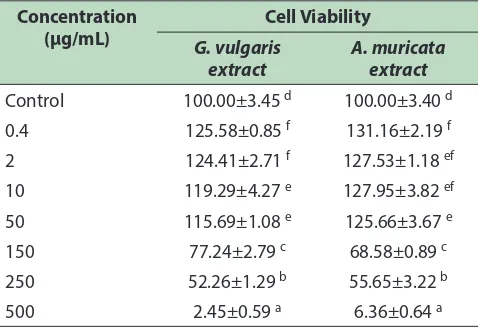
[image:3.612.316.555.508.673.2]As shown in Table 1, cell treated with GVE and AME at concentration of 150, 250, and 500 µg/mL resulted low viability which indicates toxicity to RAW264.7, whereas concentration of 0.4, 0.2, 10 and 50 µg/mL of both GVE and AME showed high viability (>90%). Viable cells obtained at concentration of 10 and 50 µg/ mL in both GVE and AME, appeared to reach normal level (control), makes such concentrations suitable for treatment of RAW264.7 cells. herefore, further analysis
Table 1: Efect of G. vulgaris and A. muricata extracts toward viability of RAW264.7 cell line
Concentration (µg/mL)
Cell Viability
G. vulgaris extract
A. muricata extract
Control 100.00±3.45 d 100.00±3.40 d
0.4 125.58±0.85 f 131.16±2.19 f
2 124.41±2.71 f 127.53±1.18 ef
10 119.29±4.27 e 127.95±3.82 ef
50 115.69±1.08 e 125.66±3.67 e
150 77.24±2.79 c 68.58±0.89 c
250 52.26±1.29 b 55.65±3.22 b
500 2.45±0.59 a 6.36±0.64 a
of GVE and AME uses concentration in range of 10 and 150 µg/mL.
3.2 Efect of
G.vulgaris
and
A. muricata
Extracts on TNF-α Level in LPS-Induced
RAW264.7 Cell Line
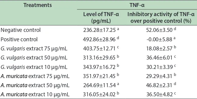
TNF-α is a multi functional cytokine which can exert regulatory, inlammatory and cytotoxic efects on a wide range of lymphoid and non-lymphoid cells and tumor cells. GVE and AME showed the inhibitory activity against TNF-α production based on the lower concentration of TNF-α compared to the positive control (LPS-stimulated cells free supernatant without extract). As shown in Table 2, GVE and AME decreased TNF-α level compared to positive control.
Treatment of AME at concentration of 50 µg/ mL in RAW264.7 resulted lowest TNF-α level 264.69 pg/mL among other treatments. Level of TNF-α in LPS-induced RAW264.7 treated with AME of 50 µg/mL, was signiicantly diferent compared to TNF-α level in positive control (Table 2). hese results indicate AME of 50 µg/mL decreased TNF-α level to play its role as anti-inlammatory. Whereas GVE at concentration of 50 µg/ mL also generated relatively low TNF-α level (313.16 pg/ mL). Both GVE and AME of 50 µg/mL showed signiicant diference compared to positive control, and resulted good inhibitory activity of TNF-α over positive control (36.46 and 46.82% respectively). AME 50 Ug/mL was
the best activity to lower TNF-A level and comparable with negative control.
3.3 Efect of
G.vulgaris
and
A. muricata
Extracts on IL-1β level in LPS-Induced
RAW264.7 Cell Line
IL-1 which refers to two proteins (IL-1α and IL-1β), is a potent immuno-modulator which mediates a wide range of immune and inlammatory responses including activation of B and T cells16. Inhibiting the production of IL-1 is important in inding the anti-inlammatory agent. GVE and AME showed the inhibitory potential against IL-1β production (Table 3). Efect of GVE and AME level on IL-1β level in LPS-induced RAW264.7 is presented in Table 3.
As shown in Table 3, GVE and AME at concentration of 10, 50 and 75 µg/mL decreased IL-1β level in LPS-induced RAW264.7, which was signiicant compared to positive control and comparable with negative control. GVE at concentration of 50 µg/mL resulted highest decreased IL-1β level of 903.53 pg/mL.
3.4 Efect of
G.vulgaris
and
A. muricata
Extracts on IL-6 Level in LPS-Induced
RAW264.7 Cell Line
[image:4.612.146.469.524.687.2]IL-6 is one of the cytokines that possess biological activities due to acute inlammation17. IL-6 along
Table 2: Efect of G. vulgaris and A. muricata extracts toward TNF-α level in RAW264.7 cell line
Treatments TNF-α
Level of TNF-α (pg/mL)
Inhibitory activity of TNF-α over positive control (%)
Negative control 236.28±17.25 a 52.06±3.50 d
Positive control 492.86±28.96 d -0.00±5.88 a G. vulgaris extract 75 µg/mL 403.75±12.71 c 18.08±2.57 b
G. vulgaris extract 50 µg/mL 313.16±29.65 b 36.46±6.01 c
G. vulgaris extract 10 µg/mL 343.97±16.72 b 30.21±3.39 c
A. muricata extract 75 µg/mL 351.97±21.45 b 29.29±4.31 b
A. muricata extract 50 µg/mL 264.69±11.54 a 46.82±2.31 d
Table 3: Efect of G. vulgaris and A. muricata extracts toward IL-1β level in RAW264.7 cell line
Treatments IL-1β
Level of IL-1β (pg/mL)
Inhibitory activity of IL-1β over positive control (%)
Negative control 888.53±8.11 a 20.09±0.73 c
Positive control 1111.93±4.67 b 0.00±0.42 a
G.vulgaris extract 75 µg/mL 954.87±16.64 a 14.13±1.50 b G.vulgaris extract 50 µg/mL 903.53±11.90 a 18.74±1.07 c
G.vulgaris extract 10 µg/mL 942.53±12.36 a 15.23±1.11 b
A. muricata extract 75 µg/mL 950.00±72.33 a 17.71±6.27 b
A. muricata extract 50 µg/mL 905.00±58.89 a 21.60±5.10 c
A. muricata extract 10 µg/mL 928.13±42.13 a 19.60±3.65 c
*Note: Data is presented as average of ± SD from 3 replications. Letter a, b, c, d in each column indicates signiicance diferent among treatments based on Duncan post hoc test with p < 0.05 is considered as signiicantly diferent.
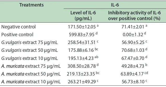
with TNF-α and IL-1 is elevated in septic or aseptic inlammation, makes it appropriate target in prevention and treatment of inlammatory disease18. In this study, GVE and AME decreased IL-6 compared to positive control, as shown in Table 4.
he results showed that LPS induced inlammation and increased IL-6 level in RAW264.7 which was indicated by high level of IL-6 in positive control (599.83 pg/mL) and signiicantly diferent compared to negative control. Levels of IL-6 in treatment of GVE and AME were lower and signiicantly diferent compared to positive control. hese results indicate both GVE and AME are able to decrease
Table 4: Efect of G. vulgaris and A. muricata extracts toward IL-6 level in RAW264.7 cell line
Treatments IL-6
Level of IL-6 (pg/mL)
Inhibitory activity of IL-6 over positive control (%)
Negative control 171.50±12.05 a 71.41±2.01 a
Positive control 599.83±7.95 d 0.00±1.32 d
G.vulgaris extract 75 µg/mL 258.54±31.51 c 56.90±5.25 c
G.vulgaris extract 50 µg/mL 175.88±6.16 bc 70.68±1.03 d
G.vulgaris extract 10 µg/mL 195.13±4.23 ab 67.47±0.70 d
A. muricata extract 75 µg/mL 308.50±28.78 d 49.28±4.73 b
A. muricata extract 50 µg/mL 219.13±23.35 bc 63.89±4.17 cd
A. muricata extract 10 µg/mL 263.21±49.29 c 56.73±8.10 c *Note: Data is presented as average of ± SD from 3 replications. Letter a, b, c, d in each column indicates signiicance diferent among treatments based on Duncan post hoc test with p < 0.05 is considered as signiicantly diferent.
IL-6 in inlammation-induced cell. GVE and AME at concentration of 50 µg/mL showed signiicant decrease in IL-6 level (175.88 pg/mL and 219.13 pg/mL respectively), and signiicantly diferent than positive control.
3.5 Efect of
G.vulgaris
and
A. muricata
Extracts on NO Level in LPS-Induced
RAW264.7 Cell Line
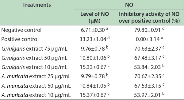
[image:5.612.149.466.339.506.2]Table 5: Efect of G. vulgaris and A. muricata extracts toward NO level in RAW264.7 cell line
Treatments NO
Level of NO (µM)
Inhibitory activity of NO over positive control (%)
Negative control 6.71±0.30 a 79.80±0.91 d
Positive control 33.23±1.04 d 0.00±3.14 a
G.vulgaris extract 75 µg/mL 9.76±0.78 b 70.63±2.37 c G.vulgaris extract 50 µg/mL 10.80±1.06 b 67.48±3.17 c
G.vulgaris extract 10 µg/mL 15.33±0.67 c 53.84±2.03 b
A. muricata extract 75 µg/mL 9.79±0.78 b 70.67±2.35 c
A. muricata extract 50 µg/mL 10.84±1.05 b 67.53±3.15 c
A. muricata extract 10 µg/mL 15.37±0.67 c 53.97±2.01 b
*Note: Data is presented as average of ± SD from 3 replications. Letter a, b, c, d in each column indicates signiicance diferent among treatments based on Duncan post hoc test with p < 0.05 is considered as signiicantly diferent.
percent of inhibitory activity was determined by the value of positive control nitrite concentration minus the nitrite concentration of treatment divided to the nitrite concentration of positive control.
Although NO level of treatment group was higher than negative control, both GVE and AME signiicantly resulted lower NO than positive control (Table 4), which indicated GVE and AME reduce NO level in inlammation-induced cell. GVE and AME at concentration of 75 µg/mL showed lower NO level (9.76 µM and 9.79 µM respectively). Decrease in NO level by GVE and AME showed both treatments supress inlammation properly, makes it promising in reducing NO to play its role in inlammation18.
4. Discussion
he result of present study showed both GVE and AME extract showed no toxicity to RAW264.7 at concentration of 0.4, 2, 10, and 50 μg/mL. Non toxicity of substrate performed with MTS assay, was recorded by over 90% of viable cells.Viability test is crucial in pharmacology to determine adverse efect of bioactive substance in living organism prior to clinical use of drug or chemical compounds19-21.
In this study, LPS was used to induce inlammation in RAW264.7 cell line. It has been reported that
bacterial LPS is able to increase cytokines production as inlammation mediator3,4. LPS compose outer
membrane of gram negative bacteria as endotoxin that induces production of proinlammatory mediators such as NO, IL-1, IL-6, TNF-α, interleukins, prostanoids and leukotrienes 3,4. LPS induces inlammation via Toll-like
receptor 4 (TLR4) binding.TLR4 is a transmembrane protein that recognizes lipopolisaccharide speciically. TLR4 signaling pathway may activates Nuclear Factor Kappa B (NF-κB) and Activation protein 1 (AP-1) which later induces the secretion of proinlammatory mediators such as NO, TNFα, IL-1 and IL-1222,23.
Anti-inlammatory activities of GVE and AME were observed through markers such as IL-1β, TNF-α, NO and IL-6 inhibitory activity assays in LPS-induced macrophage cell line (RAW264.7). Both GVE and AME extract of 50 µg/mL resulted low TNF-α level in LPS-induced RAW264.7, with lowest level generated from
AME. hese results indicate bothGVA and AME of 50 µg/mL play its role as anti-inlammatory, yet it did not exceed normal level. he TNF-α is an important cytokine involved in inlammatory response via activation of NF-κB, cytokine and adhesion molecule inducer24,25.
he TNF-α is an important target of anti-inlammatory agent screening5. In presence of anti-inlammatory,
TNF-α that exists in cascades is blocked26. Endogenous
cause fever during inlammation, following up-regulated inlammatory responses that later triggers production of acute phase reactants27.
GVE and AME at concentration of 50 μg/mL reduced IL-1β level in RAW264.7. IL-1β is prototypic proinlammatory cytokine that exert pleiotrophic efects on a variety of cells and play key roles in acute and chronic inlammatory as well as autoimmune disorders.
IL-1β is produced mainly by blood monocytes. IL-1β, TNF-α and IL-6, simultaneously promote fever during inlammation due to up-regulated inlammatory responses that later triggers production of acute phase reactants5.
In this study, GVE and AME at concentration of 50 μg/mL reduced IL-6 level in RAW264.7, with lowest IL-6 level was obtained in treatment of GVE. he IL-6 production has been detected in many cell types. Macrophages and monocytes are the primary source of cytokine during acute inlammation. IL-6 is pleiotropic cytokine to modulate inlammatory response 26, 27. IL-6 along with TNF-α and IL-1 is elevated in condition of septic or aseptic inlammation, makes it efective in prevention and treatment of inlammatory disease18.
he result of present study showed GVE and AME at concentration of 50 μg/mL reduced NO level in RAW264.7 cell, with lowest NO was obtained in treatment of GVE. Additional inlammatory pathways promoted by TNF-α, results nitric oxide (NO)26, 27. NO inhibitory
activity is frequently used as appropriate target in anti-inlammatory agent screening. NO is responsible in host immune defense, vascular regulation, neurotransmission and other system in normal condition. Excess inducible NO Synthase (iNOS) is especially asssociated with various human diseases including inlammation15,17.
It has been reported that active compounds from plants play important role in prevention and treatment of various diseases9,28. Anti-inlamatory activity of leaf
extract of G. vulgaris has been documented in previous studies. Phytochemical analysis of G. vulgaris extract revealed the presence of lavonoids glycosides, saponins, steroids, tannins and polyphenols. Anti-inlammatory efects are present due to inhibition of mediators in inlammation by glycosides or steroids9. According to
Jothimanivannan et al. (2010), lavonoid content also
play key role in anti-inlammatory acitivity of GVE10. Kim et al. (2004) reported lavonoid contained in plants possess cellular mechanism in anti-inlammatory activity by inhibiting eicosanoid that produces phospholipase A2, cyclooxigenase and lypoxigenase. Inhibition of these enzymes will reduce prostanoid and leucotrien level11.
AME is efective for both acute and chronic inlammation. It signiicantly decreases both TNF-α and IL-1β levels in CFA-induced arthritis model29.
Phytochemical test conducted on ethanolic extract of A. muricata indicates presence of alkaloids, saponins, lavonoids, tannins, triterpenes and steroid. Flavonoids have a great potential as anti-inlammatory agents. Flavonoids and tannins have been reported to inhibit prostaglandin synthesis29-32. Recent study showed certain lavonoids such as lavon, posses anti-inlammatory properties that regulates pro-inlammatory genes
cyclooxigenase-2(COX-2), nitrite oxide synthase (NOS), and cytokines11. Other substances present in extract such as tannins, may give the synergistic efect to the lavonoids.
5. Conclusion
his research revealed that ethanol extracts of G.vulgaris
and A. muricata possess the anti-inlammatory potential indicated by inhibition of inlammatory mediators including IL-1β, TNF-α, NO and IL-6.
6. Conlict of Interest
he authors declare that they have no competing interests.
7. Acknowledgement
8. References
1. Fang SC. Anti-inlammatory efects of phenolic
compounds isolated from the fruits of Artocarpus
heterophyllus. J Agric Food Chem. 2008; 56:4463–8.
2. Jung CH. Eleutherococcus senticosus extract attenuates
LPS-induced iNOS expression through the inhibition of Akt and JNK pathways in murine macrophage. J Ethnopharmacol. 2007; 113:183–7.
3. Kim AR. Flavonoids diferentially modulate nitric oxide production pathways in lipopolysaccharide-activated RAW264.7 cells. Arch Pharmacal Res. 2005; 28:297–304.
4. Kim KM. Methanol extract of Cordyceps pruinosa
inhibits in vitro and in vivo inlammatory mediators by surpressing NF-kB activation. Toxicol Applied Pharmacol. 2003; 190:1–8.
5. Boots AW. In vitro and ex vivo anti-inlammatory activity of quercetin in healthy volunteers. Nutrition. 2008; 24:703–10.
6. Rusmana D, Elizabeth M, Widowati W, Fauziah N, Maesaroh M. Inhibition of inlammatory agent production
by ethanol extract and eugenol of Syzygium aromaticum
(L.) lower bud (clove) in LPS-stimulated RAW264.7 cells. Res J Med Plant. 2015; 9(6):264–74.
7. Dewi K, Widyarto B, Erawijantari PP, Widowati W. In
vitro study of Myristica fragrans seed (Nutmeg) ethanolic
extract and quercetin compound as anti-inlammatory agent. Int J Res Med Sci. 2015; 3(9):2303–10.
8. Mehta RG, Murillo G, Naithani R, Peng X. Cancer chemoprevention by natural products: How far have we come? Pharm Res. 2010; 27(6):950–61.
9. Agnihotri SS. An overview on anti-inlammatory properties and chemo-proiles of plants used in traditional medicine. Indian J Nat. 2010; 1:150–67.
10. Jothimanivannan C, Kumar RS, Subramanian N. Anti-Inlammatory and analgesic activities of ethanol extract of
aerial parts of Justicia gandarussa Burm. J Int Pharmacol.
2010; 6(3):273-83.
11. Kim HP, Son KH, Chang HW, Kang SS. Critical review anti-inlammatory plant lavonoids and cellular action mechanisms. J Pharmacol Sci. 2004;96:229–45.
12. Bhaskar V, Balakrishnan N. Analgesic, Anti-inlammatory
and antipyretic activities of Pergularia daemia and Carissa
carandas. J Pharm Sci. 2009; 17(3):168–74.
13. Kossouoh C. Essential oil chemical composition of
Annona muricata L. leaves from Benin. J Essent Oil Res. 2007; 307–9.
14. Widowati W, Mozef T, Risdian C, Yelliantty Y. Anticancer
and free radical scavenging potency of Catharanthus
roseus, Dendrophthoe petandra, Piper betle, and Curcuma mangga extracts in breast cancer cell lines. Oxid Antioxid Med Sci 2013b; 2(2):137-42.
15. Widowati W, Wijaya L, Wargasetia TL, Bachtiar I, Yelliantty Y, Laksmitawati DR. Antioxidant, anticancer
and apoptosis-inducing efects of Piper extracts in HeLa
cells. J Exp Integr Med. 2013b; 3:225–30.
16. Mahajna SM. In vitro evaluations of cytotoxicity and antiinlammatory efects of Peganum harmala seed extracts in THP-1-derived macrophages. Eur J Med Plants. 2014; 5:165–75.
17. Gabay C. Interleukin-6 and chronic inlammation. BioMed. 2006; 8 (Suppl 2):S3.
18. Kang CH. Inhibition of lipopolysaccharide-induced iNOS, COX-2 and TNF-a expression by aqueous extract of orixa japonica in RAW264.7 cells via supression of NF-kB activity. Trop J Pharmaceut Res. 2011; 10:161–8. 19. Jothy SL. Acute oral toxicity of methanolic seed extract of
Cassia istula in mice. Molecules. 2011; 16:5268–82.
20. Lalitha PK. Acute toxicity study of extracts of Eichhornia
crassipes (MART.) solms. Asian J Pharm Clin Res. 2012; 5:59–61.
21. Rajalakshmi AA. Toxicity analysis of diferent medicinal plant extracts in Swiss Albino mice. BioMed Res. 2014; 1:1–6.
22. Abbas AK, Lichtman AH, Shiv P. Cellular and molecular
immunology. 8th ed. Canada: Elsevier Saunders; 2012. p.
59.
23. Rapsinski GJ, Wynosky-Doli MA, Oppong GO, Tursi SA, Wilson RP, Brodsky IE et al. Toll-like receptor 2 and NLRP3 cooperate to recognize a functional bacterialamyloid, curli. Infect Immun. 2015; 83:693–701.
24. Tak PP. NF-kB: a key role in inlammatory disease. J Clin Invest. 2001; 170:7–11.
25. De Cassia da Silveira e Sa RL. A review on anti-inlammatory activity of phenylpropanoids found in essential oils. Molecules. 2014; 19:1459–80.
27. Damte, DM. Anti-inlammatory activity of
dichloromethane extract of Auricularia-judae in
RAW264.7 cells. Toxicol. Res. 2011; 27:11-14.
28. Leontowicz HM. Bioactive properties of snake fruit (Salacca edulis Reinw) and Mangosteen (Garcinia mangostana) and their inluence on plasma lipid proile and antioxidant activity in rats fed cholesterol. Eur Food Res Technol. 2006; 223:697–703.
29. Foong CP, Hamid RA. Evaluation of anti-inlammatory activities of ethanolic extract of Annona muricata
leaves. Revista Brasileira de Farmacognosia. 2012; 22(6):1301–7.
30. Tapas AR. Flavonoids as nutraceuticals: A review. Trop J Pharm Res. 2008; 7:1089–99.
31. Tunon MJ. Potential of lavonoids as anti-inlammatory agents: modulation of pro-inlammatory gene expressions and signal transduction pathways. Curr Drug Metab. 2009; 10:256–71.
32. Seraini M. Flavonoids as anti-inlammatory agents. P