The African buffalo parasite
Theileria.
sp. (buffalo) can infect and
immortalize cattle leukocytes and encodes divergent orthologues
of
Theileria parva
antigen genes
R.P. Bishop
a,*,1, J.D. Hemmink
b,1,2, W.I. Morrison
b, W. Weir
d, P.G. Toye
a, T. Sitt
a,
P.R. Spooner
a, A.J. Musoke
a, R.A. Skilton
a, D.O. Odongo
a,caInternational Livestock Research Institute (ILRI), PO Box 30709, Nairobi, 00100, Kenya
bThe Roslin Institute, Royal (Dick) School of Veterinary Studies, University of Edinburgh, Easter Bush, Midlothian, EH25 9RG Scotland, UK cSchool of Biological Sciences, The University of Nairobi, PO Box 30197, Nairobi, 00100, Kenya
dCollege of Medical Veterinary and Life Sciences, University of Glasgow Glasgow, G61 1QH, UK
a r t i c l e
i n f o
Article history: Received 26 May 2015 Received in revised form 24 August 2015 Accepted 25 August 2015
Keywords: Theileria parva Theileriasp. (buffalo) Reverse line blot
Cape buffalo (Syncerus caffer) 18S rDNA
Deep sequencing
Polymorphic immunodominant molecule (PIM)
a b s t r a c t
African Cape buffalo (Syncerus caffer) is the wildlife reservoir of multiple species within the apicom-plexan protozoan genusTheileria, including Theileria parvawhich causes East coast fever in cattle. A parasite, which has not yet been formally named, known asTheileriasp. (buffalo) has been recognized as a potentially distinct species based on rDNA sequence, since 1993. We demonstrate using reverse line blot (RLB) and sequencing of 18S rDNA genes, that in an area where buffalo and cattle co-graze and there is a heavy tick challenge,T.sp. (buffalo) can frequently be isolated in culture from cattle leukocytes. We also show thatT.sp. (buffalo), which is genetically very closely related toT. parva, according to 18s rDNA sequence, has a conserved orthologue of the polymorphic immunodominant molecule (PIM) that forms the basis of the diagnostic ELISA used forT. parvaserological detection. Closely related orthologues of several CD8 T cell target antigen genes are also shared withT. parva. By contrast, orthologues of the T. parvap104 and the p67 sporozoite surface antigens could not be amplified by PCR fromT.sp. (buffalo), using conserved primers designed from the correspondingT. parvasequences. Collectively the data re-emphasise doubts regarding the value of rDNA sequence data alone for defining apicomplexan species in the absence of additional data.‘Deep 454 pyrosequencing’of DNA from twoTheileriasporozoite stabilates prepared fromRhipicephalus appendiculatusticks fed on buffalo failed to detectT.sp. (buffalo). This strongly suggests thatR. appendiculatusmay not be a vector forT.sp. (buffalo).Collectively, the data provides further evidence thatT.sp. (buffalo). is a distinct species fromT. parva.
©2015 The Authors. Published by Elsevier Ltd on behalf of Australian Society for Parasitology. This is an
open access article under the CC BY-NC-ND license (http://creativecommons.org/licenses/by-nc-nd/4.0/).
1. Introduction
In certain regions of East Africa, such as those bordering the Maasai Mara game reserve, the Laikipia district in central Kenya and Lake Mburo National park in Uganda, cattle graze in areas that are also frequented by African Cape buffalo (Syncerus caffer). Afri-can buffalo are hosts of at leastfive species ofTheileria(Allsopp et al., 1999; Norval et al., 1992; Bishop et al., 2004; Oura et al.,
2011a). Some species can be transmitted to cattle by ixodid ticks in the genusRhipicephalus,including the economically important pathogenTheileria parva, which is mainly transmitted by Rhipice-phalus appendiculatus, whereas otherTheileria species are trans-mitted by ticks of the genusAmblyomma. The heterogeneity among buffalo-derivedTheileriaparasites that infect leukocytes wasfirst identified in a study using a panel of monoclonal antibodies to type isolates from naturally infected buffalo in Kenya (Conrad et al., 1987). This revealed two very distinct patterns of anti-schizont monoclonal antibody reactivity using immunofluorescence (IFAT) staining of Theileria schizont-infected cell lines. Further studies using multicopy DNA sequences as probes to analyse restriction fragment length polymorphism revealed similar diversity and identified several cloned cell lines from one buffalo (6834) that
*Corresponding author.
E-mail address:r.bishop@cgiar.org(R.P. Bishop).
1 These two authors made equal contributions to this work.
2 Current address: The Pirbright Institute, Pirbright, Woking, GU24 0NF, United
Kingdom.
Contents lists available atScienceDirect
International Journal for Parasitology:
Parasites and Wildlife
j o u r n a l h o m e p a g e :w w w . e l s e v i e r . c o m / l o c a t e / i j p p a w
http://dx.doi.org/10.1016/j.ijppaw.2015.08.006
gave very weak hybridisation with one of the probes employed (Conrad et al., 1989). A subsequent study revealed that the 18S ri-bosomal RNA gene sequence of the parasites in these cloned lines was distinct fromT. parva, although it had the closest similarity to T. parvaamong the sixTheileriaspecies analyzed (Allsopp et al., 1993). This parasite was provisionally designatedT. sp. (buffalo) (Allsopp et al., 1993). Subsequent studies have found thisTheileria genotype to be well conserved and widespread in buffalo pop-ulations in both Uganda and South Africa, frequently in co-infections with T. parva (Chaisi et al., 2013; Mans et al., 2011a; Mans et al., 2015; Oura et al., 2011a,b; Pienaar et al., 2011b; Pienaar et al. 2014).
Serological detection ofT. parvainfection in cattle is based on an ELISA test that measures antibody responses to a schizont antigen that has been named the polymorphic immunodominant molecule, (PIM) (Toye et al., 1991; Katende et al., 1998). PIM is encoded by a single copy gene with relatively conserved N and C terminal ends and a hyper-polymorphic central domain that is rich in the amino acids glutamine and proline (Toye et al., 1995; Geysen et al., 2004). Most of the available murine anti-schizont monoclonal antibodies have been shown to be specific for this antigen (Toye et al., 1996). We describe herein the genotype of parasites withinTheileria schizont-infected leukocyte lines and in blood samples isolated from cattle at a ranch in the Rift Valley Province of Kenya, which is home to a resident herd of approximately 1000 buffalo that co-graze with cattle (Pelle et al., 2011). Analysis of the sequences of the p67 gene sequences from those leukocyte cultures containing T. parva(Obara et al., 2015) strongly suggest that they originate from ticks that had fed on buffalo (Nene et al., 1996). Several of the isolates were identified asT.sp. (buffalo) or mixed infections of this genotype together withT. parva. The genotyping was extended to orthologues of genes encoding antigens that are the target of antibody and cellular immune responses duringT. parvainfections in cattle. We further describe an analysis of two buffalo derived Theileriastabilates using high-throughput sequencing and discuss implications of the data with respect to the status ofTheileriasp. (buffalo) as a species that is distinct fromT. parva.
2. Materials and methods
2.1. Animals
Samples from a field study conducted at Marula farm near Naivasha in the Rift Valley province of central Kenya were used in the experiments. The study usedBos taurus(Friesian) cattle aged 6e8 months purchased from Marula farm where strict acaricidal
control of ticks was practised. All cattle were initially shown to be free of antibodies to theilerial antigens using an indirect ELISA. Three groups of cattle were immunised against T. parva by the infection and treatment method (Radley et al., 1975) with the following stabilates: (1) 30 animals with T.parvaMarikebuni sta-bilate 3014 (Morzaria et al., 1995); (2) 29 animals with T. parva Marikebuni stabilate 316 (Payne, 1999); (3) 27 animals withT. parva composite trivalent Muguga cocktail stabilate FAO 1 (Morzaria et al., 1999), containing the Muguga, Kiambu 5 and Serengeti stocks. An additional group of 27 animalserepresenting the
non-immunised controlsewere also included. Six weeks after
immu-nisation, a total of 113 cattle, comprising 86 immunised and 27 control animals, were exposed to tick challenge in areas co-grazed by buffalo, other species of wild ruminants and cattle. Cattle were exposed during the day and returned to an enclosure at night. All cattle were monitored for temperature daily from day 17 after exposure. Any cattle showing enlarged lymph glands or with a fever of 39.5 C or above were sampled by taking smears of aspirates
from the affected lymph node. If schizonts were detected, then a
contralateral lymph node was also sampled for schizont detection and blood smears were taken for detection of piroplasms. Lymph node aspirates were also collected in culture medium containing anticoagulant for establishment of cell cultures (see below). Blood samples for serology were taken weekly from the first day of immunisation and this was continued during the exposure period. These sera were screened for antibodies toT. parvausing the in-direct antibody detection ELISA based on the PIM antigen (Katende et al., 1998). All experimental procedures involving cattle were approved by the ILRI Institutional Animal Care and Use Committee (IACUC).
2.2. Sporozoite stabilates
Genomic DNA was prepared from sporozoite stabilates after removal of excess tick material by centrifugation for 10 min. Genomic DNA was extracted from the supernatant containing sporozoites using the DNeasy®
TisKit (Qiagen, Dusseldorf Germany) according to the manufacturer's instructions. The sporozoite sta-bilates used were stabilate 3081 derived from buffalo 7014 captured in the Laikipia District (Morzaria et al., 1995) and stabilate 4110 from buffalo 7752. These were analysed by 18S rDNA 454 deep sequencing. Two additional DNA samples, one prepared from blood from a buffalo from the Kruger National park (SC03) and one from the Marula bovine cell line N79, both known to contain T. sp. (buffalo), were included as positive controls.
2.3. Theileria-infected cell lines
Samples for cell culture were transported to the laboratory within 24 h. Mononuclear cells were isolated from lymph node aspirates and/or blood samples by density gradient centrifugation, re-suspended in RPMI growth medium supplemented with 15% foetal calf serum and cultured at 37C in 5% CO
2in 24-well plates containing adherent cells from a foetal thymic cell line as feeder cells. The resultant established cell lines were cryopreserved using previously reported techniques (Brown, 1987). In some cases cloned parasitized cell lines derived from the cultures were used to increase the resolution of the 454 sequence analysis. . The cloned lines were derived from established parasitized lines originally isolated from cattle and buffalo from different regions of Kenya. They included cell lines from cattle in the Marula farm study (Table 1) and buffalo lines isolated previously from the Maasai Mara and Laikipia district (Table 2). Specifically, there were sevenT. parva clones from the Marikebuni-cattle-derived stock and one from each of four other cattle stocks, eight clones from two buffalo-associated cattleT. parvastocks from Marula, nineT. parvacloned lines from four stocks of buffalo origin and nine clones from twoT.sp. (buffalo) lines. Further details are provided inTable 2. The cell lines were cloned by limiting dilution in 96-well U-bottom plates in a mixture of 50% filtered supernatant from established buffalo-derived T. parvacell lines and 50% cell culture medium (RPMI 1640 me-dium enriched with glutamine, 2-mercaptoethanol, and 12.5% foetal calf serum) and 2105cells/per well
g
-irradiated bovinePBMC asfiller cells.
2.4. T. parva schizont characterisation with monoclonal antibodies
A panel of monoclonal antibodies was used to characterise theilerial isolates in an immunofluorescence test (IFAT) as previ-ously described (Minami et al., 1983:Conrad et al., 1987).
2.5. Processing of DNA from blood samples and infected cell lines
(1995)using 200 ul aliquots of blood. Prior to PCR, the samples were heated for 10 min at 100C to inactivate the proteinase K and
centrifuged for 2 min. Genomic DNA was prepared from frozen pellets of 107 cultured schizont-infected lymphocytes using the DNeasy®
Tissue Kit (Qiagen, Germany) according to the manufac-turer's instructions.
2.6. Reverse line blot
Two sets of primers were separately used to amplify a 460e520 bp fragment of 18S and 16S SSU rRNA spanning the V4
variable region as originally described byGubbels et al. (1999), but with slight modifications. All primers were manufactured by MWG (Germany). For the amplification of the 18S rRNA encoding gene of Theileria/Babesia, the forward primer used was (50
-GACA-CAGGGAGGTAGTGACAAG) and the reverse primer was (biotin-50
-CTAAGAATTTCACCTCTGACAGT). The reactions were performed in an automated DNA thermal cycler (MJ research), with an enabled hot lid.
The preparation, hybridization and subsequent removal of hybridised DNA from the membrane were performed as already
described byGubbels et al. (1999)as modified byOura et al. (2004).
2.7. Nested 18s PCR assay
To confirm the data from the RLB, a semi-nested PCR assay was developed to differentiate betweenT.parvaandT. sp (buffalo) using specific reverse primers in the second round (For T. parva (CTTATTTCGGACGGAGTTCG) and forT. sp. (buffalo) (CGCTTATTT-CAGACGGAGTTA). The first round of PCR employed Theileria/ Babesia-specific primers (50GAGGTAGTGACAAGAAATAACAATA and
50CTAAGAATTTCACCTCTGACAGT) (Gubbels et al., 1999; Oura et al.,
2004). 1
m
l of PCR products was then used for a second round of PCRusing the same forward primer and the species specific reverse primers. The presence ofT.sp. (buffalo) was confirmed for some of the cell lines by sequencing of PCR products amplified using the species-specific primer, which were cloned using the pGEM-T Easy vector system (Promega Corporation, USA) according to the man-ufacturer's instructions and purified using the Promega PureYield Plasmid Miniprep System.
2.8. PCR amplification and sequencing of parasite antigen genes
For p104, p67, and PIM loci: PCR amplification was performed using Taq Expand™highfidelity PCR reagents (Roche Diagnostics, Mannheim, Germany) in 100
m
l reactions volumes containing 1XPCR buffer, 200 mM of each dNTP, 100 ng of each forward and reverse primer, 1.5 mM MgCl2, 0.5 units of Expand Highfidelity Taq DNA polymerase and 50 ng of DNA. PCR conditions and primers used in the p104 PCR were as previously described (Skilton et al., 2002). Primers ILO 6464 (50CGC GGA TCC AAG ATC TTT CCC TTT
TTA30) and ILO 8413 (50CGG GGT ACC TTA ACA ACA ATC TTC GTT
AAT GCG30) were used to amplify approximately 1.5 kb of the PIM
full length gene. Primers ILO 8222 (50CGA CAC TGA ACG ATG CAA
ATA30) and ILO 8223 (50GAG TTA TTG TTA GTG GAC GAT30) were
used to amplify approximately 2.2 kb encompassing the full length p67 gene (Nene et al., 1992, 1996). All PCR reactions were per-formed in a hot-lid MJ PCR thermo cycler (PTC-100, MJ research, CA). Amplicons were subsequently purified using the QIAquick PCR purification kit (QIAGEN, Germany), according to the manufac-turer's instructions, and sequenced on an ABI 377 automated sequencer (Applied Biosystems Inc), using big dye terminator Table 1
Theileria-infected cell lines from Marula farm and their reactivity with anti-PIM MAbs raised againstT. parvaMonoclonal Antibody number.
Cell line 1 2 3 4 7 10 12 15 20 21 22 23
N6 þ e e þ þ(<1%) þ þ e þ þ þ e
N13 þ e e þ þ þ þ þ(1%) þ þ þ(10%) þ(<1%)
N18 þ(<1%) e e þ(<5%) þ(<5%) þ þ þ(3%) þ(5%) þ þ e
N20 þ e e þ þ þ þ þ þ þ þ e
N33 þ e e þ þ þ þ þ(2%) þ þ þ e
N36 þ(<1%) e e þ þ(20%) þ þ þ(10%) þ þ þ e
N38 þ e e þ þ þ þ þ þ þ þ e
N43 þ e e þ þ þ þ þ(2%) þ þ þ e
N50 þ e e þ þ þ þ þ(5%) þ þ(40%) þ e
N55 þ e e þ þ(5%) þ þ þ(1%) þ þ þ e
N69 þ e e þ þ(10%) þ þ þ(5%) þ þ þ e
N76 þ e e þ þ þ þ þ(<1%) þ þ þ e
N77 þ e e þ þ þ þ þ(2%) þ þ(30%) þ e
N79 þ e e þ þ(30%) þ þ þ(5%) þ þ þ e
N86 þ(<1%) e e þ(<1%) þ(<1%) þ þ þ(<1%) þ(<1%) þ þ e
N88 þ e e þ þ þ þ þ(1%) þ þ(10%) þ e
N99 þ e e þ þ þ þ þ(2%) þ þ þ e
N100 þ e e þ þ þ þ þ(4%) þ þ þ e
N102 þ(<1%) e e þ(1%) þ(3%) þ þ þ(<1%) þ þ þ e
N103 þ e e þ þ þ þ þ(1%) þ þ þ e
N106 þ e e þ(70%) þ(70%) þ þ þ(5%) þ þ þ e
N107 e e e e e þ þ þ(2%) þ þ þ e
Table 2
Stocks and clones used for the phylogenetic analysis ofTheileria sp(buffalo) and T. parva.
Name/Animal Clone Location
Cattle derivedT. parva
Muguga Reference genome Kenya
Marikebuni A3, A7, B12, E43, F44, F53, I8, I38, J17 Kenya Mariakani St 3231 clone 2/3 Kenya Boleni St3230 clone 1:1 Zimbabwe
Uganda St 3645clone 1/2 Uganda
Cattle associated with African buffaloT. parva
Marula N33 Clone 2, 4, 5, 7 Nakuru, Kenya Marula N43 Clone 1, 2, 5, 6 Nakuru, Kenya Buffalo-derivedT .parva
Mara 3 Clone 3 Maasai Mara, Kenya
Mara 30 Clone 5, 8, 11 Maasai Mara, Kenya Mara 42 Clone 2,5 Maasai Mara, Kenya Buffalo 6998 Clone 2,4,9 Kenya
Buffalo-derivedT. sp(buffalo)
chemistry. The twelve PIM sequences were submitted to GenBank
with accession numbers KT258708; KT258709; KT258710;
KT258711; KT258712; KT258713 KT258714; KT258715; KT258716; KT258717; KT258718; KT258719.
Tp6, Tp7 and Tp8 loci:PCR primers designed to amplify seg-ments of three genes encodingT. parvaantigens recognized by CD8 T cells (Tp6, Tp7 and Tp8) were used to analyse the sequences of these genes and the orthologues inT.sp. (buffalo). The primers and amplicon sizes were as follows: Tp6 - forward primer 50
GTCCAA-TAATTTACGATGTGAG), reverse primer 50CTTGTTTAGCCTCTACAGC
e 376 bp amplicon; Tp7 - forward primer 50TGAAGAAGGA
CGACTCGCAC, reverse primer 50TAAGCATTTCCCACTCACGC
e
amplicon 364 bp; Tp8 forward primer_50ATCCACAACCA
AGTGCCCAG, reverse primer 50ACTGCGAAGGAGGTCAATCC
e
348 bp amplicon. Each PCR reaction comprised 20 pmol of each primer, 0.5 units BIOTAQ polymerase (Bioline, UK), 2.0
m
l 10 x PCRmaster mix and 50 ng genomic DNA in a total volume of 20 ul. A G-storm thermal cycler (Genetic Research instrumentation, Braintree, UK) with enabled hot lid was used for amplification. For the PCR reactions the annealing temperature was adjusted according to the primer pair used Tp6)54C; Tp7),58C and Tp8)59C. The PCR products were purified for Sanger sequencing using the Promega Wizard Gel and PCR product purification System (Prom-ega Corporation, USA). The Tp antigen sequences were submitted to
GenBank and assigned accession numbers: Tp6 (T. parva)
KT246308-KT246324; Tp6 orthologuesT.sp. (buffalo) KT246325-KT246331; Tp7 (T. parva) KT246332-KT246348; Tp7 orthologues T. sp. (buffalo) KT246349-KT246352; Tp8 (T. parva) KT246353-KT246369; Tp8 orthologuesT.sp. (buffalo) KT246370-KT24637. _ 2.9. Sample preparation for Roche 454 sequencing of 18s rRNA amplicons.
Primers used previously for amplification of a 375 bp product of the V4 variable region of the 18s rDNA gene (Gubbels et al., 1999; Oura et al., 2004).) were adapted for the preparation of samples for 454 sequencing by the addition of a sequencing adaptor and a multiplex identifier tag (MID) to form the so called fusion primer. The forward primer used was (50
CGTATCGCCTCCCTCGCGCCATCAG-MID GAGGTAGTGACAAGAAATAACAATA) and the reverse primer
was (50 CTATGCGCCTTGCCAGCCCGCTCAG
eMID-CTAAGAATTT
CACCTCTGACAGT 30). A unique combination of Multiplex Identi
fier
(MID) tags was used for the amplification of each of the samples to assign reads to some of the samples. The sequences of the MID's were according the 454 (Roche) Technical Bulletin (TCB013-2009). The raw sequencing data was processed using a bioinformatics pipeline. Briefly, reads for individual PCR reactions were obtained using the sequences of the Multiplex identifiers (MIDs). Sequencing noise was reduced using algorithms available on the MOTHUR platform (Schloss et al., 2009) to maximise detection and rejection of chimeric sequences, three different algorithms were used: Uchime, Perseus, and Chimera slayer (Edgar et al., 2011; Haas et al., 2011; Quince et al., 2011). Only sequences detected at a minimum fre-quency of 0.5% of total reads were maintained in thefinal dataset.
2.9. Bioinformatics and phylogenetic analysis
Sequence alignments and phylogenetic analysis for the 18srDNA sequences, used the MEGA5 software package (Tamura et al., 2011). Sequences were aligned using the MUSCLE algorithm (Edgar, 2004). Maximum composite likelihood trees were constructed using 1000 bootstrap replicates as implemented in MEGA5; the optimal nucleotide substitution model was identified using data monkey. Some of the trees were rooted using orthologues of the gene of interest identified using BLAST. For the PIM sequences a Maximum Likelihood Tree constructed with RAxML (Stamatakis et al., 2014) using a GTR/G/I model with 100 bootstrap iterations, and visualised
using FigTree. (http://tree.bio.ed.ac.uk/software/figtree/). The tree was arbitrarily rooted between the T. parva and T. sp. (buffalo) clades.
3. Results
3.1. 1. Clinical reactions of cattle exposed to tick challenge at Marula farm
Fifty-three of the 113 tick-exposed cattle developed clinical disease and were treated with buparvaquone,: 40 were immunized animals and 13 were controls (Obara et al., 2015). Among the 22 animals from which lymph node isolates were made, all exhibited severe reactions, 14 were treated and either recovered or were euthanized. The other eight died overnight. Fourteen of these iso-lates were from vaccinated and eight from control animals. Most animals exhibited clinical and parasitological features typical of those induced by buffalo-derivedT. parva(Norval et al., 1992), with low schizont parasitosis and low or, in some cases, no piroplasm parasitaemia. Piroplasm parasitaemia was detectable in only 26 of the 113 tick-exposed cattle. The clinical reactions of all 113 cattle are summarized inSupplementary Table S1. There were two cattle, N86 and N102, from which schizont-infected cell lines, were isolated that appeared to contain onlyT.sp.buffalo. Both were vaccinated with different Marikebuni stabilities, numbers 316 and 3014, respectively.
3.2. Parasite isolation and monoclonal antibody (MAb) typing
The results obtained from analyses of the established cell lines with the panel of 12 MAbs using an IFAT test, are presented in Table 1. The profiles were individually variable but the buffalo-derivedT. parvaisolates reacted (although sometimes giving only a % reaction) with all MAbs except number 23 (Table 1). TheT.sp. (buffalo) parasites reacted strongly with only MAbs 10,12 and 20 as reported previously (Conrad et al., 1987), plus MAb number 21, which was not used in the earlier study. On the basis of observed partial reactions, a third category of isolates gave profiles suggest-ing a mixture of theilerial types in the cultures. The schizont-infected lymphocyte culture isolates that were pure T. sp. (buf-falo), were maintained in culture for period of at least six months. Subsequent to this they were cryopreserved for further analysis.
3.3. Identification of T. sp. (buffalo) parasites in cattle by reverse line blot and analysis of 18s ribosomal subunit sequences
The 22 schizont-infected lymphocyte isolates were charac-terised by reverse line blot using PCR primers designed to detect all TheileriaandBabesiaspecies known to occur in cattle in East Africa, coupled with use of species-specific oligonucleotide probes. The data indicated that 17 lines hybridised only with aT. parvaprobe, two hybridised with bothT. parvaandT.sp. (buffalo) and three lines hybridised specifically withT.sp. (buffalo) (Fig. 1). The three lines that hybridised specifically toT.sp. (buffalo) were N107, N86 and N102 (Fig 1, lanes 20, 21 and 22), while N106 and N18 hybridised to oligonucleotides from both species. Three whole blood DNA sam-ples from cattle N106, N69 and N86 were also tested by line blotting and all three hybridised only with theT.sp. (buffalo) probe (Fig. 1, lanes 23, 24 and 25).
T. parvainfected lines and 3 cloned buffalo lines previously shown to be infected withT.sp. (buffalo) (Fig. 2). Fifteen of the 18 lines from cattle at Marula farm generated detectable products for T. parva.The 3T. parva-negative lines (N18, N86 and N102) and a further 4T. parvapositive lines yielded PCR products with theT.sp. (buffalo) primers (N79, N88, N99, N107). Sequencing of the prod-ucts from two of these samples confirmed that they originated from T.sp. (buffalo).
3.4. PCR amplification of genes encoding antigens that induce antibody responses in cattle
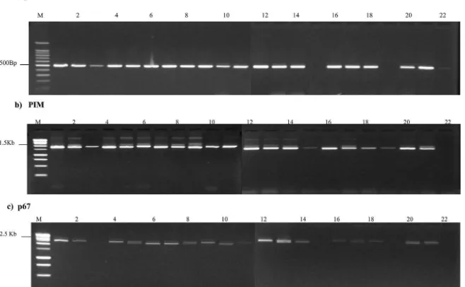
When DNA isolated from the 22 schizont-infected lymphocyte cell lines was amplified with the p104 primers that have previously been demonstrated to be specific forT. parva(Skilton et al., 2002) all except two generated a PCR product of the predicted size (Fig. 3, panel A). Consistent with RLB data, the two lines from which no product was generated were lines N86 and N102 (Fig. 3, panel A lanes 15 and 19), suggesting that these lines contained onlyT.sp. (buffalo). A faint band of the correct size was generated from N107 (Fig. 3, panel A, lane 22), suggesting the presence of a low level of T. parvaschizont-infected cells that could not be detected by RLB.
When PCR was performed using primers designed to amplify the central variable region and part of the relatively conserved N and C terminal sections of PIM, a PCR product of approximately 1.5 kb was generated from all 22 schizont-infected lines (Fig. 3, panel B). A clearly visible PCR product was generated from lines N86 and N102 (Fig. 3, panel B, lanes 15 and 19), suggesting the
presence of a PIM orthologue inT.sp. (buffalo).
The twenty two samples were also subjected to PCR with primers that amplified the near full length gene encoding p67 the major sporozoite surface antigen of T. parva (Nene et al., 1992, 1996). Nineteen samples produced a detectable PCR product of the predicted size (Fig. 3, panel C). The exceptions were lines N107 (Fig 3, panel c, lane 22) which appeared to contain a very low percentage ofT. parva-infected cells that were only detectable using p104-PCR and lines N86 and N 102 (Fig 3, panel C, lanes 15 and 19, respectively). The absence of a PCR product from the latter two cell lines suggested that the specific PCR primers employed in this study do not amplify a p67 orthologue inT.sp. (buffalo). A similar failure to amplify p104 and p67 from T. sp. (buffalo) using primers designed from theT. parvagenes encoding these antigens was re-ported byPienaar et al. (2011b), using buffalo-derived blood sam-ples from South Africa.
3.5. Cloning and sequencing of the PIM gene
. Twelve PIM nucleotide sequences were determined, two from N18 and one from each of ten other cell lines from different infected animals and predicted protein alignments, including three refer-ence sequrefer-ences were generated (data not shown). Sequrefer-ences ob-tained from N86 and N102, in which onlyT. sp. buffalowas detected, exhibited a significant level of amino acid identity withT. parvain the relatively conserved N and C termini of the protein, indicating that the PCR product represented the orthologue of PIM. The N-terminal 103 amino acids inT. parvaMuguga andT. sp. buffaloPIM T.annulata
T.parva T. mutans T.velifera T. taurotragi T. buffeli T.buffalo sp B.bigemina B.bovis
Theileria/Babesia catch all
a b c d e f g h i 1 2 3 4 5 6 7 8 9 10 1112 13 1415 16 1718 19 20 2122 23 2425
Fig. 1.Reverse line blot analysis of schizont cultures containing parasites isolated from Marula farm. The following species-specific oligonucleotide probes were used (a)T.annulata, (b)T.parva, (c)T.mutans, (d)T.velifera, (e)T.taurotragi, (f)T.buffeli, (g)T.sp. (buffalo). (h)B.bigemina, (i)B.bovis. The order of the experimental samples hybridized is DNA from cell culture isolates in lanes 1e22 was lane 1; (1) N6, (2) N13, (3) N18, (4) N20, (5) N33, (6) N36, (7) N38 (8) N43, (9) N50 (10) N55, (11) N69, (12) N76, (13) N77, (14) N79, (15) N88, (16) N99, (17) N100 (18) N103, (19) N106, (20) N107, (21) N86, (22) N102 and DNA extracted from whole cattle blood (23) N106 (24) N69 (25) N86.
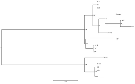
exhibited 61% identity at the amino acid level, as compared to 94% between the equivalent section of theT. parvaMuguga andT. parva Buffalo 7014 PIM. TheT. sp. buffaloPIM orthologue sequences from N86 and N102 were identical at the amino acid level. The PIM se-quences obtained from two isolates derived from animal N18, designated, (N18 and N18a) were much more closely related to N86 and N102 in the conserved sections of the protein than to the other eight sequences, all of which were derived from different animals, suggesting that they represented aT.sp. (buffalo) PIM orthologue fortuitously selected from a parasite population that appeared mixed according to RLB (Fig. 1). However, there were significant differences in the central variable region between the predicted N18 PIM protein as compared to the N86 and N 102 sequences. This suggests that the PIM orthologue ofT. sp. buffalocan vary between isolates in a similar manner to that of T. parvaPIM. The level of sequence identity of the other eight PIM gene sequences to three T. parvareference PIM sequences (derived from theT. parvastocks Muguga, Marikebuni and Kiambu V) indicated that they likely originated from T. parva. Phylogenetic analysis of the relatively conserved N-terminal PIM coding sequences using a maximum likelihood algorithm revealed a clear separation of theT.sp. (buf-falo) andT. parvasequences into two distinct clades in a tree, with high bootstrap support (Fig. 4). The C-terminal and concatenated N and C-terminal regions of the molecule generated trees with similar topology (data not shown). The alignment of the full length PIM sequences is available in Supplementary Fig. 1. The PIM se-quences have been deposited in GenBank (see materials and methods for accession numbers).
3.6. Potential immunological cross reactivity between T. sp. (buffalo) and T. parva in cattle
We have already described in section3.2characterisation of the Marula schizont-infected lymphocyte isolates with a panel of anti-schizont MAbs raised againstT. parva(Table 1). The N86 and N106T. sp. (buffalo) cell lines, in which the PIM orthologous was sequenced (see previous section), stained strongly with MAbs, 10, 12, 21 and 22, as did the T. parva isolates from Marula farm. Epitope 10 is located in the relatively conserved N-terminal region of PIM, while
epitope 12 is conformational and recognises non-contiguous resi-dues within both the N-terminal and hyper variable glutamine/ proline rich central variable regions. By contrast epitope 21 was mapped to the conserved C-terminal section of the protein, while the location of epitope 22 is unknown (Toye et al., 1996). Epitope 12 has been mapped to peptides 81e87 and 181e192 in theT. parva
Muguga PIM protein sequence (Toye et al., 1996). Since both epi-topes 10 and 12 of PIM are contained within theT. parva recom-binant sequence (the N-terminus and part of the central variable region) that forms the basis of the indirect ELISA used for sero-logical detection of exposure toT. parva, this suggests the potential for cross-reactivity between cattle exposed toT. parvaandT. sp. (buffalo).
3.7. Phylogenetic relationship between T. parva and T. sp (buffalo) based on sequences of conserved genes encoding CD8 T cell antigens
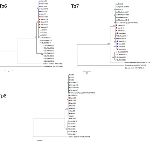
In separate studies involving 454 sequencing of genes encoding T. parvaantigens recognised by CD8 T cells (Graham et al., 2006), we identified 3 genes, Tp6, Tp7 and Tp8, that contained small numbers of synonymous point mutations but were almost completely conserved in their translated amino acid sequence. Amplicons of a segment of the orthologues of each of these 3 genes were generated from cloned buffalo cell lines confirmed as being infected withT.sp. (buffalo) and also from cloned cell lines infected with T. parva, originating from either cattle or buffalo (Table 2).
sequences separate from theT. parkasequences, with high levels of bootstrap support at the nodes separating the species (Fig. 5).
3.8. Screening of buffalo-derived stabilates of Theileria sporozoites for the presence of T. sp (buffalo)
In an attempt to determine whether available cryopreserved stabilates ofT. parvaprepared by feedingR. appendiculatusticks on buffalo also containT.sp. (buffalo), PCR amplicons of the 18s rRNA subunit from two buffalo-derived sporozoite stabilates were sub-jected to 454 sequencing (stabilates 3081 and 4110). Samples from stabilates 3081 and 4110 yielded 1479 and 607 sequence reads respectively, similar to or identical to the reference T. parva sequence; noT. sp. (buffalo) sequences were identified. The se-quences obtained included a variant present in both stabilates, which differed by one nucleotide from theT. parva18S rRNA gene reference sequence (111 and 58 reads from stabilates 3081 and 4110 respectively) and a further variant, also differing by a single nucleotide, in stabilate 4110 (45 reads). These sequences differed by two or three nucleotides from the T. sp (buffalo) reference sequence.
4. Discussion
T.sp. (buffalo), a parasite with a wide distribution in eastern and southern Africa has previously been detected at a high frequency in buffalo populations (Conrad et al., 1987; Allsopp et al., 1993; Pienaar et al., 2011a, b; Pienaar et al., 2014; Mans et al., 2015), and less frequently, also in cattle (Pienaar et al., 2011b; Githaka et al., 2014). However detection in cattle has previously been based only on PCR data and not parasite isolation. Herein we demonstrate for thefirst time thatT. sp. (buffalo) schizont cultures can also be isolated from cattle in regions of Kenya where cattle and buffalo
co-graze. The presence of T.sp. (buffalo) in cell lines isolated from cattle demonstrates that, likeT. parva,T.taurotragiand Theileria annulata,T.sp. (buffalo) is able to transform cattle cells.
Work on isolates from South African buffalo confirms that the species-specific section of the 18S rRNA gene is very similar in T. parvaand T.sp. (buffalo), as originally shown for East African animals (Allsopp et al., 1993). A further variantT. sp. (bougasvlei) also appears to be present in South African buffalo (Zweygarth et al., 2009; Pienaar et al., 2011a; Mans et al., 2015), but has not yet been detected in East Africa. Additionally, use ofTheileria cy-tochrome Oxidase III as a marker has also demonstrated that the two parasites frequently occur as co-infections in buffalo (Chaisi et al., 2013), although there is a high degree of inter-species vari-ability in this sequence, making interpretation of data from some animals difficult. Interestingly, studies in South Africa have docu-mented intermediate 18S sequences betweenT. parva andT. sp. (buffalo) (Mans et al., 2011a,b), suggesting thatTheileriamolecular epidemiology may differ regionally and that rDNA sequences alone are not definitive for speciation (Mans et al., 2015). The present study has identified polymorphism in genes encoding antigens that distinguishTheileriasp. (buffalo) fromT. parva, including p104 and p67, as noted previously (Pienaar et al., 2011b), and also PIM. Given the exceptional hyper-variability of the central polymorphic domain of PIM betweenT. parvaisolates (Toye et al., 1995; Geysen et al., 2004) it is interesting that T. sp. (buffalo) encodes an ortho-logue of the PIM gene, which could be amplified using primers derived from the conserved section of theT. parvaPIM gene and is closer in sequence to that of T. parva than those previously described from other Theileria species. Theileria sp. (buffalo) exhibited 61% amino acid identity with theT. parvareference in the conserved N-terminal domain as compared to 40% with the T. annulataorthologue, (Knight et al., 1996). A high level of con-servation of the genes encoding the Tp6, Tp7 and Tp8 antigens that 0.03
N38 N20
N107
N106 N36
N86
N77 Muguga
N18a
N102 N69
N103
N18 8
32
32 21
100
90 100
99
100
72 99
are targets of bovine CD8 T cell responses inT. parvawith theT.sp. (buffalo) orthologues also confirms the close relationship of these parasites. These genes are highly conserved within each species showing nucleotide substitutions in only a few residues. Never-theless, phylogenetic analysis demonstrates a clear demarcation of the nucleotide sequences obtained forT. parvaandT.sp. (buffalo), supporting the view that the two parasites indeed represent two separate species.
T. parvadiagnostic primers derived from the p104 gene (Skilton et al., 2002) did not generate a product from theT. sp. (buffalo) schizont-infected lymphocyte lines N86 and N102, re-confirming their specificity and usefulness forT. parvasurveillance. Primers
that amplify the p67 gene from allT. parvavariants that have been tested, whether of cattle or buffalo origin, did not generate a detectable PCR product, from theT.sp. (buffalo) lines N86 or N102, although a product was amplified from other infected cell lines, containing eitherT. parvaalone or a mixed infection with T. sp. (buffalo). This suggests that the p67 orthologue in (T. sp. buffalo) may be relatively divergent in sequence as a result of isolation and speciation. However, the differences may also be the result of minor differences in the primers derived fromT. parvaresulting in their failure to amplify theT.sp. (buffalo) orthologue. It is however very likely that there a positionally equivalent protein inT. sp. (buffalo)
that may also be antigenic, since T. annulata has an
immunologically cross reactive orthologue known as SPAG1 (Knight et al., 1996) and there is a positional homologue present in the genome of the much more distantly related apicomplexan Babesi bovisthat was identified by synteny analysis (Brayton et al., 2007). The divergence of the p67 and p104 antigen gene sequences relative to the similarity in the 18s gene, which is a widely used phylogenetic marker, suggests that complete genome sequencing would be informative in understanding the evolution of the Api-complexa and would shed further light on the reliability of 18s rDNA as a phylogenetic marker in this group of protozoa. The genome sequence would also provide insight into the evolution of the more variable Tp CD8 T cell target antigens in two closely related, but apparently distinct populations that share the same buffalo host.
The extensive amino acid sequence identity between the N-terminal sections ofT. parvaPIM and theT.sp. (buffalo) homologue, coupled with the reactivity of some of the PIM-specific MAbs with T.sp. (buffalo), suggest that the presence ofT. sp. (buffalo) could confound the results of serological analyses using the PIM-based ELISA in cattle in areas of heavy buffalo challenge. Furthermore, the schizonts of the two parasites are morphologically similar and thus parasitological analysis may also not be definitive. Therefore, in areas where buffalo are present, a real-time or conventional PCR assay (Odongo et al., 2010; Chaisi et al., 2013) may be required to confirm parasite identity. In South Africa a real time diagnostic test based on amplification of the 18s gene has been developed. The high degree of sequence similarity of the 18s rRNA gene ofT. parva andT.sp (buffalo) can occasionally lead to mis-diagnosis ofT. parva (Sibeko et al., 2008; Pienaar et al., 2011a). In the current study, the demonstration that PCR assays for p104 and p67 distinguish the two species and that the Tp6, Tp7 and Tp8 gene orthologues contain a number of nucleotide substitutions that were unique toT. sp. (buffalo) identify additional candidate genes that could be developed for differential diagnosis of these parasites. Given the high level of sequence similarity between the two species in the genes encoding the CD8 T cell target antigens, Tp6, Tp7 and Tp8 (Graham et al., 2006), it would be worthwhile to determine whetherT.sp. (buffalo) induces immune responses in cattle that cross-react withT. parvaand might convey a degree of immunity. It would also be interesting to investigate whether T. sp. (buffalo) contains orthologues of the more variable CD8 T cell target antigens (Graham et al., 2006; Pelle et al., 2011).
In common with certain otherTheileriaspecies in East Africa, for exampleT. buffeli, the tick vector involved in transmission ofT.sp. (buffalo) still needs to be identified. High-throughput sequencing failed to detectT.sp. (buffalo) in two sporozoite stabilates, produced by feedingR. appendiculatusticks on buffalo naturally infected with Theileriaparasites. Unfortunately, samples were not available from the buffalo from which these stabilates were prepared to verify whether or not they were infected withT.sp. (buffalo). One of these buffalo had been captured from the Ol Pejeta ranch in the Laikipia district of Kenya; a recent study of samples from buffalo on the same site (now a Game Conservancy), demonstrated the presence ofT. sp. (buffalo) as well as T. parva in all 8 animals examined (Hemmink H, unpublished data). Hence, the absence of the parasite in the two stabilates examined, although not conclusive, suggests thatR. appendiculatus may well not be a suitable vector for the transmission ofT.sp. (buffalo). The identification of the tick vector involved in transmission represents an area for future research. One approach would be to use PCR or RLB assays to survey dissected salivary glands from ticks collected from areas whereT.sp. (buffalo) is known to occur.T.sp. (buffalo) has not been detected in cattle in regions where buffalo are absent, as indicated by RLB studies (Njiiri et al., 2015), suggesting that it is not transmissible between cattle.
More targeted epidemiological studies to examine cattle
undergoing the acute phase of infection withTheileriaparasites in regions where buffalo are present, will be required to determine the incidence of infection with this parasite in cattle under naturalfield conditions. However, the possible role ofT.sp. (buffalo) in inducing pathogenesis in cattle will only be resolved definitively by obtain-ing T. sp. (buffalo) sporozoite isolates free ofT. parva, to enable experimental testing in cattle. In addition to identification of the tick vector, it will be important to formally name this buffalo Theileriawhose existence has been known for more than twenty years. The evidence thatT.sp. (buffalo) is a species distinct from T. parva, as has been suggested previously (Pienaar et al., 2014; Mans et al., 2015), is strong. If these twoTheileria populations were not reproductively isolated, their loci would rapidly be ho-mogenized through sexual recombination.
Acknowledgements
We are grateful to the World Bank and CGIAR consortium research project CRP3.7 for funding. Some of the work was sup-ported by a grant awarded jointly by the Department for Interna-tional Development (UK Government) and the Biotechnology and Biological Sciences Research Council (BBSRC) UK [grant number BB/ H009515/1]. Also Johanneke Hemmink was supported by a BBSRC CASE PhD studentship awarded by Genesis Faraday in partnership with The Global Alliance for Livestock Veterinary Medicines We appreciate the generous gift of South African buffalo DNA from Dr. Nick Juleff. We thank Dr. Lucilla Steinaa for her critical reading and valuable comments on the manuscript.
Appendix A. Supplementary data
Supplementary data related to this article can be found athttp:// dx.doi.org/10.1016/j.ijppaw.2015.08.006.
References
Allsopp, B.A., Baylis, H.A., Allsopp, M.T.E.P., Cavalier-Smith, T., Bishop, R.P., Carrington, D.M., Sohanpal, B.K., Spooner, P.R., 1993. Discrimination between six species of Theileria using oligonucleotide probes which detect small subunit ribosomal RNA sequences. Parasitology 107, 157e165.
Allsopp, M.T., Theron, J., Coetzee, M.L., Dunsterville, M.Y., Allsopp, B.A., 1999. The occurrence of Theileria and Cowdria parasites in African buffalo (Syncerus caffer)and their associated amblyomma hebraeum. Onderstepoort. J. Vet. Res. 66, 245e249.
Bishop, R., Musoke, A., Morzaria, S., Gardner, M., Nene, V., 2004. Theileria: intra-cellular protozoan parasites of wild and domestic ruminants transmitted by ixodid ticks. Parasitology 129, S271eS283.
Brayton, K., Lau, A.O.T., Herndon, D.R., Lao, A.O.T., Herndon, D.R., Hannick, L., Kappmeyer, L.S., Berens, S.J., Bidwell, S.L., Brown, W.C., Crabtree, J., Fadrosh, D., Feldblum, T., Forberger, A., Haas, B.J., Howell, J.M., Khouri, H., Koo, H., Mannv, D.J., Ntrominie, J., Paulsen, I.T., Radune, D., Ren, Q., Smith, R.K., Suarez, C.E., White, O., Wortmann, J.R., Knoels, D.P., McElwain, T.F., Nene, V.N., 2007. Genome sequence of babesia bovis and comparative analysis of api-complexan hemoprotozoa. Plos Pathog. 3, 1401e1411.
Brown, C.G.D., 1987. Theileridae. In: Taylor, A.E.R., Baker, H.R. (Eds.), In Vitro Methods for Parasite Cultivation. Academic Press, London, pp. 250e253.
Chaisi, M.E., Janssens, M.E., Vermeiren, L., Oosthuizen, M.C., Collins, N.E., Geysen, D., 2013. Evaluation of a real time PCR test for the detection and discrimination of Theileria species in the African buffalo (Syncerus caffer). Plos One 18, 1e11.
Conrad, P., Stagg, D.A., Grootenhuis, J.G., Irvin, A.D., Newson, J., Njamunggeh, R., Rossiter, P.B., Young, A.S., 1987. Isolation of Theileria parasites from African buffalo (Syncerus caffer) and characterization with anti-schizont monoclonal antibodies. Parasitology 94, 413e423.
Conrad, P., ole-MoiYoi, O., Baldwin, C., Dolan, T., O'Callaghan, C., Njamunggeh, R., Grootenhuis, J., Stagg, D., Leitch, B., Young, A., 1989. Characterization of buffalo-derived Theilerial parasites with monoclonal antibodies and DNA probes. Parasitology 98, 179e188.
D'Oliviera, C., van der Weide, M., Habela, M.A., Jacquiet, P., Jongejan, F., 1995. Detection of Theileria annulata in blood samples of carrier cattle by PCR. J. Clin. Microbiol. 33, 2665e2669.
Edgar, R.C., 2004. MUSCLE: multiple sequence alignment with high accuracy and high throughput. Nucleic Acids Res. 32, 1972e1979.
Geysen, D., Bazarusanga, T., Brandt, J., Dolan, T.T., 2004. An unusual mosaic gene structure of the PIM gene of Theileria parva and its relationship to allelic di-versity. Mol. Biochem. Parasitol. 133, 163e173.
Githaka, N., Konnai, S., Bishop, R., Odongo, D., Lekolool, I., Kariuki, E., Gakuya, S., Kamau, L., Isezaki, M., Murat, S., Ohashi, K., 2014. Identification and sequence characterization of novel Theileria genotypes from the waterbuck (kobus defassa) in a Theileria parva-endemic area in Kenya. Vet. Parasitol. 202, 180e193.
Graham, S., Pelle, R., Honda, Y., Mwangi, D., Nyerhovwo, M., Tonukari, J., Yamage, M., Glew, E., de Villiers, E., Shah, T., Bishop, R., Abuya, E., Awino, E., Gachanja, J., Luyai, A., Mbwika, F., Muthiani, A., Ndegwa, D., Njahira, M., Nyanjui, J., Onono, F., Osaso, J., Saya, R., Wildmann, C., Fraser, C., Maudlin, M., Gardner, M., Morzaria, M., Loosmore, F., Gilbert, S., Audonnet, J.C., van der Bruggen, P., Nene, V., Taracha, E., 2006. Theileria parva candidate vaccine antigens recog-nized by immune bovine cytotoxic T lymphocytes. Proc. Nat. Acad. Sci. U. S. A. 103, 3286e3291.
Gubbels, J.M., de Vos, A.P., van der Weide, M., Viseras, J., Schouls, L.M., de Vries, E., Jongejan, F., 1999. Simultaneous detection of bovine Theileria and Babesia species by reverse line blot hybridization. J. Clin. Microbiol. 37, 1782e1789.
Haas, B.J., Gevers, D., Earl, A.M., Feldgarden, M., Ward, D.V., Giannoukos, G., Ciulla, D., Tabbaa, D., Highlander, S.K., Sodergren, E., Methe, B., deSantis, T.Z., 2011. Chimeric 16S rRNA sequence formation and detection in sanger and 454-pyrosequenced PCR amplicons. Genome Res. 21, 494e504.
Katende, J., Morzaria, S., Toye, P., Skilton, R.,, Nene, V., Nkonge, C., Musoke, A., 1998. An enzyme-linked immunosorbent assay for detection of Theileria parva anti-bodies in cattle using a recombinant polymorphic immunodominant molecule. Parasitol. Res. 84, 408e416.
Knight, P.A., Musoke, A.J., Gachanja, J.N., Nene, V., Katzer, F., Boluter, N., Hall, R., Brown, C.G.D., Williamson, S., Kirvar, E., Bell-Skayi, L., Hussain, A., Tait, A., 1996. Conservation of neutralizing determinants between the sporozoite surface antigens of Theileria annulata and Theileria parva. Exp. Parasitol. 82, 229e241.
Mans, B.J., Pienaar, R., Latif, A.A., Potgieter, F.T., 2011a. Diversity in the 18S SSU rRNA V4 hyper-variable region of Theileria spp. in Cape buffalo (Syncerus caffer) and cattle from southern Africa. Parasitology 1e14.
Mans, B.J., Pienaar, R., Potgieter, F.T., Latif, A.A., 2011b. Theileria parva, T. sp. (buffalo) and T. sp. (bougasvlei) 18S variants. Vet. Parasitol. 182, 382e383.
Mans, B.J., Pienaar, R., Latif, A.A., 2015. A review of Theileria diagnostics and epidemiology. Int. J. Parasitol. Parasites Wildl. 4, 104e118.http://dx.doi.org/10.
1016//j.ijppaw.2014.12.006.
Minami, T., Spooner, P., Irvin, A., Ocama, J., Dobbelaere, D., Fujinaga, T., 1983. Characterisation of stocks of Theileria parva by monoclonal antibody profiles. Res. Vet. Sci. 35, 334e340.
Morzaria, S.P., Dolan, T.T., Norval, R.A.I., Bishop, R.P., Spooner, P.R., 1995. Generation and characterisation of cloned Theileria parva parasites. Parasitology 111, 39e49.
Morzaria, S., Spooner, P., Bishop, R., Mwaura, S., 1999. The preparation of a com-posite stabilate for immunisation against East Coast fever. In: Morzaria, S., Williamson, S. (Eds.), Live Vaccines for Theileria Parva: Deployment in Eastern, Central and Southern Africa. International Livestock Research Institute, Kenya, pp. 56e61.
Nene, V., Iams, K., Gobright, E., Musoke, A.J., 1992. Characterization of the gene encoding a candidate vaccine antigen of Theileria parva sporozoites. Mol. Bio-chem. Parasitol. 51, 17e27.
Nene, V., Musoke, A., Gobright, E., Morzaria, S., 1996. Conservation of the sporozoite p67 vaccine antigen in cattle-derived Theileria parva stocks with different cross-immunity profiles. Infect. Immun. 64, 2056e2061.
Njiiri, N.E., Bronsvoort, B.M., Collins, N.E., Collins, N.E., Steyn, H.C., Troskie, M., Vorster, I., Thumbi, S.M., Sibeko, K.P., Jennings, A., van Vyk, I.C., Mbole-Kariuki, M., Kiara, H., Poole, E.J., Haotte, H., Coetzer, K., Oosthuizen, M.C., Woolhouse, M., Toye, P., 2015. The Epidemiology of tick-borne haemopar-asasites as determined by reverse line blot hybridization assay in an intensively studied cohort of calves in Western Kenya. Vet. Parasitol. 210, 69e76.http://
dx.doi.org/10.1016/vetpar.2015.02.020.
Norval, R.A.I., Perry, B.D., Young, A.S., 1992. The Epidemiology of Theileriosis in Africa. Academic Press, London.
Obara, I., Seitzer, U., Musoke, A., Spooner, P.R., Ahmed, J., David, D.O., Kemp, S., Silva, J.C., Bishop, R.P., 2015. Molecular evolution of a central region containing B-cell epitopes in the gene encoding the p67 sporozoite antigen within afield population of Theileria parva. Parasitol. Res. 114, 1729e1737.http://dx.doi.org/
10.1007/s00436-015-4358-6.
Odongo, D.O., Sunter, J.D., Kiara, H.S., Skilton, R.A., Bishop, R.P., 2010. A nested PCR assay exhibits enhanced sensitivity for detection of Theileria parva infections in
bovine blood samples from carrier cattle. Parasitol. Res. 106, 357e365.
Oura, C.A., Bishop, R.P., Wampande, E.M., Lubega, G.W., Tait, A., 2004. Application of a reverse line blot assay to the study of haemoparasites in cattle in Uganda. Int. J. Parasitol. 34, 603e613.
Oura, C.A., Tait, A., Asiimwe, B., Lubega, G.W., Weir, W., 2011a. Haemoparasite prevalence and Theileria parva strain diversity in Cape buffalo (Syncerus caffer) in Uganda. Vet. Parasitol. 175, 212e219.
Oura, C.A., Tait, A., Asiimwe, B., Lubega, G.W., Weir, W., 2011b. Theileria parva ge-netic diversity and haemoparasite prevalence in cattle and wildlife in and around lake Mburo National Park in Uganda. Parasitol. Res. 108, 1365e1374.
Payne, R., 1999. Preparation of stabilates for immunisation against East Coast fever at NVRC, Muguga, Kenya. 1999. In: Morzaria, S., Williamson, S. (Eds.), Live Vaccines for Theileria Parva: Deployment in Eastern, Central and Southern Africa. International Livestock Research Institute, Kenya, pp. 62e65.
Pelle, R., Graham, S.P., Njahira, M., Osaso, J., Saya, R.M., Odongo, D.O., Toye, P.G., Spooner, P.R., Musoke, A.J., Mwangi, D.M., Taracha, E.L.N., Morrison, I.W., Weir, W., Silva, J.C., Bishop, R.P., 2011. Two Theileria parva CD8 T cell antigen genes are more variable in buffalo than cattle and differ in pattern of sequence diversity. Plos One 6, e19015, 1-/10.1371.
Pienaar, R., Potgieter, F.T., Latif, A.A., Thekisoe, O.M., Mans, B.J., 2011a. The hybrid II assay; a sensitive and specific real-time hgridization assay for the diagnosis of Theileria parva infection in Cape buffalo (Syncerus caffer). Parasitology 138, 1935e1944.http://dx.doi.org/10.1017/S0031182011001454.
Pienaar, R., Potgieter, F.T., Latif, A.A., Thekisoe, O.M.M., Mans, B.J., 2011b. Mixed Theileria infections in free-ranging buffalo herds: implications for diagnosing Theileria parva infections in Cape buffalo (Syncerus caffer). Parasitology 138, 884e895.
Pienaar, R., Latif, A.A., Thekisoe, O.M., Mans, B.J., 2014. Geographic distribution of Theileria sp. (buffalo) and Theileria sp. (bougasvelei) in southern Africa: im-plications for speciation. Parasitology 141, 411e424.
Quince, C., Lanzen, A., Davenport, R.J., Turnbaugh, P.J., 2011. Removing noise from pyrosequenced amplicons. BMC Bioinforma. 12, 38.http://dx.doi.org/10.1186/ 1471-2105-12.
Radley, D.E., Brown, C.G.D., Cunningham, M.P., Kimber, C.D., Musisi, F.L., Payne, R.C., Purnell, R.E., Stagg, S.M., Young, A.S., 1975. East Coast fever: 3.Chemoprophy-lactic immunization of cattle using oxytetracycline and a combination of thei-lerial strains. Vet. Parasitol. 1, 51e60.
Schloss, P.D., Westcott, S.L., Ryabin, T., Hall, J.R., Hartmann, M., Hollister, E.B., Lesniewski, R.A., Oakley, B.B., Parks, D.H., Robinson, C.J., Stahl, J.W., Stres, B., Thallinger, G.G., Van Horn, D.J., Weber, C.F., 2009. Introducing mothur: open-source, platform-independent, community-supported software for describing and comparing microbial communities. Appl. Env. Microbiol. 75, 7537e7541.
Sibeko, K.P., Oosthuizen, M.C., Collins, N.E., Geysen, D., Rambritch, N.E., Latif, A.A., Groeneveld, H.T., Potgieter, F.T., Coetzer, J.A., 2008. Development and evaluation of a real-time polymerase chain reaction test for the detection of Theileria parva infections in Cape buffalo (Syncerus caffer) and cattle. Vet. Parasitol. 155, 37e48.
Skilton, R.A., Bishop, R.P., Katende, J.M., Mwaura, S., Morzaria, S.P., 2002. The persistence of Theileria parva infection in cattle immunised using two stocks which differ in their ability to induce a carrier state: analysis using a novel blood spot PCR assay. Parasitology 124, 265e276.
Stamatakis, A., 2014. RAxML version 8: a tool for phylogenetic analysis and post analysis of large phylogenies. Bioinformatics 30, 1312e1313.http://dx.doi.org/
10.1903/bioinformatics/btu033.
Tamura, K., Peterson, D., Peterson, N., Stecher, G., Nei, M., Kumar, S., 2011. MEGA5: molecualr evolutionary genetics analysis using maximum likelihood, evolu-tionary diatance, and maximum parsimony methods. Mol. Biol. Evol. 28, 2731e2739.
Toye, P.G., Goddeeris, B.M., Iams, K., Musoke, A.J., Morrison, W.I., 1991. Character-ization of a polymorgphic immunodominant molecular in sporozoites and schizonts of Theileria parva. Parasite Immunol. 13, 49e52.
Toye, P., Gobright, E., Nyanjui, J., Nene, V., Bishop, R., 1995. Structure and sequence variation of the genes encoding the polymorphic immunodominant molecule (PIM), an antigen of Theileria parva recognised by inhibitory monoclonal an-tibodies. Mol. Biochem. Parasitol. 73, 165e178.
Toye, P., Nyanjui, J., Goddeeris, B., Musoke, A.J., 1996. Identification of neutralization and diagnostic epitopes on PIM, the polymorphic immunodominant molecule of Theileria parva. Inf. Immun. 64, 1832e1838.
Zweygarth, E., Koekemoer, O., Josemans, A.I., Rambritch, N., Pienaar, R., Putterill, J., Latif, A., Potgieter, F.T., 2009. Theileria-infected cell line from an African Buffalo (Syncerus caffer). Parasitol. Res. 105, 579e581. http://dx.doi.org/10.1007/


