Nicotine and Amyloid Formation
Hong Zeng, Yongbo Zhang, Li-Jun Peng, Haiyan Shao, Nanda K. Menon,
Jing Yang, Arthur R. Salomon, Robert P. Freidland, and Michael G. Zagorski
The major protein constituents of amyloid deposits in
Alzheimer’s disease (AD) are the 40-residue
b
-amyloid
(A
b
) (1– 40) peptide and the 42-residue A
b
(1– 42) peptide.
The A
b
(1– 42) is more pathogenic and produced in
greater quantities in familial forms of AD. A major goal of
research is to uncover a suitable inhibitor that either
slows down or inhibits A
b
formation (
b
-amyloidosis).
During
b
-amyloidosis, structural changes associated with
the conversion of monomeric A
b
peptide building blocks
into the aggregated fibrillar
b
-sheet structures occur
(
a
-helix
3
b
-sheet or random, extended chain
3
b
-sheet).
In previous work, we and others established that nicotine,
a major component of cigarette smoke, inhibits
b
-amy-loidosis of the A
b
(1– 42), which may result from nicotine
binding to the
a
-helical structure. These conclusions were
based on solution nuclear magnetic resonance (NMR)
spectroscopic studies with the nonnative 28-residue A
b
(1–
28). This information suggests that, when administered
therapeutically to AD patients, nicotine may not only
affect cholinergic activation, but could also conceivably
alter amyloid deposition. In this report, NMR studies were
augmented with the naturally occurring A
b
(1– 42), under
conditions where the peptide folds into a predominantly
a
-helical or random, extended chain structure. The major
result is that nicotine shows only modest binding to these
conformations, indicating that the nicotine inhibition to
b
-amyloidosis probably results from binding to a small,
soluble
b
-sheet aggregate that is NMR invisible. Biol
Psychiatry 2001;49:248 –257 © 2001 Society of
Biologi-cal Psychiatry
Key Words: Amyloid, A
b
peptide, nicotine, NMR,
Alz-heimer’s disease
Introduction
A
lzheimer’s disease (AD) is the major cause of
adult-onset dementia and is characterized by an abundance
of intraneuronal neurofibrillary tangles and extracellular
amyloid plaques (Iqbal et al 1999; Selkoe 2000). The
major component of the amyloid plaques is the
b
-amyloid
(A
b
), a normally secreted small peptide that exists in two
predominant forms: 1) the 40-residue A
b
(1– 40) and 2) the
42-residue A
b
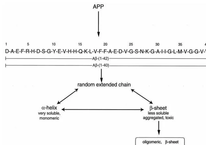
(1– 42) (Figure 1). The A
b
(1– 40) and
A
b
(1– 42) peptides differ in the absence or presence of
two extra C-terminal residues. Both peptides result from
the processing of a larger amyloid precursor protein (APP)
(Hardy 1997) (Figure 1). The amyloid deposits are
char-acterized by distinct tinctorial properties and fibrils of
7–10 nm in diameter, in which the primary components
are aggregated proteins with antiparallel cross–
b
-pleated
sheet structures (Malinchik et al 1998; Teplow 1998).
The biological functions of both the APP and the A
b
are
currently unknown, although they are believed to play
roles in neuronal homeostasis, cell adhesion, G protein
coupling, and/or oxidative stress. Numerous genetic and
cell viability studies support a key role for the A
b
peptide
in AD neurodegeneration. The A
b
peptide becomes
neu-rotoxic to cortical cell cultures when aggregated as
amy-loidlike
b
-sheet structures (Geula et al 1998; Iversen et al
1995; Pike et al 1995; Simmons et al 1994). Biochemical
studies also suggest that the longer, 42-residue A
b
(1– 42)
is more pathogenic than the shorter 40-residue A
b
(1– 40)
(Younkin 1995), due to its greater in vitro tendency to
aggregate and precipitate as amyloid (Barrow and
Zagor-ski 1991). Recent studies demonstrated that the A
b
(1– 42)
is significantly elevated in cerebrospinal fluid of AD
patients (Andreasen et al 1999; Jensen et al 1999; Kuo et
al 1999; Wang et al 1999).
On the basis of extensive biophysical measurements, in
solution the A
b
peptide can fold into
a
-helical or random,
extended chain structures, as well as soluble
b
-sheet
structures that eventually precipitate as amyloid (Huang et
al 2000; Teplow 1998; Zagorski 1999). The relative ratios
of the structures are strongly dependent on the solution
conditions, with hydrophobic lipidlike environments
en-couraging
a
-helical structure and high ionic strength and
midrange pH 4 –7 values favoring
b
-sheet. The
a
-helical
and random, extended chain structures are monomeric,
whereas the late-formed
b
-sheet structures in solution are
aggregated (oligomeric) and neurotoxic (Lambert et al
1998; Sayre et al 1997). Because the
b
-sheet structure is
From the Department of Chemistry (HZ, YZ, L-JP, HS, JY, ARS, MGZ) and the Department of Neurology, School of Medicine (RPF), Case Western Reserve University, Cleveland, Ohio, and the Department of Biochemistry, University of Georgia, Athens (NKM).
Address reprint requests to Michael G. Zagorski, Case Western Reserve University, Department of Chemistry, Cleveland OH 44106-7078.
Received May 30, 2000; revised November 10, 2000; accepted November 21, 2000.
neurotoxic, a therapeutically useful inhibitor should bind
or stabilize the
a
-helical, random, extended chain, or early
formed
b
-sheet structures, which are very soluble and
nontoxic.
Nicotine is a predominant component of cigarette
smoke and is currently being used in pilot clinical studies
for the treatment of AD (Emilien et al 2000; Wilson et al
1995). The beneficial effects of this treatment have been
attributed to an upregulation of nicotine receptors that are
deficient in the AD brain, or possibly a protection from the
A
b
-induced neurotoxicity (Kihara et al 1997; Zamani et al
1997). We and others have shown that nicotine may have
a dual effect, in that besides promoting the upregulation of
receptors or being neuroprotective, it may also inhibit
b
-amyloidosis. These conclusions were formulated on in
vitro studies that established that nicotine slows down
A
b
(1– 42) fibril formation (Moore et al 2000; Salomon et
al 1996) and that this inhibition may be due to binding to
the
a
-helical structure (Zagorski 1999). The latter was
based on nuclear magnetic resonance (NMR) studies of
the homologous A
b
(1–28) peptide that contains residues
1–28 of the A
b
(1– 42). Here we report an extension of this
work with the A
b
(1– 42) and show that the inhibition does
not result from binding to either the
a
-helical or random,
extended chain structures.
Methods and Materials
Sample Preparation
Distilled/deionized water, nicotine, and cotinine were of the highest grade possible from commercial sources and were used without further purification. The organic solvents trifluoroacetic acid (TFA) and hexafluoroisopropanol (HFIP) were distilled under an inert atmosphere of nitrogen and stored in opaque bottles at 5°C. The perdeuterated sodium dodecyl sulfate (SDS-d25), EDTA (Na2EDTA-d12),
3-(trimethylsilyl)propionate-2,2,3,3-d4(TSP), and the solvent deuterium oxide (D2O) were
obtained from Cambridge Isotopes (Andover, MA).
The synthetic Ab peptides were obtained from commercial sources (Anaspec, San Jose, CA) or prepared using standard Fmoc chemistry on an automated (PerSeptive Biosystems [Foster City, CA] 9050-Plus) synthesizer. Uniformly (.95%) 15
N-labeled Ab(1– 42) were prepared biosynthetically from
Esche-richia coli as a recombinant fusion protein in minimal media
containing15
NH4Cl as the sole nitrogen source. The peptide was
cleaved from the fusion protein at pH 8.3 using restriction protease Factor Xa. To isolate the cleaved Ab(1– 42) from the fusion protein components, aggregation of the Ab(1– 42) was induced by stirring in water (12 hours, pH 4 –7). The aggregated Ab(1– 42) was collected by centrifugation (10,000 g, 20 min) and purified by reverse-phase high-performance liquid chroma-tography, with either a Zorbax-300 Bonded Silica (Rockland Technologies) or a Vydaq-259VHP822 column (Separation Group). The solvent system consisted of a linear gradient of Figure 1. Overview of the formation of theb-amyloid (Ab) peptide from the amyloid precursor protein (APP), including the amino acid sequences for the Ab(1– 40) and Ab(1– 42) peptides. Depending upon conditions, the Abpeptide exists in distinct conformations in solution, but in the amyloid deposit, only the oligomericb-pleated sheet structure is present. Both peptides can aggregate into soluble
b-sheet structures, which are neurotoxic and eventually precipitate as amyloid.
Nicotine Binding tob-Amyloid BIOL PSYCHIATRY 249
20 – 80% acetonitrile in water that contained either 0.1– 0.08% TFA or a sodium acetate buffer (5 mmol/L) at pH 8.0, which was heated to 55– 60°C to improve peak resolution and purity (Boyes 1995). Peptide identity, verified by mass spectrometry and NMR, had purity levels greater than 90%.
To disaggregate the Ab(1– 42) and generate exclusively mo-nomeric random, extended chain structure, the purified peptides were predissolved in neat TFA solution (Jao et al 1997; Zagorski et al 1999). Trace amounts of TFA were removed by redissolving in HFIP (5:1 [mg:mL]) and then removing the HFIP with N2and
vacuum (0.5 mm Hg, 2 hours). The disaggregation afforded by TFA allows the dry, unstructured, and monomeric Ab to fold into its nativelike conformation in water, without potential interferences from trace organic cosolvents or small aggregates (“seeds”) of the Ab.
The Ab(1– 42) peptide solutions (0.60 mL) were prepared in
5-mm NMR tubes at concentrations of 0.1–1.4 mmol/L. The solvent-free, TFA-pretreated peptides were dissolved in either SDS-d25solution in H2O (for studies examining the effects of
nicotine on the a-helical structure) or in 9:1 H2O:D2O (for
studies examining the effects of nicotine on the random, ex-tended chain structure). Stock solutions of (S)-(2)-cotinine, (R)-(1)-nicotine, and (S)-(2)-nicotine were prepared in buffered water (pH 7.2) at 20 mmol/L concentrations, after which aliquots were added to the NMR solutions containing the Ab(1– 42). All solutions contained 20 mmol/L sodium phosphate buffer at pH 7.2, 0.05 mmol/L Na2EDTA-d12, 0.05 mmol/L sodium azide
(NaN3), and 0.05 mmol/L TSP. The latter three components were
included to remove trace metal ions, prevent microbial growth, and provide an internal chemical shift reference at 0 ppm, respectively. The SDS-d25concentration was kept high relative
to the peptide concentration so that it was well above 8 mM (the
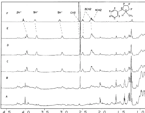
Figure 2. Upfield regions of the proton nuclear magnetic resonance (1
H NMR) spectra (600 MHz) for theb-amyloid (Ab) (1– 42) (A) alone (0.5 mM) and with (B) 0.25 mM, (C) 0.50 mM, (D) 1.0 mM, and (E) 2.0 mM nicotine. The upper spectrum (F) corresponds to nicotine without the Ab(1– 42). All solutions contained 80 mM sodium dodecyl sulfate (SDS-d25) and 20 mM phosphate buffer at pH
7.2 and 25°C. The NMR peaks for nicotine are shown with the assignments.
250 BIOL PSYCHIATRY H. Zeng et al
critical micelle concentration) and also above the average aggrega-tion number (Henry and Sykes 1994). To ensure that the buffer maintained pH 7.2 during the titrations, the pH of the solutions was checked with a special pH electrode (Microelectrodes) that fit inside the 5-mm NMR tube. The pH values were measured at room temperature and corrections for isotope effects or for the presence of SDS-d25were not performed, since control experiments showed that
these substances did not significantly alter the pH.
Nuclear Magnetic Resonance Spectroscopy
All NMR spectra were acquired at 600 MHz using a Varian Inova-600 spectrometer. The NMR data were transferred to Indigo XS24 (Silicon Graphics) computer workstations and processed using the FELIX (version 97, Biosym) program.
Chemical shifts were referenced to an internal standard of TSP. The one-dimensional NMR spectra were acquired with a pre-saturation pulse that was applied during the recycle delay (2 sec) to suppress the H2O signal. Typically, most one-dimensional
spectra had 8000-Hz spectral widths and 32-K complex data points. To further enhance the digital resolution and the signal to noise, before Fourier transformation the data were zero filled once and multiplied by a Lorentzian-to-Gaussian window function.
The two-dimensional 1
H-15
N heteronuclear single-quantum coherence (HSQC) experiments were recorded with a uniformly
15
N-labeled Ab(1– 42) sample with 32 scans and 1024 complex points and the transmitter placed on the water signal (Bax and Grzesiek 1993). The sweep widths were 6373.5 and 2000.0 Hz in the F2 and F1 dimensions, respectively. Phase-sensitive data
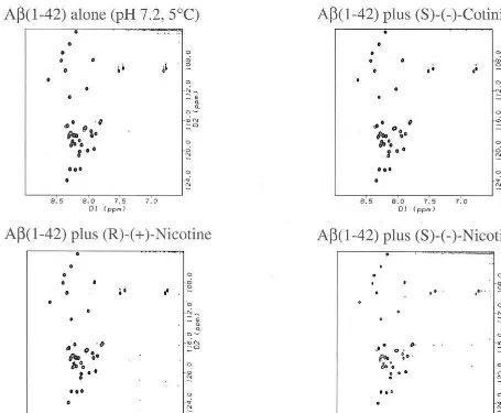
Figure 3. The two-dimensional1H-15N heteronuclear single-quantum coherence spectra of recombinant15N-labeledb-amyloid (Ab) (1– 42) (0.20 mM) in 9:1 H2O:D2O phosphate buffer at pH 7.2 and 5°C. A single sample was separated into four parts to which 0.40
mmol/L of (S)-(2)-cotinine, (R)-(1)-nicotine (the inactive enantiomer), or (S)-(2)-nicotine (the active enantiomer) was added. The spectra for 0.40 mmol/L of (S)-(2)-cotinine and (R)-(1)-nicotine were identical to that of the Ab(1– 42) alone; however, the spectra containing (S)-(2)-nicotine showed minor perturbations (see expanded plot in Figure 4).
Nicotine Binding tob-Amyloid BIOL PSYCHIATRY 251
were collected using time-proportional phase incrementation in the F1dimension, and pulsed field gradients were employed to
minimize the artifact content of the spectra, suppress the intense H2O resonance, and select for the coherence transfer pathway,
whereby magnetization passes from 15N to1H for observation (Kay 1995).
Results
The NMR approach has the distinct advantage of being
able to sequence-specifically determine where a particular
secondary structure is located within the primary
se-quence. Furthermore, for drug design the NMR method
can provide valuable information about protein dynamics
and specific data about the individual amino acid
side-chains that bind to a particular ligand (Craik 1996; Hajduk
et al 1999; Shuker et al 1996). With sufficient distance and
dihedral angle constraints, the NMR approach is also
capable of rendering a complete three-dimensional
structure.
Our sample preparation protocol ensures that the A
b
(1–
42) peptide adopts a well-defined, monomeric state before
starting the NMR measurements. A major difficulty relates
to the ease in which the A
b
(1– 42) aggregates and
precip-itates, which in turn creates problems in the reiteration of
results. Because the peptide is very prone to
time-depen-dent aggregation (Zagorski et al 1999), all preaggregated
peptide material was carefully removed from the samples
before analysis (Jao et al 1997). After this disaggregation
was performed, the A
b
solutions were stable for several
days at pH 7.2 and 5°C, with no precipitation or other
spectral changes detected by NMR. Nuclear magnetic
resonance diffusion and sedimentation equilibrium
exper-iments confirmed that the A
b
(1– 42) adopts intact,
nonde-graded monomeric states (Shao et al, submitted).
NMR Studies with the
a-Helical Structure
Previous studies established that the A
b
(1–28), A
b
(1– 40),
and A
b
(1– 42) peptides fold into predominantly
mono-meric
a
-helical structures in lipid environments (Coles et
al 1998; Marcinowski et al 1998; Shao et al 1999;
Talafous et al 1994; Terzi et al 1997). Because solution
NMR studies with high-molecular-weight lipids (i.e.,
bi-layers) are not feasible (due to slow tumbling rates),
detergents such as the negatively charged SDS micelle are
commonly used. Detergent micelles adequately mimic a
membranelike environment and are frequently used in the
structural studies of peptides and proteins (Henry and
Sykes 1994). Studies of the A
b
in lipidlike environments
are important and may be pertinent to the native state,
when bound to lipoproteins and albumin in human plasma
(Biere et al 1996). The present studies were undertaken to
explore the effect of nicotine on the
a
-helical structure of
the A
b
(1– 42) peptide in SDS solution.
Our previous NMR studies of the A
b
(1–28)
demon-Figure 4. The expanded1
H-15
N heteronuclear single-quantum coherence spectra of recombinant15
N-labeledb-amyloid (Ab) (1– 42) [0.20 mmol/L alone and with (S)-(2)-nicotine (0.60 mmol/L)] (for complete spectrum, see Figure 3). Note that peaks of Tyr10, Glu11, Val12, and His13 become reduced in intensity with nicotine and the Val12 peaks splits into a doublet.
252 BIOL PSYCHIATRY H. Zeng et al
strated that nicotine binds to the His side-chains when
folded in an
a
-helical conformation (Salomon et al 1996;
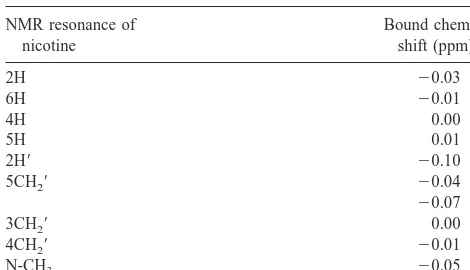
Zagorski 1999). Figure 2 shows the upfield spectral
regions in SDS solution of the A
b
(1– 42) alone (0.5
mmol/L) (Figure 2A), nicotine alone (Figure 2E), and the
A
b
(1– 42) with varying amounts of nicotine (0.25 mmol/L
[Figure 2B], 0.50 mmol/L [Figure 2C], and 1.0 mmol/L
[Figure 2D]). The observed chemical shift changes
of nicotine when mixed with the A
b
(1– 42) are
summa-rized in Table 1. The signals showing the most pronounced
changes in chemical shifts are for the 2
9
H, 5
9
CH
2, and
N-CH
3of nicotine, which are significantly less in
magni-tude than in previous NMR studies of the A
b
(1–28)
(Salomon et al 1996). The binding is in fairly rapid
exchange, as shown by single averaged NMR peaks and
the lack of any linearity between the chemical shift and
nicotine concentration. Most of the signals show upfield
chemical shift changes, and the largest shifts occur in the
5-membered N-methylpyrrolidine ring.
To further explore the possibility of binding, we
ob-tained two-dimensional HSQC NMR data with uniformly
15
N-labeled A
b
(1– 42) in SDS solution. The HSQC
con-tains cross-peaks for all protonated nitrogens (
15N-H
bonds) and thus provides a fingerprint map of a protein.
The binding of a ligand induces movement of specific
cross-peaks indicative of particular binding locations in
the protein. Recently, the HSQC experiment has emerged
as a powerful tool for the rapid screening of compounds
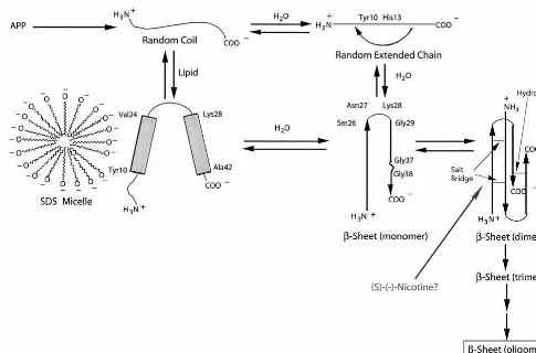
Figure 5. Presumed mechanism ofb-amyloidosis for theb-amyloid (Ab) (1– 42), which involves formation of a largely monomeric
a-helical structure when bound to lipids (Shao et al 1999) and a predominantly monomeric random, extended chain structure in water solution. Thea-helices are depicted with darkened cylinders, and the random coil or extended strand regions are drawn with wavy lines. The initially formed monomeric b-sheet structure is presumptive and is based on previous studies showing a reverse turn at Ser26 –Asn27–Lys28 –Gly29 (Hilbich et al 1991; Lee et al 1995). In the brain, after proteolytic cleavage of the amyloid precursor protein (APP), the Ab(1– 42) presumably monomeric structures that can aggregate intob-sheet–like structures as a result of possible brain microenvironmental changes such as localized regions of high peptide concentration or possibly small amounts of preformed
b-sheet (“seed”) material. Thea-helical structure remains on the sodium dodecyl sulfate (SDS) or lipid surface and does not become imbedded into the hydrophobic interior. On the basis of the present nuclear magnetic resonance data, nicotine does not bind to the
a-helical or random extended chain structures, suggesting that the inhibition may result from binding to a smallb-sheet aggregate. A more effective nicotinelike amyloid inhibitor could be designed to bind to either the random, extended chain structure ora-helical structure, thereby slowing down aggregation to the toxicb-sheet structures that eventually produce amyloid.
Nicotine Binding tob-Amyloid BIOL PSYCHIATRY 253
that can facilitate the design of more specific drug
candi-dates (Hajduk et al 1999; Shuker et al 1996). When
compared to conventional assays, the high-throughput
HSQC-based screening approach can identify
high-affin-ity ligands for protein targets with no known function.
The HSQC spectra were obtained in SDS solution at
neutral pH with the A
b
(1– 42) at 0.25 mM concentration.
Incremental titrations of (R)-(
1
)-nicotine (the inactive
isomer) and (S)-(
2
)-nicotine (the active isomer) were
added to separate peptide solutions, in which the peptide
concentration was constant (0.25 mM) and the nicotine
concentration varied (0.050 mM, 0.15 mM, 0.25 mM, and
0.50 mM). The HSQC data were acquired within 10 min
after each addition of the nicotine, and the pH was checked
to ensure adequate buffering capacity. The NMR data
showed no changes in chemical shifts, thus establishing
that (R)-(
1
)-nicotine and (S)-(
2
)-nicotine do not bind to
the
a
-helical structure of the A
b
(1– 42) in a manner that
induces changes in the backbone secondary structure.
NMR Studies with the Random, Extended
Chain Structure
We have recently completely assigned the
1H and
15N
NMR assignments of the A
b
(1– 42) in water solution at
pH 7.2 (Shao et al, submitted). Overall, the NMR data
demonstrate that the peptide adopts a predominantly
ran-dom, extended chainlike structure. These results suggest
that by itself the A
b
(1– 42) peptide may be soluble and
that brain microenvironmental changes in the AD brain
may be required to induce a random, extended chain
3
b
-sheet conversion.
To analyze whether or not nicotine binds to the random,
extended chainlike structure, we obtained
two-dimen-sional HSQC data NMR with uniformly
15N-labeled
peptide, in an analogous manner as done in the SDS
solution. The HSQC spectra were obtained at neutral pH
with gradual titration of (R)-(
1
)-nicotine (the inactive
isomer) and (S)-(
2
)-nicotine (the active isomer) to
sepa-rate A
b
(1– 42) solutions; the peptide concentration was
maintained constant (0.20 mM), whereas the nicotine
concentration varied (0.050 mM, 0.15 mM, 0.25 mM, and
0.50 mM). A representative HSQC is shown in Figure 3
for the A
b
(1– 42) alone and with 3 molar equivalents of
(S)-(
2
)-cotinine, (R)-(
1
)-nicotine, and (S)-(
2
)-nicotine.
Cotinine is the major metabolite of nicotine and is readily
excreted in the urine (Benowitz 1996). The HSQC spectra
show a narrow NH chemical shift dispersion, in support of
a predominantly random, extended chain structure. The
spectra for 0.40 mM of (S)-(
2
)-cotinine and (R)-(
1
)-nicotine were identical to that of the A
b
(1– 42) alone,
without any major chemical shift changes. These data
suggest that (S)-(
2
)-cotinine and (R)-(
1
)-nicotine do not
bind to monomeric A
b
(1– 42) when folded as a random,
extended chain structure. Still, the spectra for A
b
(1– 42)
with (S)-(
2
)-nicotine showed minor perturbations, and
expanded plots are shown in Figure 4. These spectra
indicate that (S)-(
2
)-nicotine induces slight chemical shift
variations of the cross-peaks for Tyr10, Glu11, Val12, and
His13, which includes weakening of the signal intensities
and a doubling of the Val12 peak. However, the
magni-tude of these variations and the lack of any
concentration-dependent chemical shift changes suggest that (R)-(
1
)-nicotine, (S)-(
2
)-nicotine, and (S)-(
2
)-cotinine do not
bind to the random extended chain structure of the
A
b
(1– 42).
Discussion
The molecular mechanisms for the accumulation of the
A
b
peptide into insoluble amyloid remain largely
un-known. A highly regarded mechanism involves an
“amy-loid-initiated cascade” pathway, where altered production,
removal, and aggregation of the A
b
peptide initiate a
sequence of events that leads to neuronal death (Selkoe
1999). Besides amyloid, other lesions such as the
neuro-fibrillary tangles are also abundant in AD brains, and it
may happen that the tangles are more important in the
pathogenesis of the disease. The tangles are intracellular
deposits consisting of twisted filaments of the cytoskeletal
Tau protein. However, since the majority of genetic and
biological data support a critical role for amyloid
forma-tion in AD, the amyloid-initiated cascade hypothesis
appears to be the most promising model for drug
discovery.
An intense area of research in AD involves identifying
potential inhibitors that either slow down or prevent the
precipitation of the
b
-peptide into amyloid. Numerous
studies indicate that A
b
amyloid inhibition is a good
Table 1. Chemical Shifts in the Complex of Nicotine and the 42-Residueb-Amyloid Peptide in Sodium Dodecyl Sulfate Solution
aObtained by subtracting the chemical shifts in nicotine from those seen in the complex. The negative shifts are upfield.
254 BIOL PSYCHIATRY H. Zeng et al
therapeutic approach for treatment of AD. Because
mono-meric and potentially toxic, proteolytic-resistant
(Garzon-Rodriguez et al 1997; Roher et al 1996), oligomeric
species of A
b
exist in equilibrium within tissue culture
medium (Esler et al 1996; Huang et al 1997; Johnstone et
al 1996; Knauer et al 1992; LeVine 1995; Pike et al 1991),
the A
b
monomer is perhaps the best therapeutic target for
binding by an amyloid-formation inhibitor. On the basis of
reports of an inverse relationship between the risk of AD
and cigarette smoking (Friedland 1994; Lerner et al 1997),
we have been studying the effects of nicotine and its
metabolite, cotinine, on the solution conformations and
aggregational properties of the
b
-peptide (Salomon et al
1996; Zagorski 1999).
A possible mechanism for
b
-amyloidosis once the
b
-pep-tide is released from the APP is outlined in Figure 5. The
various pathways shown connect the in vitro biophysical
studies to a natural situation that may exist in the brain. The
model emphasizes a conformationally driven mechanism in
which the three major solution structures of the
b
-peptide
coexist in equilibrium: random coil (monomeric, water
solu-tion),
a
-helix (monomeric, in membranelike conditions
sim-ilar to the SDS micelle), and the
b
-sheet (oligomeric). The
aggregated
b
-sheet is the predominant structural motif in the
amyloid plaques. When the aggregation reaches a critical
mass, the
b
-sheet structure precipitates as amyloid, and
reconversion back to soluble random coil or
a
-helical
struc-tures is no longer possible.
From the NMR data, we can conclude that nicotine does
not show any significant binding to either the
a
-helical or
the random extended chain structures. Thus, the nicotine
inhibition to amyloid formation may result from binding to
a small-sized soluble
b
-sheet aggregate, which is not yet
detectable by NMR methods. Further studies with the
A
b
(1– 42)
b
-sheet structure as well as with additional
nicotine analogs are required to support this binding mode.
Potential analogs could be synthesized with
electron-donating substituents in the 4 position of the pyridine ring
of nicotine, since this would increase the basicity of the
N-1 nitrogen and perhaps increase binding. An analogous
structural motif may be involved in the binding of the A
b
peptide to transthyretin, a normal protein component of
plasma (Schwarzman et al 1994). More recent work
established that the A
b
(1– 42) binds to the
a
7 nicotinic
acetylcholine receptor site, and the critical epitope is
located with the Val12–Lys28 region (Wang et al 2000).
Although it is not yet known whether nicotine does indeed
bind to the A
b
in the human brain, the possibility of such
an event is intriguing. We anticipate that additional studies
(directed at monomeric A
b
) may facilitate the
develop-ment of more selective nicotinelike inhibitors to prevent
amyloid plaque formation in AD.
Supported in part by grants from the National Institutes of Health (No. AG-14363-04), the Smokeless Tobacco Research Council, and Philip Morris, USA.
The authors thank Frank So¨nnichsen, Mark Smith, and Larry Sayre for helpful discussions.
Aspects of this work were presented at the symposium “Nicotine Mechanisms in Alzheimer’s Disease,” March 16 –18, 2000, Fajardo, Puerto Rico. The conference was sponsored by the Society of Biological Psychiatry through an unrestricted educational grant provided by Janssen Pharmaceutica LP.
References
Andreasen N, Hesse C, Davidsson P, Minthon L, Wallin A, Winblad B, et al (1999): Cerebrospinal fluid beta-amyloid(1-42) in Alzheimer disease: differences between early- and late-onset Alzheimer disease and stability during the course of disease. Arch Neurol 56:673– 680.
Barrow CJ, Zagorski MG (1991): Solution structures of b
peptide and its constituent fragments: relation to amyloid deposition. Science 253:179 –182.
Bax A, Grzesiek S (1993): Methodological advances in protein NMR. Acc Chem Res 26:131–138.
Benowitz NL (1996): Pharmacology of nicotine: Addition and therapeutics. Annu Rev Pharmacol Toxicol 36:597– 613. Biere AL, Ostaszewski B, Stimson ER, Hyman BT, Maggio JE,
Selkoe DJ (1996): Amyloid b-peptide is transported on lipoproteins and albumin in human plasma. J Biol Chem 271:32916 –32922.
Boyes BE (1995): Efficient high yield reversed-phase HPLC separations of amyloid precursor polypeptide C-terminal fragments. Presented at the 14th American Peptide Sympo-sium, Columbus, Ohio.
Coles M, Bicknell W, Watson AA, Fairlie DP, Craik DJ (1998): Solution structure of amyloid beta-peptide(1-40) in a water-micelle environment. Is the membrane-spanning domain where we think it is? Biochemistry 37:11064 –11077. Craik DJ (1996): NMR in drug design. In: Hancock WS, editor.
CRC Series in Analytical Biotechnology. Boca Raton, FL:
CRC Press.
Emilien G, Beyreuther K, Masters CL, Maloteaux JM (2000): Prospects for pharmacological intervention in Alzheimer disease. Arch Neurol 57:454 – 459.
Esler WP, Stimson ER, Ghilardi JR, Vinters HV, Lee JP, Mantyh PW, Maggio JE (1996): In vitro growth of Alzheimer’s diseaseb-amyloid plaques displays first-order kinetcis.
Bio-chemistry 35:749 –757.
Friedland RP (1994): Epidemiology and neurobiology of the multiple determinants of Alzheimer’s disease. Neurobiol
Aging 15:239 –241.
Garzon-Rodriguez W, Sepulveda-Becerra M, Milton S, Glabe CG (1997): Soluble amyloid ab-(1-40) exists as a stable dimer at low concentrations. J Biol Chem 272:21037–21044. Geula C, Wu CK, Saroff D, Lorenzo A, Yuan M, Yankner BA (1998): Aging renders the brain vulnerable to amyloid beta-protein neurotoxicity. Nat Med 4:827– 831.
Hajduk PJ, Gerfin T, Boehlen JM, Haberli M, Marek D, Fesik
Nicotine Binding tob-Amyloid BIOL PSYCHIATRY 255
SW (1999): High-throughput nuclear magnetic resonance-based screening. J Med Chem 42:2315–2317.
Hardy J (1997): Amyloid, the presenilins and Alzheimer’s disease. Trends Neurosci 20:154 –159.
Henry GD, Sykes BD (1994): Methods to study membrane protein structure in solution. Methods Enzymol 239:520 –534. Hilbich C, Kisters-Woike B, Reed J, Masters CL, Beyreuther K (1991): Aggregation and secondary structure of synthetic amyloid ba4 peptides of Alzheimer’s disease. J Mol Biol 218:149 –163.
Huang TH, Yang DS, Plaskos NP, Go S, Yip CM, Fraser PE, Chakrabartty A (2000): Structural studies of soluble oli-gomers of the Alzheimer beta-amyloid peptide. J Mol Biol 297:73– 87.
Huang THJ, Fraser PE, Chakrabartty A (1997): Fibrillogenesis of Alzheimer Abpeptides studied by fluorescence energy trans-fer. J Mol Biol 269:214 –224.
Iqbal K, Swaab DF, Winblad B, Wisniewski HM (1999):
Alzheimer’s Disease and Related Disorders: Etiology, Patho-genesis, and Therapeutics. New York: Wiley.
Iversen LL, Mortishire-Smith RJ, Pollack SJ, Shearman MS (1995): The toxicity in vitro ofb-amyloid protein. Biochem J 311:1–16.
Jao S-C, Ma K, Talafous J, Orlando R, Zagorski MG (1997): Trifluoroacetic acid pretreatment reproducibly disaggregates the amyloidb-peptide. Amyloid Int J Exp Clin Invest 4:240 – 252.
Jensen M, Schroder J, Blomberg M, Engvall B, Pantel J, Ida N, et al (1999): Cerebrospinal fluid A beta42 is increased early in sporadic Alzheimer’s disease and declines with disease progression. Ann Neurol 45:504 –511.
Johnstone EM, Babbey LE, Stepenson D, Paul DC, Santerre RF, Clemens JA, et al (1996): Nuclear and cytoplasmic localiza-tion of theb-amyloid peptide (1-43) in transfected 293 Cells.
Biochem Biophys Res Commun 220:710 –718.
Kay LE (1995): Pulsed field gradient multi-dimensional nmr methods for the study of protein structure and dynamics in solution. Prog Biophys Mol Biol 63:277–299.
Kihara T, Shimohama S, Sawada H, Kimura J, Kume T, Kochiyama H, et al (1997): Nicotinic receptor stimulation protects neurons against b-amyloid toxicity. Ann Neurol 42:159 –163.
Knauer M, Soreghan B, Burdick D, Kosmoski J, Glabe C (1992): Intracellular accumulation and resistance to degradation of the Alzheimer amyloid a4/b protein. Proc Natl Acad Sci
U S A 89:7437–7441.
Kuo YM, Emmerling MR, Lampert HC, Hempelman SR, Kok-john TA, Woods AS, et al (1999): High levels of circulating Ab42 are sequestered by plasma proteins in Alzheimer’s disease. Biochem Biophys Res Commun 257:787–791. Lambert MP, Barlow AK, Chromy BA, Edwards C, Freed R,
Liosatos M, et al (1998): Diffusible, nonfibrillar ligands derived from Abeta1– 42 are potent central nervous system neurotoxins. Proc Natl Acad Sci U S A 95:6448 – 6453. Lee JP, Stimson ER, Ghilardi JR, Mantyh PW, Lu YA, Felix
AM, et al (1995): 1H NMR of abamyloid peptide congeners in water solution. Conformational changes correlate with plaque competence. Biochemistry 34:5191–5200.
Lerner A, Koss E, Debanne S, Rowland D, Smyth K, Friedland
R (1997): Smoking and oestrogen-replacement therapy as protective factors for Alzheimer’s disease. Lancet 349:403. LeVine H (1995): Soluble multimeric Alzheimer b(1-40)
pre-amyloid complexes in dilute solution. Neurobiol Aging 16: 755–764.
Malinchik SB, Inouye H, Szumowski KE, Kirschner DA (1998): Structural analysis of Alzheimer’s b(1-40) amyloid: proto-filament assembly of tubular fibrils. Biophys J 74:537–545. Marcinowski KJ, Shao H, Clancy EL, Zagorski MG (1998):
Solution structure model of residues 1-28 of the amyloid
b-peptide when bound to micelles. J Am Chem Soc 120: 11082–11091.
Moore S, Davies Y, Gibson G (2000): Do inhibitors ofb -loid aggregation also inhibit the aggregation of other amy-loid-forming peptides? Neurobiol Aging 21:S54.
Pike C, Walencewicz A, Glabe C, Cotman C (1991): In vitro aging ofb-amyloid protein causes peptide aggregation and neurotoxicity. Brain Res 563:311–314.
Pike CJ, Walencewicz-Wasserman AJ, Kosmoski J, Cribbs DH, Glabe CG, Cotman CW (1995): Structure-activity analysis of
b-amyloid peptides: contributions of the b25-35 region to aggregation and neurotoxicity. J Neurochem 64:253-265. Roher AE, Chaney MO, Kuo Y-M, Webster SD, Stine WB,
Haverkamp LJ, et al (1996): Morphology and toxicity of ab-(1-42) dimer derived from neuritic and vascular amyloid deposits of Alzheimer’s disease. J Biol Chem 271:20631– 20635.
Salomon AR, Marcinowski KJ, Friedland RP, Zagorski MG (1996): Nicotine inhibits amyloid formation by theb-peptide.
Biochemistry 35:13568 –13578.
Sayre LM, Zagorski MG, Surewicz WK, Krafft GA, Perry G (1997): Mechanisms of neurotoxicity with amyloidb depo-sition and the role of free radicals in the pathogenesis of Alzheimer’s disease. A critical appraisal. Chem Res Toxicol 10:518 –526.
Schwarzman AL, Gregori L, Vitek MP, Lyubski S, Strittmatter WJ, Enghilde JJ, et al (1994): Transthyretin sequesters amyloidbprotein and prevents amyloid formation. Proc Natl
Acad Sci U S A 91:8368 – 8372.
Selkoe DJ (1999): Translating cell biology into therapeutic advances in Alzheimer’s disease. Nature 399:A23–A31. Selkoe DJ (2000): The origins of Alzheimer disease: A is for
amyloid. JAMA 283:1615–1617.
Shao H, Jao S-C, Ma K, Zagorski MG (1999): Solution structures of micelle-bound amyloidb-(1-40) andb-(1-42) peptides of Alzheimer’s disease. J Mol Biol 285:755–773.
Shao H, Menon NK, Neuhaus EB, et al (submitted): Amyloid Ab(1-40) and Ab(1-42) adopt remarkably stable and random extended structures in water solution at physiological ph. Nat
Struct Biol.
Shuker SB, Hajduk PJ, Meadows RP, Fesik SW (1996): Discov-ering high-affinity ligands for proteins: SAR by NMR.
Science 274:1531–1534.
Simmons LK, May PC, Tomaselli KJ, Rydel RE, Fuson KS, Brigham EF, et al (1994): Secondary structure ofb-amyloid peptide correlates with toxicity in vitro. Mol Pharmacol 45:373–379.
Talafous J, Marcinowski KJ, Klopman G, Zagorski MG (1994): Solution structure of residues 1-28 of the amyloidb-peptide.
Biochemistry 33:7788 –7796.
256 BIOL PSYCHIATRY H. Zeng et al
Teplow DB (1998): Structural and kinetic features of amyloid
b-protein fibrillogenesis. Amyloid Int J Exp Clin Invest 5:121–142.
Terzi E, Ho¨lzemann G, Seelig J (1997): Interaction of Alzheimer
b-amyloid peptide (1-40) with lipid membranes.
Biochemis-try 36:14845–14852.
Wang HY, Lee DH, D’Andrea MR, Peterson PA, Shank RP, Reitz AB (2000): Beta-amyloid (1-42) binds to alpha7 nico-tinic acetylcholine receptor with high affinity. Implications for Alzheimer’s disease pathology. J Biol Chem 275:5626 – 5632.
Wang J, Dickson DW, Trojanowski JQ, Lee VM (1999): The levels of soluble versus insoluble brain abeta distinguish Alzheimer’s disease from normal and pathologic aging. Exp
Neurol 158:328 –337.
Wilson AL, McCarten JR, Langley LK, et al (1995):
Transder-mal Nicotine Administration in Alzheimer’s Disease: Effects
on Cognition, Behavior, and Cardiac Function. New York:
Wiley.
Younkin SG (1995): Evidence that Ab42 is the real culprit in Alzheimer’s disease. Ann Neurol 37:287–288.
Zagorski MG (1999): The Molecular mechanisms of amyloidosis in Alzheimer’s disease. In: Snyder JK, Cooper R, editors. The
Biology-Chemistry Interface, Vol 14. New York: Marcel
Dekker, 397– 430.
Zagorski MG, Yang J, Shao H, Ma K, Zeng H, Hong A (1999): Methodological and chemical factors affecting amyloid b
peptide amyloidogenicity. In: Wetzel R, editor. Amyloid,
Prions, and Other Protein Aggregates, Vol 309. New York:
Academic Press, 189 –204.
Zamani MR, Allen YS, Owen GP, Gray JA (1997): Nicotine modulates the neurotoxic effect ofb-amyloid protein (25-35) in hippocampal cultures. Neuroreport 8:513–517.
Nicotine Binding tob-Amyloid BIOL PSYCHIATRY 257
![Figure 4. The expanded 1H-15N heteronuclear single-quantum coherence spectra of recombinant 15N-labeled �-amyloid (A�) (1–42)[0.20 mmol/L alone and with (S)-(�)-nicotine (0.60 mmol/L)] (for complete spectrum, see Figure 3)](https://thumb-ap.123doks.com/thumbv2/123dok/3142591.1383347/5.612.71.544.78.327/figure-expanded-heteronuclear-coherence-recombinant-nicotine-complete-spectrum.webp)