Volume 2013, Article ID 162750,16pages http://dx.doi.org/10.1155/2013/162750
Review Article
Chemistry and Biological Activities of Flavonoids: An Overview
Shashank Kumar and Abhay K. Pandey
Department of Biochemistry, University of Allahabad, Allahabad 211002, India
Correspondence should be addressed to Abhay K. Pandey; [email protected]
Received 24 August 2013; Accepted 7 October 2013
Academic Editors: K. P. Lu and J. Sastre
Copyright © 2013 S. Kumar and A. K. Pandey. This is an open access article distributed under the Creative Commons Attribution License, which permits unrestricted use, distribution, and reproduction in any medium, provided the original work is properly cited.
There has been increasing interest in the research on flavonoids from plant sources because of their versatile health benefits reported in various epidemiological studies. Since flavonoids are directly associated with human dietary ingredients and health, there is need to evaluate structure and function relationship. The bioavailability, metabolism, and biological activity of flavonoids depend upon the configuration, total number of hydroxyl groups, and substitution of functional groups about their nuclear structure. Fruits and vegetables are the main dietary sources of flavonoids for humans, along with tea and wine. Most recent researches have focused on the health aspects of flavonoids for humans. Many flavonoids are shown to have antioxidative activity, free radical scavenging capacity, coronary heart disease prevention, hepatoprotective, anti-inflammatory, and anticancer activities, while some flavonoids exhibit potential antiviral activities. In plant systems, flavonoids help in combating oxidative stress and act as growth regulators. For pharmaceutical purposes cost-effective bulk production of different types of flavonoids has been made possible with the help of microbial biotechnology. This review highlights the structural features of flavonoids, their beneficial roles in human health, and significance in plants as well as their microbial production.
1. Introduction
Flavonoids consist of a large group of polyphenolic
com-pounds having a benzo-𝛾-pyrone structure and are
ubiq-uitously present in plants. They are synthesized by phenyl-propanoid pathway. Available reports tend to show that sec-ondary metabolites of phenolic nature including flavonoids
are responsible for the variety of pharmacological activities [1,
2]. Flavonoids are hydroxylated phenolic substances and are
known to be synthesized by plants in response to microbial
infection [3]. Their activities are structure dependent. The
chemical nature of flavonoids depends on their structural class, degree of hydroxylation, other substitutions and
con-jugations, and degree of polymerization [4]. Recent interest
in these substances has been stimulated by the potential health benefits arising from the antioxidant activities of these polyphenolic compounds. Functional hydroxyl groups in flavonoids mediate their antioxidant effects by scavenging
free radicals and/or by chelating metal ions [5, 6]. The
chelation of metals could be crucial in the prevention of
radical generation which damage target biomolecules [7,8].
As a dietary component, flavonoids are thought to have health-promoting properties due to their high antioxidant
capacity bothin vivoandin vitrosystems [9,10]. Flavonoids
have ability to induce human protective enzyme systems. The number of studies has suggested protective effects of flavonoids against many infectious (bacterial and viral diseases) and degenerative diseases such as cardiovascular
diseases, cancers, and other age-related diseases [2,8–10]. The
mechanisms involved in protection provided by flavonoids are described separately in this review. Flavonoids also act as a secondary antioxidant defense system in plant tissues exposed to different abiotic and biotic stresses. Flavonoids are located in the nucleus of mesophyll cells and within centers of ROS generation. They also regulate growth factors in plants
such as auxin [11]. Biosynthetic genes have been assembled
in several bacteria and fungi for enhanced production of
flavonoids [12]. This review deals with the structural aspects
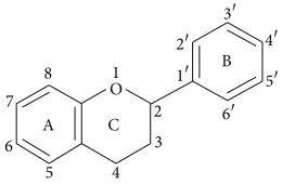
A C
Figure 1: Basic flavonoid structure.
2. Chemistry of Flavonoids
Flavonoids are a group of natural compounds with variable phenolic structures and are found in plants. In 1930 a new substance was isolated from oranges. At that time it was believed to be a member of a new class of vitamins and was designated as vitamin P. Later on it became clear that this substance was a flavonoid (rutin) and till now more than 4000
varieties of flavonoids have been identified [13].
Chemically flavonoids are based upon a fifteen-carbon skeleton consisting of two benzene rings (A and B as
shown in Figure 1) linked via a heterocyclic pyrane ring
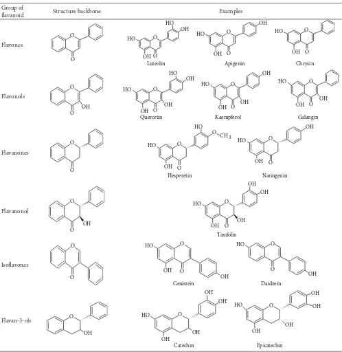
(C). They can be divided into a variety of classes such as flavones (e.g., flavone, apigenin, and luteolin), flavonols (e.g., quercetin, kaempferol, myricetin, and fisetin), flavanones (e.g., flavanone, hesperetin, and naringenin), and others.
Their general structures are shown in Table 1. The various
classes of flavonoids differ in the level of oxidation and pattern of substitution of the C ring, while individual compounds within a class differ in the pattern of substitution of the A and
B rings [13].
Flavonoids occur as aglycones, glycosides, and methy-lated derivatives. The basic flavonoid structure is aglycone (Figure 1). Six-member ring condensed with the benzene
ring is either a 𝛼-pyrone (flavonols and flavanones) or its
dihydroderivative (flavonols and flavanones). The position of the benzenoid substituent divides the flavonoid class into flavonoids (2-position) and isoflavonoids (3-position). Flavonols differ from flavanones by hydroxyl group at the
3-position and a C2–C3 double bond [40]. Flavonoids are often
hydroxylated in positions 3, 5, 7, 2, 3, 4, and 5. Methyl ethers
and acetyl esters of the alcohol group are known to occur in nature. When glycosides are formed, the glycosidic linkage is normally located in positions 3 or 7 and the carbohydrate can be L-rhamnose, D-glucose, glucorhamnose, galactose, or
arabinose [41].
2.1. Spectral Characteristics of Flavonoids. Studies on
flavonoids by spectroscopy have revealed that most flavones and flavonols exhibit two major absorption bands: Band I (320–385 nm) represents the B ring absorption, while Band II (250–285 nm) corresponds to the A ring absorption. Functional groups attached to the flavonoid skeleton may cause a shift in absorption such as from 367 nm in kaempferol
(3,5,7,4-hydroxyl groups) to 371 nm in quercetin (3,5,7,3,4
-hydroxyl groups) and to 374 nm in myricetin (3,5,7,3,4,5
-hydroxyl groups) [42]. The absence of a 3-hydroxyl group in
flavones distinguishes them from flavonols. Flavanones have a saturated heterocyclic C ring, with no conjugation between the A and B rings, as determined by their UV spectral
characteristics [43]. Flavanones exhibit a very strong Band
II absorption maximum between 270 and 295 nm, namely, 288 nm (naringenin) and 285 nm (taxifolin), and only a shoulder for Band I at 326 and 327 nm. Band II appears as one peak (270 nm) in compounds with a monosubstituted B ring, but as two peaks or one peak (258 nm) with a shoulder
(272 nm) when a di-, tri-, oro-substituted B ring is present.
As anthocyanins show distinctive Band I peak in the 450– 560 nm region due to hydroxyl cinnamoyl system of the B ring and Band II peaks in the 240–280 nm region due to the benzoyl system of the A ring, the colour of the anthocyanins varies with the number and position of the hydroxyl groups
[44].
3. Flavonoid Rich Food and Medicinal Plants
Flavonoids are the most common and widely distributed group of plant phenolic compounds, occurring virtually in all plant parts, particularly the photosynthesising plant cells. They are a major coloring component of flowering plants. Flavonoids are an integral part of human and animal diet. Some food sources containing different classes of flavonoids
are given inTable 2. Being phytochemicals, flavonoids cannot
be synthesized by humans and animals [45]. Thus flavonoids
found in animals are of plant origin rather than being biosyn-thesized in situ. Flavonols are the most abundant flavonoids in foods. Flavonoids in food are generally responsible for colour, taste, prevention of fat oxidation, and protection of
vitamins and enzymes [46]. Flavonoids found in the highest
amounts in the human diet include the soy isoflavones, flavonols, and the flavones. Although most fruits and some legumes contain catechins, the levels vary from 4.5 to
610 mg/kg [47]. Preparation and processing of food may
decrease flavonoid levels depending on the methods used. For example, in a recent study, orange juices were found to contain 81–200 mg/L soluble flavanones, while the content in the cloud was 206–644 mg/L which suggest that the flavanones are concentrated in the cloud during processing
and storage [48]. Accurate estimation of the average dietary
intake of flavonoids is difficult, because of the wide varieties of available flavonoids and the extensive distribution in various
plants and also the diverse consumption in humans [49].
Recently there has been an upsurge of interest in the therapeutic potential of medicinal plants which might be due to their phenolic compounds, specifically to flavonoids
[50, 51]. Flavonoids have been consumed by humans since
the advent of human life on earth, that is, for about 4 million years. They have extensive biological properties that promote human health and help reduce the risk of diseases. Oxidative modification of LDL cholesterol is thought to play a key role during atherosclerosis. The isoflavan glabridin, a
major polyphenolic compound found inGlycyrrhiza glabra
(Fabaceae), inhibits LDL oxidation via a mechanism
involv-ing scavenginvolv-ing of free radicals [52]. Several epidemiologic
Table 1: Structure of flavonoids.
Group of
flavanoid Structure backbone Examples
Flavones
tea may lower blood cholesterol concentrations and blood pressure, thereby providing some protection against cardio-vascular disease. Flavonoids are also known to influence the quality and stability of foods by acting as flavorants,
colorants, and antioxidants [53, 54]. Flavonoids contained
in berries may have a positive effect against Parkinson’s disease and may help to improve memory in elderly people. Antihypertensive effect has been observed in total flavonoid
fraction ofAstragalus complanatusin hypertensive rats [55].
Intake of antioxidant flavonoids has been inversely related to
the risk of incidence of dementia [56].Table 3summarizes
Table 2: Classification, structure, and food sources of some dietary flavonoids.
Class Flavonoid Dietary source References
Flavanol
(+)-Catechin (−)-Epicatechin Epigallocatechin
Tea [14]
Flavone
Chrysin, apigenin Rutin, luteolin, and luteolin glucosides
Fruit skins, red wine, buckwheat, red pepper, and tomato skin [15–18]
Flavonol Kaempferol, quercetin,
myricetin, and tamarixetin Onion, red wine, olive oil, berries, and grapefruit. [17]
Flavanone Naringin, naringenin, taxifolin,
and hesperidin Citrus fruits, grapefruits, lemons, and oranges [19,20]
Isoflavone Genistin, daidzin Soyabean [21]
Anthocyanidin Apigenidin, cyanidin Cherry, easberry, and strawberry [17,18]
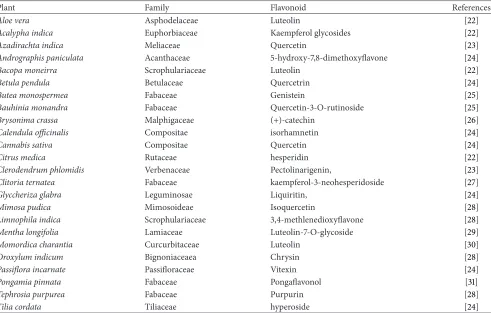
Table 3: Medicinal plants rich in flavonoids contents.
Plant Family Flavonoid References
Aloe vera Asphodelaceae Luteolin [22]
Acalypha indica Euphorbiaceae Kaempferol glycosides [22]
Azadirachta indica Meliaceae Quercetin [23]
Andrographis paniculata Acanthaceae 5-hydroxy-7,8-dimethoxyflavone [24]
Bacopa moneirra Scrophulariaceae Luteolin [22]
Betula pendula Betulaceae Quercetrin [24]
Butea monospermea Fabaceae Genistein [25]
Bauhinia monandra Fabaceae Quercetin-3-O-rutinoside [25]
Brysonima crassa Malphigaceae (+)-catechin [26]
Calendula officinalis Compositae isorhamnetin [24]
Cannabis sativa Compositae Quercetin [24]
Citrus medica Rutaceae hesperidin [22]
Clerodendrum phlomidis Verbenaceae Pectolinarigenin, [23]
Clitoria ternatea Fabaceae kaempferol-3-neohesperidoside [27]
Glyccheriza glabra Leguminosae Liquiritin, [24]
Mimosa pudica Mimosoideae Isoquercetin [28]
Limnophila indica Scrophulariaceae 3,4-methlenedioxyflavone [28]
Mentha longifolia Lamiaceae Luteolin-7-O-glycoside [29]
Momordica charantia Curcurbitaceae Luteolin [30]
Oroxylum indicum Bignoniaceaea Chrysin [28]
Passiflora incarnate Passifloraceae Vitexin [24]
Pongamia pinnata Fabaceae Pongaflavonol [31]
Tephrosia purpurea Fabaceae Purpurin [28]
Tilia cordata Tiliaceae hyperoside [24]
water-soluble flavonoids, for example, hydroxyethylrutosides and inositol-2-phosphatequercetin, has been implicated for
the treatment of hypertension and microbleeding [57].
4. Metabolism of Flavonoids in Humans
The absorption of the dietary flavonoids liberated from the food by chewing will depend on its physicochemical properties such as molecular size, configuration, lipophilicity, solubility, and pKa. The flavonoid can be absorbed from the small intestine or has to go to the colon before absorption.
It may depend upon structure of flavonoid, that is, whether it is glycoside or aglycone. Most flavonoids, except for the subclass of catechins, are present in plants bound to sugars
asb-glycosides (Figure 2). Aglycans can be easily absorbed
by the small intestine, while flavonoid glycosides have to be
converted into aglycan form [58].
The hydrophilic flavonoid glucoside such as quercetin are transported across the small intestine by the
intesti-nal Na+-dependent glucose cotransporter (SGLT1) [58].
O O
O O
OH
OH
OH
OH OH
OH HO
HO
(a)
O O
OH
OH
OH
OH HO
(b)
Figure 2: Structure of (a) flavonoid glycoside and (b) aglycone flavonoid.
a𝛽-glucosidase on the outside of the brush border membrane
of the small intestine. Subsequently, the liberated aglycone
can be absorbed across the small intestine [59]. The
sub-strate specificity of this LPH enzyme varies significantly in a broad range of glycosides (glucosides, galactosides,
arabinosides, xylosides, and rhamnosides) of flavonoids [60].
The glycosides which are not substrates for these enzymes are transported toward the colon where bacteria have ability to hydrolyze flavonoid glycosides, but simultaneously they
will also degrade the liberated flavonoid aglycones [61]. Since
absorption capacity of the colon is far less than that of the small intestine, only trivial absorption of these glycosides is to be expected.
After absorption, the flavonoids are conjugated in the liver by glucuronidation, sulfation, or methylation or
metab-olized to smaller phenolic compounds [62]. Due to these
conjugation reactions, no free flavonoid aglycones can be
found in plasma or urine, except for catechins [63].
Depend-ing on the food source bioavailability of certain flavonoids differs markedly; for example, the absorption of quercetin from onions is fourfold greater than that from apple or tea
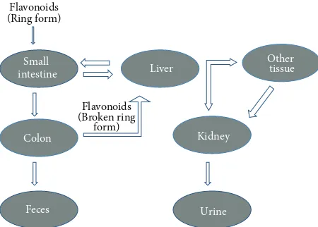
[64]. The flavonoids secreted with bile in intestine and those
that cannot be absorbed from the small intestine are degraded in the colon by intestinal microflora which also break down
the flavonoid ring structure (Figure 3). Oligomeric flavonoids
may be hydrolyzed to monomers and dimers under influence of acidic conditions in the stomach. Larger molecules reach the colon where they are degraded by bacteria. The sugar moiety of flavonoid glycosides is an important determinant of their bioavailability. Dimerization has been shown to reduce bioavailability. Among all the subclasses of flavonoids,
isoflavones exhibit the highest bioavailability [65]. After
ingestion of green tea, flavonoid content is absorbed rapidly as shown by their elevated levels in plasma and urine. They enter the systemic circulation soon after ingestion and cause
a significant increase in plasma antioxidant status [66].
5. Biological Activities of Flavonoids
5.1. Antioxidant Activity. Flavonoids possess many
biochemi-cal properties, but the best described property of almost every
Flavonoids (Ring form)
Colon Small intestine
Feces
Liver Other tissue
Kidney
Urine Flavonoids
(Broken ring form)
Figure 3: Compartments involved in the metabolism of flavonoid.
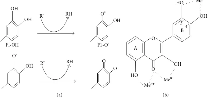
group of flavonoids is their capacity to act as antioxidants. The antioxidant activity of flavonoids depends upon the arrangement of functional groups about the nuclear struc-ture. The configuration, substitution, and total number of hydroxyl groups substantially influence several mechanisms of antioxidant activity such as radical scavenging and metal
ion chelation ability [4, 67]. The B ring hydroxyl
configu-ration is the most significant determinant of scavenging of ROS and RNS because it donates hydrogen and an electron to hydroxyl, peroxyl, and peroxynitrite radicals, stabilizing them and giving rise to a relatively stable flavonoids radical
[68].
Mechanisms of antioxidant action can include (1) sup-pression of ROS formation either by inhibition of enzymes or by chelating trace elements involved in free radical gener-ation; (2) scavenging ROS; and (3) upregulation or protection
of antioxidant defenses [69,70]. Flavonoid action involves
OH OH
Fl-OH
R∘ RH
O∘
OH
∘ F1-O
O∘
OH
R∘ RH O
O
(a)
HO A
O
O
Men+ Me
n+ OH
B
3
4
HO Me
n+
OH
(b)
Figure 4: (a) Scavenging of ROS (R∘) by flavonoids (Fl-OH) and (b) binding sites for trace metals where Me𝑛+indicates metal ions.
S-transferase, mitochondrial succinoxidase, NADH oxidase,
and so forth [71].
Lipid peroxidation is a common consequence of oxidative stress. Flavonoid protect lipids against oxidative damage
by various mechanisms [5, 51]. Free metal ions enhance
ROS formation by the reduction of hydrogen peroxide with generation of the highly reactive hydroxyl radical. Due to their lower redox potentials flavonoids (Fl-OH) are ther-modynamically able to reduce highly oxidizing free radicals (redox potentials in the range 2.13–1.0 V) such as superoxide, peroxyl, alkoxyl, and hydroxyl radicals by hydrogen atom
donation (Figure 4(a)). Because of their capacity to chelate
metal ions (iron, copper, etc.), flavonoids also inhibit free
radical generation [70,72]. Quercetin in particular is known
for its iron-chelating and iron-stabilizing properties. Trace metals bind at specific positions of different rings of flavonoid
structures [73]. The binding sites are shown inFigure 4(b).
A 3,4-catechol structure in the B ring firmly enhances
inhibition of lipid peroxidation. This trait of flavonoids makes them most effective scavengers of peroxyl, superoxide, and
peroxynitrite radicals [4]. Epicatechin and rutin are strong
radical scavengers and inhibitors of lipid peroxidationin vitro
[74]. Because of oxidation on the B ring of flavonoids having
catechol group a fairly stable orthosemiquinone radical is formed which is strong scavengers. Flavones lacking catechol system on oxidation lead to formation of unstable radicals
exhibit weak scavenging potential [75]. The literature shows
that flavonoids having an unsaturated 2-3 bond in conju-gation with a 4-oxo function are more potent antioxidants than the flavonoids lacking one or both features. Conjugation between the A and B rings allows a resonance effect of the aromatic nucleus that provides stability to the flavonoid radical. Free radical scavenging by flavonoids is potentiated by the presence of both the elements besides other structural
features [76].
The flavonoid heterocycle contributes to antioxidant activity by permitting conjugation between the aromatic rings and the presence of a free OH. Removal of a 3-OH annuls coplanarity and conjugation which compromises
scavenging ability [77]. It is proposed that B ring OH groups
form hydrogen bonds with the 3-OH, aligning the B ring with the heterocycle and A ring. Due to this intramolecular hydrogen bonding, the influence of a 3-OH is enhanced by
the presence of a 3,4-catechol, elucidating the potent
antiox-idant activity of flavan-3-ols and flavon-3-ols that possess the latter feature. Generally O-methylation of hydroxyl groups of
flavonoids decreases their radical scavenging capacity [76].
Occurrence, position, structure, and total number of sugar moieties in flavonoid (flavonoids glycosides) play an important role in antioxidant activity. Aglycones are more potent antioxidants than their corresponding glyco-sides. There are reports that the antioxidant properties of flavonol glycosides from tea declined as the number of
glycosidic moieties increased [78]. Though glycosides are
usually weaker antioxidants than aglycones, bioavailability is sometimes enhanced by a glucose moiety. In the diet, flavonoid glycosidic moieties occur most frequently at the
3- or 7-position [79]. Increasing degree of polymerization
enhances the effectiveness of procyanidins against a vari-ety of radical species. Procyanidin dimers and trimers are more effective than monomeric flavonoids against superoxide anion. Tetramers exhibit greater activity against peroxyni-trite and superoxide mediated oxidation than trimers, while heptamers and hexamers demonstrate significantly greater superoxide scavenging properties than trimers and tetramers
[80].
5.2. Hepatoprotective Activity. Several flavonoids such as
catechin, apigenin, quercetin, naringenin, rutin, and
venoru-ton are reported for their hapatoprotective activities [81].
Different chronic diseases such as diabetes may lead to devel-opment of hepatic clinical manifestations. glutamate-cysteine ligase catalytic subunit (Gclc) expression, glutathione, and ROS levels are reported to be decreased in liver of diabetic mice. Anthocyanins have drawn increasing attention because of their preventive effect against various diseases. Zhu et
al. [82] demonstrated that anthocyanin cyanidin-3-O-𝛽
in turn upregulates cAMP response element binding protein (CREB) phosphorylation to promote CREB-DNA binding and increase Gclc transcription. Increased Gclc expression results in a decrease in hepatic ROS levels and proapop-totic signaling. Furthermore, C3G treatment lowers hepatic lipid peroxidation, inhibits the release of proinflammatory cytokines, and protects against the development of hepatic
steatosis [82].
Silymarin is a flavonoids having three structural com-ponents silibinin, silydianine, and silychristine extracted
from the seeds and fruit of milk thistleSilybum marianum
(Compositae). Silymarin has been reported to stimulate enzymatic activity of DNA-dependent RNA polymerase 1 and subsequent biosynthesis of RNA and protein, resulting in DNA biosynthesis and cell proliferation leading to liver
regeneration only in damaged livers [83]. Silymarin increases
proliferating hepatocytes in response to FB1 (Fumonisin B1,
a mycotoxin produced byFusarium verticillioides) induced
cell death without modulation of cell proliferation in normal livers. The pharmacological properties of silymarin involve the regulation of cell membrane permeability and integrity, inhibition of leukotriene, ROS scavenging, suppression of
NF-𝜅B activity, depression of protein kinases, and collagen
production [84]. Silymarin has clinical applications in the
treatment of cirrhosis, ischemic injury, and toxic hepatitis induced by various toxins such as acetaminophen, and toxic
mushroom [85].
Hepatoprotective activities were observed in flavonoids
isolated from Laggera alata against carbon-tetrachloride
(CCl4-) induced injury in primary cultured neonatal rat
hepatocytes and in rats with hepatic damage. Flavonoids
at a concentration range of 1–100𝜇g/mL improved cell
via-bility and inhibited cellular leakage of hepatocyte aspartate aminotransferase (AST) and alanine aminotransferase (ALT)
caused by CCl4 [86]. Similarly in an in vivo experiment
flavonoids at of 50, 100, and 200 mg/kg oral doses significantly reduced the levels of AST, ALT, total protein, and albumin in serum and the hydroxyproline and sialic acid levels in liver. Histopathological examinations also revealed the
improve-ment in damaged liver with the treatimprove-ment of flavonoid [86].
Several clinical investigations have shown the efficacy and safety of flavonoids in the treatment of hepatobiliary dysfunc-tion and digestive complaints, such as sensadysfunc-tion of fullness,
loss of appetite, nausea, and abdominal pain. Equisetum
arvenseflavonoids as well as hirustrin and avicularin isolated
from some other sources is reported to provide protection against chemically induced hepatotoxicity in HepG2 cells
[87,88].
5.3. Antibacterial Activity. Flavonoids are known to be
syn-thesized by plants in response to microbial infection; thus
it should not be surprising that they have been found in
vitroto be effective antimicrobial substances against a wide
array of microorganisms. Flavonoid rich plant extracts from different species have been reported to possess antibacterial
activity [70, 72, 89, 90]. Several flavonoids including
api-genin, galangin, flavone and flavonol glycosides, isoflavones, flavanones, and chalcones have been shown to possess potent
antibacterial activity [91].
Antibacterial flavonoids might be having multiple cellular targets, rather than one specific site of action. One of their molecular actions is to form complex with proteins through nonspecific forces such as hydrogen bonding and hydrophobic effects, as well as by covalent bond formation. Thus, their mode of antimicrobial action may be related to their ability to inactivate microbial adhesins, enzymes, cell envelope transport proteins, and so forth. Lipophilic
flavonoids may also disrupt microbial membranes [92,93].
Catechins, the most reduced form of the C3 unit in flavonoid compounds, have been extensively researched due to their antimicrobial activity. These compounds are reported
for theirin vitroantibacterial activity againstVibrio cholerae,
Streptococcus mutans, Shigella, and other bacteria [94, 95].
The catechins have been shown to inactivate cholera toxin
inVibrio choleraand inhibit isolated bacterial
glucosyltrans-ferases inS. mutans,probably due to complexing activities
[94,96]. Robinetin, myricetin, and (−)-epigallocatechin are
known to inhibit DNA synthesis inProteus vulgaris. Mori
et al. [97] suggested that the B ring of the flavonoids may
intercalate or form hydrogen bond with the stacking of nucleic acid bases and further lead to inhibition of DNA and RNA synthesis in bacteria. Another study demonstrated
inhibitory activity of quercetin, apigenin, and 3,6,7,3,4
-pentahydroxyflavone against Escherichia coli DNA gyrase
[98].
Naringenin and sophoraflavanone G have intensive
antibacterial activity against methicilline resistant
Staphy-lococcus aureus (MRSA) and streptococci. An alteration
of membrane fluidity in hydrophilic and hydrophobic regions may be attributed to this effect which suggests that these flavonoids might reduce the fluidity of outer and
inner layers of membranes [99]. The correlation between
antibacterial activity and membrane interference supports the theory that flavonoids may demonstrate antibacterial activity by reducing membrane fluidity of bacterial cells.
The 5,7-dihydroxylation of the A ring and 2,4-or 2,6
-dihydroxylation of the B ring in the flavanone structure is
important for anti-MRSA activity [100]. A hydroxyl group
at position 5 in flavanones and flavones is important for their activity against MRSA. Substitution with C8 and C10 chains may also enhance the antistaphylococcal
activ-ity of flavonoids belonging to the flavan-3-ol class [101].
Osawa et al. have shown that hydroxyflavanones and 5-hydroxyisoflavanones with one, two, or three additional
hydroxyl groups at the 7, 2 and 4 positions inhibited the
growth ofS. mutansandStreptococcus sobrinus[102].
Haraguchi and colleagues [100] studied antibacterial
activity of two flavonoids, licochalcones A and C, isolated
from the roots of Glycyrrhiza inflata against S. aureus
andMicrococcus luteus. They observed that licochalcone A
inhibited incorporation of radioactive precursors into macro-molecules (DNA, RNA, and protein). This activity was similar to the mode of action of antibiotics inhibiting respiratory chain, since energy is required for active uptake of various metabolites as well as for biosynthesis of macromolecules. After further studies it was suggested that the inhibition site of these flavonoids was between CoQ and cytochrome
There are many examples that lend support to the prowess of phytoconstituents derived from edible and medicinal plants
as potent antibacterial agents [103–105].
5.4. Anti-Inflammatory Activity. Inflammation is a normal
biological process in response to tissue injury, microbial pathogen infection, and chemical irritation. Inflammation is initiated by migration of immune cells from blood vessels and release of mediators at the site of damage. This process is followed by recruitment of inflammatory cells, release of ROS, RNS, and proinflammatory cytokines to eliminate foreign pathogens, and repairing injured tissues. In general, normal inflammation is rapid and self-limiting, but aber-rant resolution and prolonged inflammation cause various
chronic disorders [106].
The immune system can be modified by diet, phar-macologic agents, environmental pollutants, and naturally occurring food chemicals. Certain members of flavonoids significantly affect the function of the immune system and
inflammatory cells [107]. A number of flavonoids such as
hesperidin, apigenin, luteolin, and quercetin are reported to possess anti-inflammatory and analgesic effects. Flavonoids may affect specifically the function of enzyme systems crit-ically involved in the generation of inflammatory processes,
especially tyrosine and serine-threonine protein kinases [108,
109]. The inhibition of kinases is due to the competitive
binding of flavonoids with ATP at catalytic sites on the enzymes. These enzymes are involved in signal transduction and cell activation processes involving cells of the immune system. It has been reported that flavonoids are able to inhibit expression of isoforms of inducible nitric oxide synthase, cyclooxygenase, and lipooxygenase, which are responsible for the production of a great amount of nitric oxide, prostanoids, leukotrienes, and other mediators of the inflammatory pro-cess such as cytokines, chemokines, or adhesion molecules
[110]. Flavonoids also inhibit phosphodiesterases involved
in cell activation. Much of the anti-inflammatory effect of flavonoid is on the biosynthesis of protein cytokines that mediate adhesion of circulating leukocytes to sites of injury. Certain flavonoids are potent inhibitors of the production of prostaglandins, a group of powerful proinflammatory
signaling molecules [111].
Reversal of the carrageenan induced inflammatory chan-ges has been observed with silymarin treatment. It has been found that quercetin inhibit mitogen stimulated
immu-noglobulin secretion of IgG, IgM, and IgA isotypesin vitro
[112]. Several flavonoids are reported to inhibit platelet
adhesion, aggregation, and secretion significantly at 1–10 mM
concentration [113]. The effect of flavonoid on platelets has
been related to the inhibition of arachidonic acid metabolism
by carbon monoxide [114]. Alternatively, certain flavonoids
are potent inhibitors of cyclic AMP phosphodiesterase, and this may in part explain their ability to inhibit platelet function.
5.5. Anticancer Activity. Dietary factors play an important
role in the prevention of cancers. Fruits and vegetables having flavonoids have been reported as cancer chemopreventive
agents [72,115]. Consumption of onions and/or apples, two
major sources of the flavonol quercetin, is inversely associated with the incidence of cancer of the prostate, lung, stomach, and breast. In addition, moderate wine drinkers also seem to have a lower risk to develop cancer of the lung, endometrium,
esophagus, stomach, and colon [116]. The critical relationship
of fruit and vegetable intake and cancer prevention has been thoroughly documented. It has been suggested that major public health benefits could be achieved by substantially
increasing consumption of these foods [117].
Several mechanisms have been proposed for the effect of flavonoids on the initiation and promotion stages of the carcinogenicity including influences on development and
hormonal activities [118]. Major molecular mechanisms of
action of flavonoids are given as follows:
(1) downregulation of mutant p53 protein, (2) cell cycle arrest,
(3) tyrosine kinase inhibition, (4) inhibition of heat shock proteins, (5) estrogen receptor binding capacity, (6) inhibition of expression of Ras proteins.
Mutations of p53 are among the most common genetic abnormalities in human cancers. The inhibition of expression of p53 may lead to arrest the cancer cells in the G2-M phase of the cell cycle. Flavonoids are found to downregulate expression of mutant p53 protein to nearly undetectable levels
in human breast cancer cell lines [119]. Tyrosine kinases are
a family of proteins located in or near the cell membrane involved in the transduction of growth factor signals to the nucleus. Their expression is thought to be involved in oncogenesis via an ability to override normal regulatory growth control. Drugs inhibiting tyrosine kinase activity are thought to be possible antitumor agents without the cytotoxic side effects seen with conventional chemotherapy. Quercetin was the first tyrosine kinase inhibiting compound tested in
a human phase I trial [120]. Heat shock proteins form a
complex with mutant p53, which allows tumor cells to bypass normal mechanisms of cell cycle arrest. Heat shock proteins also allow for improved cancer cell survival under different bodily stresses. Flavonoids are known to inhibit production of heat shock proteins in several malignant cell lines, including
breast cancer, leukemia, and colon cancer [119].
Recently it has been shown that the flavanol epigallocate-chin-3-gallate inhibited fatty acid synthase (FAS) activity and lipogenesis in prostate cancer cells, an effect that is strongly
associated with growth arrest and cell death [116, 121]. In
contrast to most normal tissues expression of FAS is markedly increased in various human cancers. Upregulation of FAS occurs early in tumor development and is further enhanced
in more advanced tumors [122].
Quercetin is known to produce cell cycle arrest in pro-liferating lymphoid cells. In addition to its antineoplastic activity, quercetin exerted growth-inhibitory effects on
sev-eral malignant tumor cell linesin vitro. These included
human breast cancer cells, human squamous and gliosarcoma
cells, and ovarian cancer cells [119]. Markaverich et al. [123]
proposed that tumor cell growth inhibition by quercetin may be due to its interaction with nuclear type II estrogen binding sites (EBS). It has been experimentally proved that increased signal transduction in human breast cancer cells is markedly reduced by quercetin acting as an antiproliferative
agent [124].
Barnes [125] has extensively reviewed the anticancer
effects of genistein on in vitro and in vivo models. In an
study to determine effects of isoflavones genistein, daidzein, and biochanin A on mammary carcinogenesis, genistein was found to suppress the development of chemically induced mammary cancer without reproductive or endocrinological toxicities. Neonatal administration of genistein (a flavonoid) exhibited a protective effect against the subsequent
develop-ment of induced mammary cancer in rats [126]. Hesperidin,
a flavanone glycoside, is known to inhibit azoxymethanol
induced colon and mammary cancers in rats [127]. The
anticancer properties of flavonoids contained in citrus fruits
have been reviewed by Carroll et al. [128]. Several flavonols,
flavones, flavanones, and the isoflavone biochanin A are
reported to have potent antimutagenic activity [129]. A
carbonyl function at C-4 of the flavone nucleus was found to be essential for their activity. Flavone-8-acetic acid has also
been shown to have antitumor effects [130]. In earlier studies
ellagic acid, robinetin, quercetin, and myricetin have been shown to inhibit the tumorigenicity of BP-7, 8-diol-9, and
10-epoxide-2 on mouse skin [131].
Higher consumption of phytoestrogens, including isoflavones and other flavonoids, has been shown to provide
protection against prostate cancer risk [132]. It is well known
that due to oxidative stress cancer initiation may take place and thus potent antioxidants show potential to combat progression of carcinogenesis. Potential of antioxidant as an anticancer agent depends on its competence as an
oxygen radical inactivator and inhibitor [70, 72, 133].
Therefore diets rich in radical scavengers would diminish the
cancer-promoting action of some radicals [134].
5.6. Antiviral Activity. Natural compounds are an important
source for the discovery and the development of novel antivi-ral drugs because of their availability and expected low side effects. Naturally occurring flavonoids with antiviral activity have been recognized since the 1940s and many reports on the antiviral activity of various flavonoids are available. Search of effective drug against human immunodeficiency virus (HIV) is the need of hour. Most of the work related with antiviral compounds revolves around inhibition of various enzymes associated with the life cycle of viruses. Structure function relationship between flavonoids and their enzyme inhibitory activity has been observed. Gerdin and Srensso
[135] demonstrated that flavan-3-o1 was more effective than
flavones and flavonones in selective inhibition of 1, HIV-2, and similar immunodeficiency virus infections. Baicalin, a
flavonoid isolated fromScutellaria baicalensis(Lamieaceae),
inhibits HIV-1 infection and replication. Baicalein and other flavonoids such as robustaflavone and hinokiflavone have
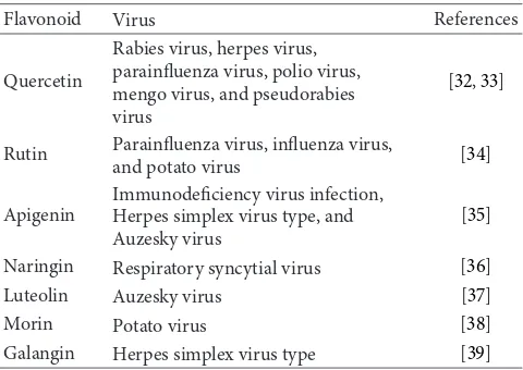
Table 4: Antiviral activity of various flavonoids.
Flavonoid Virus References
Quercetin
Rabies virus, herpes virus, parainfluenza virus, polio virus, mengo virus, and pseudorabies virus
[32,33]
Rutin Parainfluenza virus, influenza virus,
and potato virus [34]
Apigenin
Immunodeficiency virus infection, Herpes simplex virus type, and Auzesky virus
[35]
Naringin Respiratory syncytial virus [36] Luteolin Auzesky virus [37]
Morin Potato virus [38]
Galangin Herpes simplex virus type [39]
also been shown to inhibit HIV-1 reverse transcriptase [136].
Another study revealed inhibition of HIV-1 entry into cells expressing CD4 and chemokine coreceptors and antagonism
of HIV-1 reverse transcriptase by the flavone O-glycoside
[137]. Catechins are also known to inhibit DNA polymerases
of HIV-1. Flavonoid such as demethylated gardenin A and
robinetin are known to inhibit HIV-1 proteinase [136]. It has
also been reported that the flavonoids chrysin, acacetin, and apigenin prevent HIV-1 activation via a novel mechanism that
probably involves inhibition of viral transcription [138].
Various combinations of flavones and flavonols have been shown to exhibit synergism. Kaempferol and luteolin show synergistic effect against herpes simplex virus (HSV). Synergism has also been reported between flavonoids and other antiviral agents. Quercetin is reported to potentiate the effects of 5-ethyl-2-dioxyuridine and acyclovir against HSV
and pseudorabies infection [136]. Studies have displayed that
flavonols are more active than flavones against herpes simplex virus type 1 and the activity order was found to be galangin,
kaempferol, and quercetin [136].
Zandi et al. [139] studied the antidengue virus properties
of quercetin, hesperetin, naringin, and daidzein at differ-ent stages of DENV-2 (dengue virus type-2) infection and replication cycle. Quercetin was found to be most effective against DENV-2 in Vero cells. Many flavonoids, namely, dihydroquercetin, dihydrofisetin, leucocyanidin, pelargoni-din chloride, and catechin, show activity against several types of virus including HSV, respiratory syncytial virus, polio virus
and Sindbis virus [135]. Inhibition of viral polymerase and
binding of viral nucleic acid or viral capsid proteins have
been proposed as antiviral mechanisms of action [139]. List
of some flavonoids and their efficacy against viruses is given inTable 4.
6. Role of Flavonoids in Plants
6.1. Combating Oxidative Stress. Flavonoids have long been
reported as serving multiple functions in plants [140]. Various
in plants is almost exclusively enhanced due to oxidative stress. They have capacity to absorb the most energetic solar wavelengths (i.e., UV-B and UV-A), inhibit the generation of
ROS, and quench ROS once they are formed [141]. Flavonoids
discharged primary UV-B screening functions when early plants moved from water to colonize the land. Extent of antioxidant capacity and ability to absorb UV-wavelengths depends upon the nature of substitution on different rings of flavonoids. Dihydroxy B ring substituted flavonoids have a greater antioxidant capacity, while their monohydroxy B ring substituted counterparts have greater ability to absorb
UV-wavelengths [141].
The most reactive hydroxyl groups (7-OH in flavones or the 3-OH in flavonols) in flavonoids are generally glycosy-lated. Glycosylation increases solubility in the aqueous cellu-lar environment, protects the reactive hydroxyl groups from
autooxidation [142], and allows the transport of flavonoids
from the endoplasmic reticulum to various cellular com-partments and their secretion to the plasma membrane and
the cell wall [143]. Recent evidence shows that antioxidant
flavonoids are located in the nucleus of mesophyll cells and within centers of ROS generation, that is, the chloroplast.
Here they can easily quench H2O2, hydroxyl radical, and
singlet oxygen [141,144].
Oxidative stress due to an excess of excitation energy in the chloroplast may be aggravated under conditions that
limit the diffusion of CO2 to the carboxylation sites and
the efficiency of carboxylation [141,145]. The environmental
limitations to CO2assimilation rate include drought/salinity,
low/high temperature, and nutrient scarcity. Under these conditions the activity of ROS detoxifying enzymes may
sig-nificantly reduce in the chloroplast [146,147], which in turn
upregulates the biosynthesis of ROS scavenging flavonoids. The reducing functions of flavonoids are of key significance in plants under severe stress conditions. These functional roles are concomitant with the very high concentration of
dihydroxy B ring substituted flavonoids [148]. Flavonoids
have been suggested as representing a secondary antioxidant defense system in plant tissues exposed to different stresses
[141]. Lipid peroxidation is the common consequence of
oxidative stress which disrupts the cell membrane integrity. Quercetin 3-O-rutinoside (rutin) may interact with the polar head of phospholipids at water lipid interface, enhancing membrane rigidity and consequently protecting membranes
from oxidative damage [149].
6.2. As Growth Regulator. Flavonoids carry out functional
roles of amazing significance in plant-environment inter-actions. Flavonoids (in the nanomolar range) may regulate auxin movement and catabolism. The ability of flavonoids to create auxin gradients translates into phenotypes with
different morphoanatomical features [150]. The control of
flavonoids on auxin movement may have immense value in the stress-induced morphogenic responses of plants such as the flight strategy of sessile organisms exposed to unfavorable
environments [151]. Species rich in dihydroxy flavonoids
exhibit phenotypes with strikingly different morphologi-cal traits as compared with those rich in monohydroxy
flavonoids [152]. Dwarf bushy phenotypes with few, small,
and thick leaves to direct sunlight irradiance are usually present in sunny environments, thus protecting leaves located deep in the canopy from light-induced severe cellular home-ostasis perturbations. On the contrary, shaded plants, which are rich in kaempferol and/or apigenin derivatives (having negligible concentrations of quercetin derivatives), have long internodes, and large leaf lamina coupled with reduced leaf
thickness [151].
Flavonoids at the plasma membrane are effective inhib-itors of PIN (pin formed) and MDR (multidrug resistance) glycoproteins that are involved in the cell to cell movement of auxin. The ability of flavonoids to inhibit the activity of the efflux facilitator PIN and MDR proteins depends on the presence of the catechol group in the B ring of the flavonoid skeleton. In addition flavonoids regulate the activity of IAA-oxidase with markedly different effects based
on their chemical structure [153]. Recent evidence of a
nuclear location of flavonoids (as well as of enzymes of flavonoid biosynthesis) supports that flavonoids are capable of modulating the activity of proteins involved in cell growth. Flavonoids may therefore act as transcriptional regulators
[154,155].
7. Microbial Production of Flavonoids
In response to the low production efficiency from plants and chemical synthesis, research groups have directed their attention to the production of flavonoids in microorganisms
using metabolic engineering and synthetic biology [156].
Chemical synthesis of flavonoids requires extreme reaction
conditions and toxic chemicals [157]. Because of the rapid
development in molecular biology tools and the flooding of genome information from a variety of organisms, combi-natorial biosynthesis offers an advantage for production of rare and expensive natural products. It can be used in both simple and complex transformations without the tiresome blocking and deblocking steps that are common in organic
synthesis [158]. Several prokaryotes and eukaryotes such as
E. coli,Saccharomyces cerevisiae,Streptomyces venezuelae, and
Phellinus igniarius,a medicinal mushroom, have been used
for production of flavonoids [12].
7.1. Phenylpropanoid Pathway. In plants naringenin chalcone
is the precursor for a large number of flavonoids produced by the phenylpropanoid synthetic pathway. Fermentative
production by E. coli carrying an artificially assembled
phenylpropanoid pathway was the first example to show that a nearly complete biosynthetic pathway in plants was established in a heterologous microorganism for production of flavanones from the amino acid precursors, phenylalanine,
and tyrosine [159]. As the first step in the phenylpropanoid
pathway in plants, phenylalanine is deaminated to yield cinnamic acid by the action of phenylalanine ammonia-lyase (PAL). Cinnamic acid is hydroxylated by
cinnamate-4-hydroxylase (C4H) to p-coumaric acid, which is then
activated top-coumaroyl-CoA by the action of 4-coumarate:
condensation of three acetate units from malonyl-CoA with p-coumaroyl-CoA to yield naringenin chalcone. Naringenin chalcone is converted to naringenin by chalcone isomerase
(CHI) or nonenzymaticallyin vitro[160].
7.2. Enhancement of Flavonoid Production. Combination of
promoter and target genes; knockout of related genes; over-expression of malonyl-CoA; and construction of artificial P450 enzymes are the key molecular biology technology pro-cedures used in the heterologous production of flavonoids. Every gene from the phenylpropanoid pathway is cloned in the host under the control of the promoter which often plays an important role in the heterologous expression of secondary metabolites. Several promoters have been used to enhance production of flavonoids according to the need of
specific host such as T7, ermE∗, and GAL1 promoter [12].
The extremely low concentration of malonyl-CoA in the microbial cell was one of the drawbacks in the microbio-logical production of flavonoids. Through the coordinated
overexpression of acetyl-CoA carboxylase genes from
Pho-torhabdus luminescens intracellular malonyl-CoA pool was
amplified leading to increased production of flavonoids [161].
Supplication of UDP-glucose is also a key effector in the biosynthesis of flavonoids. It was proved in an experiment in which researchers knocked out the udg gene encoding for UDP-glucose dehydrogenase. This resulted in elimination of endogenous UDP-glucose consumption pathway leading to increase in intracellular concentration of UDP-glucose and as a consequence increment in the production of flavanones
and anthocyanins was observed [162].
One of the barriers to the production of flavonoids and their related compounds in microorganisms by means of assembling biosynthetic genes to form an artificial pathway is the difficulty in expression of active, membrane-bound
cinnamate-4-hydroxylase [163]. This enzyme is not expressed
efficiently in bacteria due to its instability and the lack of its cognate cytochrome P450 reductase in the host. An advantage of flavonoid production in yeasts or fungi is their ability to express functionally active microsomal cytochrome P450 enzymes, which are usually difficult to be expressed in an active form in bacterial cells. There are various microsomal cytochrome P450 enzymes that are involved in the flavonoid
biosynthesis pathway [164]. Combining bacterial cells and
eukaryotic cells in a pot enabled researchers to generate a wider library of natural and unnatural products than any
of the previously reported systems.De novo production of
the key flavonoid intermediate naringenin has been
demon-strated for the first time from glucose using an engineeredS.
cerevisiaestrain which led to fourfold higher concentrations
than reported in earlier studies on de novo biosynthesis [165,
166].
8. Conclusion
Prevention and cure of diseases using phytochemicals espe-cially flavonoids are well known. Fruits and vegetables are natural sources of flavonoids. Variety of flavonoids found in the nature possesses their own physical, chemical, and
physiological properties. Structure function relationship of flavonoids is epitome of major biological activities. Medicinal efficacy of many flavonoids as antibacterial, hepatoprotective, anti-inflammatory, anticancer, and antiviral agents is well established. These substances are more commonly used in the developing countries. Therapeutic use of new compounds must be validated using specific biochemical tests. With the use of genetic modifications, it is now possible to produce flavonoids at large scale. Further achievements will provide newer insights and will certainly lead to a new era of flavonoid based pharmaceutical agents for the treatment of many infectious and degenerative diseases.
Conflict of Interests
The authors declare that they do not have any conflict of interests.
Acknowledgment
Shashank Kumar acknowledges financial support from UGC, India, in the form of Rajiv Gandhi National Senior Research fellowship.
References
[1] M. F. Mahomoodally, A. Gurib-Fakim, and A. H. Subratty, “Antimicrobial activities and phytochemical profiles of endemic medicinal plants of Mauritius,”Pharmaceutical Biology, vol. 43, no. 3, pp. 237–242, 2005.
[2] A. K. Pandey, “Anti-staphylococcal activity of a pan-tropical aggressive and obnoxious weedParihenium histerophorus: anin vitrostudy,”National Academy Science Letters, vol. 30, no. 11-12, pp. 383–386, 2007.
[3] R. A. Dixon, P. M. Dey, and C. J. Lamb, “Phytoalexins: enzymology and molecular biology,”Advances in Enzymology and Related Areas of Molecular Biology, vol. 55, pp. 1–136, 1983. [4] E. H. Kelly, R. T. Anthony, and J. B. Dennis, “Flavonoid
antiox-idants: chemistry, metabolism and structure-activity relation-ships,”Journal of Nutritional Biochemistry, vol. 13, no. 10, pp. 572–584, 2002.
[5] S. Kumar, A. Mishra, and A. K. Pandey, “Antioxidant mediated protective effect ofParthenium hysterophorusagainst oxidative damage using in vitro models,” BMC Complementary and Alternative Medicine, vol. 13, article 120, 2013.
[6] S. Kumar and A. K. Pandey, “Phenolic content, reducing power and membrane protective activities ofSolanum xanthocarpum
root extracts,”Vegetos, vol. 26, pp. 301–307, 2013.
[7] M. Leopoldini, N. Russo, S. Chiodo, and M. Toscano, “Iron chelation by the powerful antioxidant flavonoid quercetin,”
Journal of Agricultural and Food Chemistry, vol. 54, no. 17, pp. 6343–6351, 2006.
[8] S. Kumar, A. Gupta, and A. K. Pandey, “Calotropis proceraroot extract has capability to combat free radical mediated damage,”
[10] C. A. Rice-Evans, N. J. Miller, P. G. Bolwell, P. M. Broamley, and J. B. Pridham, “The relative antioxidant activities of plant-derived polyphenolic flavonoids,”Free Radical Research, vol. 22, no. 4, pp. 375–383, 1995.
[11] G. Agati, E. Azzarello, S. Pollastri, and M. Tattini, “Flavonoids as antioxidants in plants: location and functional significance,”
Plant Science, vol. 196, pp. 67–76, 2012.
[12] F. Du, F. Zhang, F. Chen et al., “Advances in microbial heterolo-gous production of flavonoids,”African Journal of Microbiology Research, vol. 5, no. 18, pp. 2566–2574, 2011.
[13] E. J. Middleton, “Effect of plant flavonoids on immune and inflammatory cell function,”Advances in Experimental Medicine and Biology, vol. 439, pp. 175–182, 1998.
[14] M. Lopez, F. Martinez, C. Del Valle, C. Orte, and M. Miro, “Analysis of phenolic constituents of biological interest in red wines by high-performance liquid chromatography,”Journal of Chromatography A, vol. 922, no. 1-2, pp. 359–363, 2001. [15] Y. Hara, S. J. Luo, R. L. Wickremasinghe, and T. Yamanishi,
“Special issue on tea,”Food Reviews International, vol. 11, pp. 371–542, 1995.
[16] S. Kreft, M. Knapp, and I. Kreft, “Extraction of rutin from buckwheat (Fagopyrum esculentumMoench) seeds and deter-mination by capillary electrophoresis,”Journal of Agricultural and Food Chemistry, vol. 47, no. 11, pp. 4649–4652, 1999. [17] A. J. Stewart, S. Bozonnet, W. Mullen, G. I. Jenkins, M. E.
Lean, and A. Crozier, “Occurrence of flavonols in tomatoes and tomato-based products,”Journal of Agricultural and Food Chemistry, vol. 48, no. 7, pp. 2663–2669, 2000.
[18] M. G. L. Hertog, P. C. H. Hollman, and M. B. Katan, “Content of potentially anticarcinogenic flavonoids of 28 vegetables and 9 fruits commonly consumed in the Netherlands,”Journal of Agricultural and Food Chemistry, vol. 40, no. 12, pp. 2379–2383, 1992.
[19] Y. Miyake, K. Shimoi, S. Kumazawa, K. Yamamoto, N. Kinae, and T. Osawa, “Identification and antioxidant activity of flavonoid metabolites in plasma and urine of eriocitrin-treated rats,”Journal of Agricultural and Food Chemistry, vol. 48, no. 8, pp. 3217–3224, 2000.
[20] R. L. Rousseff, S. F. Martin, and C. O. Youtsey, “Quantitative survey of narirutin, naringin, hesperidin, and neohesperidin in citrus,”Journal of Agricultural and Food Chemistry, vol. 35, no. 6, pp. 1027–1030, 1987.
[21] K. Reinli and G. Block, “Phytoestrogen content of foods: a compendium of literature values,”Nutrition and Cancer, vol. 26, no. 2, pp. 123–148, 1996.
[22] M. L. L´azaro, “Distribution and biological activities of the flavonoid luteolin,”Mini-Reviews in Medicinal Chemistry, vol. 9, no. 1, pp. 31–59, 2009.
[23] E. Tripoli, M. L. Guardia, S. Giammanco, D. D. Majo, and M. Giammanco, “Citrus flavonoids: molecular structure, biological activity and nutritional properties: a review,”Food Chemistry, vol. 104, no. 2, pp. 466–479, 2007.
[24] K. K. Gupta, S. C. Taneja, K. L. Dhar, and C. K. Atal, “Flavonoids ofAndrographis paniculata,”Phytochemistry, vol. 22, no. 1, pp. 314–315, 1983.
[25] A. Murlidhar, K. S. Babu, T. R. Sankar, P. Redenna, G. V. Reddy, and J. Latha, “Antiinflammatory activity of flavonoid fraction isolated from stem bark ofButea monosperma(Lam): a mecha-nism based study,”International Journal of Phytopharmacology, vol. 1, pp. 124–132, 2010.
[26] M. A. Aderogba, A. O. Ogundaini, and J. N. Eloff, “Isolation of two flavonoids fromBauhinia monandraleaves and their antioxidative effects,”The African Journal of Traditional, Com-plementary and Alternative Medicines, vol. 3, no. 4, pp. 59–65, 2006.
[27] S. Sankaranarayanan, P. Bama, J. Ramachandran et al., “Ethnob-otanical study of medicinal plants used by traditional users in Villupuram district of Tamil Nadu, India,”Journal of Medicinal Plant Research, vol. 4, no. 12, pp. 1089–1101, 2010.
[28] M. Sannomiya, V. B. Fonseca, M. A. D. Silva et al., “Flavonoids and antiulcerogenic activity from Byrsonima crassa leaves extracts,”Journal of Ethnopharmacology, vol. 97, no. 1, pp. 1–6, 2005.
[29] K. Kogawa, K. Kazuma, N. Kato, N. Noda, and M. Suzuki, “Biosynthesis of malonylated flavonoid glycosides on basis of malonyl transferase activity in the petals ofClitoria ternatea,”
Journal of Plant Physiology, vol. 164, no. 7, pp. 886–894, 2007. [30] S. Ghoulami, A. I. Idrissi, and S. Fkih-Tetouani, “Phytochemical
study ofMentha longifoliaof Morocco,”Fitoterapia, vol. 72, no. 5, pp. 596–598, 2001.
[31] M. Agarwal and R. Kamal, “Studies on flavonoid production usingin-vitrocultures ofMomordica charantia,”Indian Journal of Biotechnology, vol. 6, no. 2, pp. 277–279, 2007.
[32] S. A. Ghazal, M. Abuzarqua, and A. M. Mahansneh, “Effect of plant flavonoids on immune and inflammatory cell function,”
Phototherapy Research, vol. 2, pp. 265–271, 1992.
[33] K. P. Kell, A. M. Manadi, Z. F. Adiyasora, R. M. Kunaera, I. Z. V. Akad, and S. S. R. Naun, “Bioflavonoids and health effects in man,”Chemical Abstracts, vol. 107, pp. 366–367, 1987.
[34] R. T. Deca, I. J. Gouzalez, T. M. V. Mactinez, J. Moreno, and S. C. A. Romo, “Soil bioI.In vitroantifungal activity of some flavonoids and their metabolites,”Biochemistry, vol. 19, pp. 223– 231, 1987.
[35] Y. Tsuchiya, M. Shimizu, Y. Hiyama et al., “Inhibitory effect of flavonoids on fungal diseases,”Chemical and Pharmaceutical Bulletin, vol. 33, pp. 3881–3890, 1985.
[36] M. Bakay, I. Mucsi, I. Beladi, and M. M. Gabor, “Antiviral flavonoids from Alkena orientalis,”Acta Microbiologica, vol. 15, pp. 223–232, 1968.
[37] K. Hayashi, T. Hayashi, M. Arisawa, and N. Morita, “In vitro
inhibition of viral disease by flavonoids,”Antiviral Chemistry and Chemotherapy, vol. 4, no. 1, pp. 49–53, 1993.
[38] V. D. Tripathi and R. P. Rastogi, “In vitro anti-HIV activity of flavonoids isolated from Garcinia multifolia,” Journal of Scientific and Industrial Research, vol. 40, pp. 116–121, 1981. [39] U. P. Singh, V. B. Pandey, K. N. Singh, and R. O. N. Singh,
“Structural and biogenic relationships of isoflavonoids,” Cana-dian Journal of Botany, vol. 166, pp. 1901–1910, 1988.
[40] K. R. Narayana, M. S. Reddy, M. R. Chaluvadi, and D. R. Krishna, “Bioflavonoids classification, pharmacological, bio-chemical effects and therapeutic potential,”Indian Journal of Pharmacology, vol. 33, no. 1, pp. 2–16, 2001.
[41] E. Middleton, “The flavonoids,” Trends in Pharmacological Sciences, vol. 5, pp. 335–338, 1984.
[42] L. H. Yao, Y. M. Jiang, J. Shi et al., “Flavonoids in food and their health benefits,”Plant Foods for Human Nutrition, vol. 59, no. 3, pp. 113–122, 2004.
[44] E. Wollenweber and V. H. Dietz, “Occurrence and distribution of free flavonoid aglycones in plants,”Phytochemistry, vol. 20, no. 5, pp. 869–932, 1981.
[45] R. Koes, W. Verweij, and F. Quattrocchio, “Flavonoids: a colorful model for the regulation and evolution of biochemical path-ways,”Trends in Plant Sciences, vol. 10, no. 5, pp. 236–242, 2005. [46] L. H. Yao, Y. M. Jiang, J. Shi et al., “Flavonoids in food and their health benefits,”Plant Foods for Human Nutrition, vol. 59, no. 3, pp. 113–122, 2004.
[47] I. C. W. Arts, V. B. Putte, and P. C. H. Hollman, “Catechin contents of foods commonly consumed in the Netherlands 1. Fruits, vegetables, staple foods and processed foods,”Journal of Agricultural and Food Chemistry, vol. 48, no. 5, pp. 1746–1751, 2000.
[48] A. Gil-Izquierdo, M. I. Gil, F. Ferreres, and F. A. Tom´as-Barber´an, “In vitroavailability of flavonoids and other phenolics in orange juice,”Journal of Agricultural and Food Chemistry, vol. 49, no. 2, pp. 1035–1041, 2001.
[49] F. A. Tom´as-Barber´an and M. N. Clifford, “Flavanones, chal-cones and dihydrochalchal-cones-nature, occurrence and dietary burden,”Journal of the Science of Food and Agriculture, vol. 80, pp. 1073–1080, 2000.
[50] F. Pourmorad, S. J. Hosseinimehr, and N. Shahabimajd, “Antiox-idant activity, phenol and flavonoid contents of some selected Iranian medicinal plants,”The African Journal of Biotechnology, vol. 5, no. 11, pp. 1142–1145, 2006.
[51] S. Kumar and A. K. Pandey, “Antioxidant, lipo-protective and antibacterial activities of phytoconstituents present inSolanum xanthocarpumroot,”International Review of Biophysical Chem-istry, vol. 3, no. 3, pp. 42–47, 2012.
[52] B. Fuhrman, S. Buch, and J. Vaya, “Licorice extract and its major polyphenol glabridin protect low-density lipoprotein against lipid peroxidation:in vitroandex vivostudies in humans and in atherosclerotic apolipoprotein E-deficient mice,”The American Journal of Clinical Nutrition, vol. 66, no. 2, pp. 267–275, 1997. [53] W. J. Craig, “Health-promoting properties of common herbs,”
The American Journal of Clinical Nutrition, vol. 70, no. 3, pp. 491–499, 1999.
[54] S. Kumar, U. K. Sharma, A. K. Sharma, and A. K. Pandey, “Pro-tective efficacy ofSolanum xanthocarpumroot extracts against free radical damage: phytochemical analysis and antioxidant effect,”Cellular and Molecular Biology, vol. 58, no. 1, pp. 171–178, 2012.
[55] J. X. Li, B. Xue, Q. Chai, Z. X. Liu, A. P. Zhao, and L. B. Chen, “Antihypertensive effect of total flavonoid fraction ofAstragalus complanatusin hypertensive rats,”The Chinese Journal of Physiology, vol. 48, no. 2, pp. 101–106, 2005.
[56] D. Commenges, V. Scotet, S. Renaud, H. Jacqmin-Gadda, P. Barberger-Gateau, and J. F. Dartigues, “Intake of flavonoids and risk of dementia,”The European Journal of Epidemiology, vol. 16, no. 4, pp. 357–363, 2000.
[57] B. H. Havsteen, “The biochemistry and medical significance of the flavonoids,”Pharmacology and Therapeutics, vol. 96, no. 2-3, pp. 67–202, 2002.
[58] P. C. H. Hollman, M. N. C. P. Buijsman, Y. van Gameren, P. J. Cnossen, J. H. M. de Vries, and M. B. Katan, “The sugar moiety is a major determinant of the absorption of dietary flavonoid glycosides in man,”Free Radical Research, vol. 31, no. 6, pp. 569– 573, 1999.
[59] A. J. Day, F. J. Canada, J. C. Diaz et al., “Dietary flavonoid and isoflavone glycosides are hydrolysed by the lactase site of lactase
phlorizin hydrolase,”FEBS Letters, vol. 468, no. 2-3, pp. 166–170, 2000.
[60] T. Walle, “Serial review: flavonoids and isoflavones (phytoestro-gens: absorption, metabolism, and bioactivity): absorption and metabolism of flavonoids,”Free Radical Biology and Medicine, vol. 36, no. 7, pp. 829–837, 2004.
[61] R. R. Scheline, “Metabolism of foreign compounds by gastroin-testinal microorganisms,”Pharmacological Reviews, vol. 25, no. 4, pp. 451–532, 1973.
[62] L. Bravo, “Polyphenols: chemistry, dietary sources, metabolism, and nutritional significance,”Nutrition Reviews, vol. 56, no. 11, pp. 317–333, 1998.
[63] P. C. H. Hollman, “Absorption, bioavailability and metabolism of flavonoids,”Pharmaceutical Biology, vol. 42, pp. 74–83, 2004. [64] P. C. H. Hollman, J. M. P. van Trijp, M. N. C. P. Buysman et al., “Relative bioavailability of the antioxidant flavonoid quercetin from various foods in man,”FEBS Letters, vol. 418, no. 1-2, pp. 152–156, 1997.
[65] J. E. Spencer, F. Chaudry, A. S. Pannala, S. K. Srai, E. Debnam, and E. C. Rice, “Decomposition of cocoa procyanidins in the gastric milieu,”Biochemical and Biophysical Research Commu-nications, vol. 272, no. 1, pp. 236–241, 2000.
[66] I. F. F. Benzie, Y. T. Szeto, J. J. Strain, and B. Tomlinson, “Consumption of green tea causes rapid increase in plasma antioxidant power in humans,”Nutrition and Cancer, vol. 34, no. 1, pp. 83–87, 1999.
[67] A. K. Pandey, A. K. Mishra, and A. Mishra, “Antifungal and antioxidative potential of oil and extracts derived from leaves of Indian spice plant Cinnamomum tamala,” Cellular and Molecular Biology, vol. 58, pp. 142–147, 2012.
[68] G. Cao, E. Sofic, and R. L. Prior, “Antioxidant and prooxidant behavior of flavonoids: structure-activity relationships,” Free Radical Biology and Medicine, vol. 22, no. 5, pp. 749–760, 1997. [69] B. Halliwell and J. M. C. Gutteridge,Free Radicals in Biology and
Medicine, Oxford University Press, Oxford, UK, 1998. [70] A. Mishra, S. Kumar, and A. K. Pandey, “Scientific validation
of the medicinal efficacy of Tinospora cordifolia,”The Scientific World Journal, vol. 2013, Article ID 292934, 2013.
[71] J. E. Brown, H. Khodr, R. C. Hider, and C. Rice-Evans, “Structural dependence of flavonoid interactions with Cu2+ ions: implications for their antioxidant properties,”Biochemical Journal, vol. 330, no. 3, pp. 1173–1178, 1998.
[72] A. Mishra, A. K. Sharma, S. Kumar, A. K. Saxena, and A. K. Pandey, “Bauhinia variegataleaf extracts exhibit considerable antibacterial, antioxidant and anticancer activities,” BioMed Research International, vol. 2013, Article ID 915436, 10 pages, 2013.
[73] A. Van, S. A. B. E. van den Berg, D. J. M. N. J. L. Tromp et al., “Structural aspects of antioxidant activity of flavonoids,”Free Radical Biology and Medicine, vol. 20, no. 3, pp. 331–342, 1996. [74] N. L. Kerry and M. Abbey, “Red wine and fractionated phenolic
compounds prepared from red wine inhibit low density lipopro-tein oxidationin vitro,”Atherosclerosis, vol. 135, no. 1, pp. 93–102, 1997.
[75] A. Sekher Pannala, T. S. Chan, P. J. O Brien, and C. A. Rice-Evans, “Flavonoid B-ring chemistry and antioxidant activity: fast reaction kinetics,” Biochemical and Biophysical Research Communications, vol. 282, no. 5, pp. 1161–1168, 2001.
[77] W. Bors, W. Heller, C. Michel, and M. Saran, “Flavonoids as antioxidants: determination of radical-scavenging efficiencies,”
Methods in Enzymology, vol. 186, pp. 343–355, 1990.
[78] A. K. Ratty and N. P. Das, “Effects of flavonoids on nonenzy-matic lipid peroxidation: structure-activity relationship,” Bio-chemical Medicine and Metabolic Biology, vol. 39, no. 1, pp. 69– 79, 1988.
[79] P. C. Hollman, M. N. Bijsman, Y. van Gameren, E. P. Cnossen, J. H. de Vries, and M. B. Katan, “The sugar moiety is a major determinant of the absorption of dietary flavonoid glycosides in man,”Free Radical Research, vol. 31, no. 6, pp. 569–573, 1999. [80] B. Vennat, M. A. Bos, A. Pourrat, and P. Bastide, “Procyanidins from tormentil: fractionation and study of the anti-radical activity towards superoxide anion,”Biological and Pharmaceu-tical Bulletin, vol. 17, no. 12, pp. 1613–1615, 1994.
[81] A. R. Tapas, D. M. Sakarkar, and R. B. Kakde, “Flavonoids as nutraceuticals: a review,”Tropical Journal of Pharmaceutical Research, vol. 7, pp. 1089–1099, 2008.
[82] W. Zhu, Q. Jia, Y. Wang, Y. Zhang, and M. Xia, “The anthocyanin cyanidin-3-O-𝛽-glucoside, a flavonoid, increases hepatic glu-tathione synthesis and protects hepatocytes against reactive oxygen species during hyperglycemia: involvement of a cAMP-PKA-dependent signaling pathway,”Free Radical Biology and Medicine, vol. 52, no. 2, pp. 314–327, 2012.
[83] J. Sonnenbichler and I. Zetl, “Biochemical effects of the flavono-lignan silibinin on RNA, protein and DNA synthesis in rat livers,” inProgress in Clinical and Biological Research, V. Cody, E. Middleton, and J. B. Karborne, Eds., vol. 213, pp. 319–331, Alan R. Liss, New York, NY, USA, 1986.
[84] Q. He, J. Kim, and R. P. Sharma, “Silymarin protects against liver damage in balb/c mice exposed to fumonisin b1 despite increasing accumulation of free sphingoid bases,”Toxicological Sciences, vol. 80, no. 2, pp. 335–342, 2004.
[85] R. Saller, R. Meier, and R. Brignoli, “The use of silymarin in the treatment of liver diseases,”Drugs, vol. 61, no. 14, pp. 2035–2063, 2001.
[86] Y. Wu, F. Wang, Q. Zheng et al., “Hepatoprotective effect of total flavonoids from Laggera alata against carbon tetrachloride-induced injury in primary cultured neonatal rat hepatocytes and in rats with hepatic damage,”Journal of Biomedical Science, vol. 13, no. 4, pp. 569–578, 2006.
[87] J. P. E. Spencer, D. Vauzour, and C. Rendeiro, “Flavonoids and cognition: the molecular mechanisms underlying their behavioural effects,” Archives of Biochemistry and Biophysics, vol. 492, no. 1-2, pp. 1–9, 2009.
[88] S. M. Kim, K. Kang, E. H. Jho et al., “Hepatoprotective effect of flavonoid glycosides fromLespedeza cuneataagainst oxidative stress induced by tert-butyl hyperoxide,”Phytotherapy Research, vol. 25, no. 7, pp. 1011–1017, 2011.
[89] A. Mishra, S. Kumar, A. Bhargava, B. Sharma, and A. K. Pandey, “Studies onin vitroantioxidant and antistaphylococcal activities of some important medicinal plants,”Cellular and Molecular Biology, vol. 57, no. 1, pp. 16–25, 2011.
[90] A. K. Pandey, A. K. Mishra, A. Mishra, S. Kumar, and A. Chandra, “Therapeutic potential ofC. zeylanicumextracts: an antifungal and antioxidant perspective,”International Journal of Biological and Medical Research, vol. 1, pp. 228–233, 2010. [91] T. P. T. Cushnie and A. J. Lamb, “Antimicrobial activity of
flavonoids,”International Journal of Antimicrobial Agents, vol. 26, no. 5, pp. 343–356, 2005.
[92] M. M. Cowan, “Plant products as antimicrobial agents,”Clinical Microbiology Reviews, vol. 12, no. 4, pp. 564–582, 1999.
[93] A. K. Mishra, A. Mishra, H. K. Kehri, B. Sharma, and A. K. Pandey, “Inhibitory activity of Indian spice plantCinnamomum zeylanicumextracts againstAlternaria solani andCurvularia lunata, the pathogenic dematiaceous moulds,”Annals of Clinical Microbiology and Antimicrobials, vol. 8, article 9, 2009. [94] R. P. Borris, “Natural products research: perspectives from a
major pharmaceutical company,”Journal of Ethnopharmacol-ogy, vol. 51, no. 1–3, pp. 29–38, 1996.
[95] D. E. Moerman, “An analysis of the food plants and drug plants of native North America,”Journal of Ethnopharmacology, vol. 52, no. 1, pp. 1–22, 1996.
[96] K. Nakahara, S. Kawabata, H. Ono et al., “Inhibitory effect of oolong tea polyphenols on glucosyltransferases of mutans streptococci,”Applied and Environmental Microbiology, vol. 59, no. 4, pp. 968–973, 1993.
[97] A. Mori, C. Nishino, N. Enoki, and S. Tawata, “Antibacterial activity and mode of action of plant flavonoids againstProteus vulgarisandStaphylococcus aureus,”Phytochemistry, vol. 26, no. 8, pp. 2231–2234, 1987.
[98] K. A. Ohemeng, C. F. Schwender, K. P. Fu, and J. F. Barrett, “DNA gyrase inhibitory and antibacterial activity of some flavones(1),”Bioorganic and Medicinal Chemistry Letters, vol. 3, no. 2, pp. 225–230, 1993.
[99] H. Tsuchiya and M. Iinuma, “Reduction of membrane fluidity by antibacterial sophoraflavanone G isolated from Sophora exigua,”Phytomedicine, vol. 7, no. 2, pp. 161–165, 2000. [100] H. Haraguchi, K. Tanimoto, Y. Tamura, K. Mizutani, and T.
Kinoshita, “Mode of antibacterial action of retrochalcones from
Glycyrrhiza inflata,”Phytochemistry, vol. 48, no. 1, pp. 125–129, 1998.
[101] L. E. Alcaraz, S. E. Blanco, O. N. Puig, F. Tomas, and F. H. Ferretti, “Antibacterial activity of flavonoids against methicillin-resistantStaphylococcus aureusstrains,”Journal of Theoretical Biology, vol. 205, no. 2, pp. 231–240, 2000.
[102] K. Osawa, H. Yasuda, T. Maruyama, H. Morita, K. Takeya, and H. Itokawa, “Isoflavanones from the heartwood of Swartzia polyphylla and their antibacterial activity against cariogenic bacteria,”Chemical and Pharmaceutical Bulletin, vol. 40, no. 11, pp. 2970–2974, 1992.
[103] A. Maurya, P. Chauhan, A. Mishra, and A. K. Pandey, “Surface functionalization of TiO2 with plant extracts and their com-bined antimicrobial activities againstE. faecalisandE. Coli,”
Journal of Research Updates in Polymer Science, vol. 1, pp. 43– 51, 2012.
[104] A. K. Mishra, B. K. Singh, and A. K. Pandey, “In vitro -antibacterial activity and phytochemical profiles of Cinnamo-mum tamala(Tejpat) leaf extracts and oil,”Reviews in Infection, vol. 1, pp. 134–139, 2010.
[105] A. K. Mishra, A. Mishra, A. Bhargava, and A. K. Pandey, “Antimicrobial activity of essential oils from the leaves of
Cinnamomumspp.,”National Academy Science Letters, vol. 31, no. 11-12, pp. 341–345, 2008.
[106] M. H. Pan, C. S. Lai, and C. T. Ho, “Anti-inflammatory activity of natural dietary flavonoids,”Food and Function, vol. 1, no. 1, pp. 15–31, 2010.
[107] E. Middleton and C. Kandaswami, “Effects of flavonoids on immune and inflammatory cell functions,”Biochemical Phar-macology, vol. 43, no. 6, pp. 1167–1179, 1992.