Case Report
Avulsion of the Levator Ani at First Pregnancy
Avulsi pada Levator Ani saat Kehamilan Pertama
Budi I. Santoso, Denny Khusen
Department of Obstetrics and Gynecology Medical Faculty of Indonesia University/ Dr. Cipto Mangunkusumo General Hospital
Jakarta
INTRODUCTION
The levator ani muscle, in particular the puborec-talis muscle which forms the levator hiatus, is thought to be of central importance for pelvic or-gan support and the maintenance of urinary and faecal continence. The prevalence of women who are pregnant for the first time to have avulsion of the levator ani is 2-7%. Then the incidence avul-sion of the levator ani muscle seems to occur in 15-30% of vaginally parous women.1,2 According
to data, in United States alone more than 200,000 pregnant women at first pregnancy had avulsion of the levator ani, thus requiring surgery in labor which requires a higher cost.3-6
Levator ani muscle damage contributes greatly to the occurrence of pelvic floor dysfunction. Until very recently, the clinical relevance of such trauma is uncertain. Undoubtedly, there are many women who have suffered an avulsion injury without being in any way symptomatic of pelvic floor dysfunc-tion.1 There are various risk factors affecting the
levator ani muscle damage: the demographic, ob-stetric and gynecologic risk factors.7 Demographic
risk factors include maternal age, race, parity, and body mass index.1,8,9 Obstetric risk factors include
maternal age at first birth, mode of delivery, dura-tion of second stage, the estimated fetal weight, episiotomy and lacerations vagina.1,8,10
Gynecol-Abstract
Objective: To investigate whether demographic and obstetric risk factors are associated with avulsion of the levator ani at first preg-nancy.
Methods: A series of case was undertaken on 4 pregnant nulli-parous women seen with a gestational age of more than 37 weeks. We analyzed with 3D/4D translabial ultrasound and digital assess-ment data of 4 women seen in a tertiary Urogynecological clinic. The use of 3D/4D ultrasound as a means of detection of trauma to the levator ani.
Result: We had 4 women who had levator ani avulsion. Diagnosis of levator trauma (avulsion) on tomographic ultrasound was corre-lated with predelivery demographic variables and ultrasound pa-rameters.
Conclusion: With a greater understanding on the function of pelvic floor muscles, risk factors for trauma and damage as a result of preg-nancy and birth, healthcare professionals will have better ability to meet the needs of women in the childbearing year. This study util-ized a scoring system that can be used to predict the occurrence of levator ani muscle damage. In the end, the decline in quality of life for women, especially after a birth, can be prevented.
[Indones J Obstet Gynecol 2013; 37-1: 51-6]
Keywords: 3D/4D translabial ultrasound, demographic, levator ani avulsion, obstetric risk factors
Abstrak
Tujuan: Untuk mengetahui apakah faktor risiko demografi dan ob-stetri berhubungan dengan avulsi pada levator ani saat kehamilan pertama.
Metode: Serial kasus diambil dari 4 perempuan hamil nulipara ngan usia kehamilan lebih dari 37 minggu. Kami menganalisis de-ngan USG translabial 3D/4D dan data penilaian digital dari 4 perem-puan tercatat di klinik Uroginekologi tersier. Penggunaan USG 3D/4D sebagai sarana deteksi trauma pada levator ani.
Hasil: Kami memiliki 4 perempuan yang mengalami avulsi pada leva-tor ani. Diagnosis trauma (avulsi) levaleva-tor pada USG tomografi ber-korelasi dengan variabel demografi sebelum kelahiran dan parameter USG.
Kesimpulan: Dengan pemahaman yang lebih dalam pada fungsi otot dasar panggul, faktor risiko untuk trauma dan kerusakan sebagai akibat dari kehamilan dan kelahiran, tenaga kesehatan profesional akan memiliki kemampuan yang lebih baik untuk mengetahui kebu-tuhan perempuan di tahun melahirkan anak. Penelitian ini menggu-nakan sebuah sistem penilaian yang dapat digumenggu-nakan untuk mem-prediksi terjadinya kerusakan pada otot levator ani. Pada akhirnya, penurunan kualitas hidup bagi perempuan, terutama setelah kela-hiran, dapat dicegah.
[Maj Obstet Ginekol Indones 2013; 37-1: 51-6]
Kata kunci: avulsi pada levator ani, demografi, faktor risiko obstetri, USG translabial 3D/4D
ogic risk factors include previous gynecologic sur-gery, menopausal status, use of hormone replace-ment therapy.
Gainey described an incidence of trauma to the pubococcygeus muscle of approximately 20-30% in primiparous women, with a preponderance of right sided trauma– exactly as we are observing today with the help of modern imaging methods.1
Pregnancy and childbirth is not only threatening the soul condition of the mother but also can cause damage to the pelvic floor muscles (levator ani) that ultimately lead to pelvic floor dysfunction. Pel-vic floor dysfunction can cause various symptoms that interfere with quality of life such as inkonti-nensia urine, alvi, pelvic organ prolapse and sexual dysfunction. Recent magnetic resonance and ultra-sound studies have shown major levator defects in 20-35% of vaginally parous women. No such de-fects have been found in women who delivered ex-clusively by caesarean section.6 Levator trauma
(avulsion) is a strong risk factor for POP. Childbirth related trauma may involve not only the pelvic floor muscle but also damage to structures such as myofascial and connective tissue.7 Most studies
evaluating the pelvic floor during pregnancy and following delivery focus on the impact of delivery on urinary or anal incontinence.8
But there has been no measurement of disability with respect to various risk factors that can be used to predict the levator ani muscle damage.
METHODS
A series of case was undertaken on 4 pregnant nul-liparous women were recruited in the Dr. Cipto Mangunkusumo Hospital (RSCM) and other hospi-tals which became partners from June 2010 through December 2011. The inclusion criteria were nulliparous women with a gestational age of more than 37 weeks complete or more; the subject has undergone levator ani muscle avulsion prena-tal; willing to be subjects in this study. The exclu-sion criteria was subjects with pregnancy compli-cations, such as the location of latitude or ante-partum hemorrhage.
Method sampling is done by using the consecu-tive method, that is every pregnant woman to com-plete 37 weeks of gestation or more who have met the study criteria to be included in the study sam-ple.
Antenatal examination and abdominal ultra-sound examination and ultraultra-sound of the pelvic floor were performed on the respondents as the initial screening for the detection of levator ani muscle damage. Ultrasound examination was done in order to establish the diagnosis of levator ani muscle damage that occurs in this antenatal exam-ination.
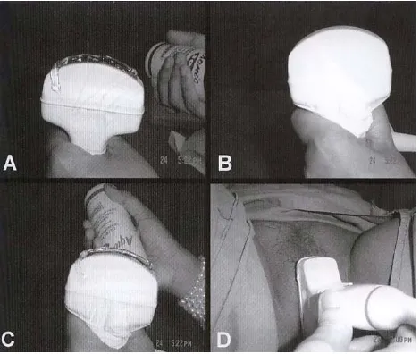
Translabial pelvic floor imaging is done using the 3D Voluson 730 Pro Ultrasound System transducer RAB 4-8 MHz. Before being placed in translabial, tranducer is wrapped with gloves or thin plastic to keep it hygienic. Do not use gloves that have been given the powder because it can damage the image quality because the gloves can cause unexpected resonance (reverberations). Im-aging is performed while the patient is in dorsal lithotomy position (hip in flexion and slight abduc-tion) or in a standing position after the bladder and rectum are being emptied. Transducer is placed translabial.
Examination steps of levator ani muscle by 3D ultrasound (Figure1):
which will be displayed in 3D. The technique is: press the button on the trackball once so that the volume that will be in 3D is now made up of the dotted line so that the size can be changed. Move the trackball upwards to raise the volume, left and right to position the volume. Once the measure is complete, press button above the trackball once again to set the broad volume so that it will not change anymore. Then press the freeze button to run the 3D facility. Press the button to split the screen into 2, so that the left side displays sagittal image piece, the right side displays the pelvic floor in 3D (rendered volume). Turn the 2D button to the left in order to darken the image a little bit so the picture of pelvic floor is more contrast. Con-sider the volume box on the left side, make sure the green box is located on the top side to make the axial cutting of pelvic floor. Shrink the size of volume box by pressing the button on the trackball once so that the box is now made up of dotted lines, move the trackball to the top so that the width of the box is reduced and create a width of
± 2 cm. Press the button above the trackball twice
so that the size does not change. Then move the trackball to move the sagittal images on the left screen, position so that the minimum hiatus plane is about a little below the edge lines of the green box. If the position is correct, hence on the right screen will be displayed the picture of the pelvic floor in 3D (volume rendered) clearly.
Instruct the patient to contract the levator ani muscle a minimum of twice, then press the freeze button. Press the save button in the form of movie so that the image of levator muscle contract twice is stored in the form of 4D (moving pictures). By moving the trackball to the left and right we can see moving pictures which had been stored and can choose the best moment of maximum contrac-tion. Measure hiatus area by pressing the point area on the touchscreen and measure hiatus area by tracing along the edge of the hiatus, lastly press the button twice to give output result of the area that we trace. Activate the Tomographic Ultra-sound Imaging (TUI) by selecting it via touch-screen, and then select plane C, then turn the Z but-ton to the left so that the image rotates 90°. Make sure that the TUI setting: the distance is 25 mm/ pieces, number of pieces is 11, the image can be enlarged by turning the zoom button. The system that describes the assessment of levator ani trauma after childbirth by measuring the Levator Urethra Gap (LUG) on three central pieces of the TUI.
Measure the distance on the left and right LUG on 3 central image of TUI. Note that if there is > 25 mm indicating levator avulsion. The levator muscle defects caused by trauma is defined when there is a LUG measurement > 25 mm on three central pieces of TUI, it shows positive results. Le-vator ani muscle defect is usually expressed as an avulsion of unilateral or bilateral avulsion. When finished, go back by pressing the rendered volume and init buttons on the touchscreen. Like when you first display the rendered volume but this time the patient is instructed strong straining at least twice and then measure the vast hiatus the same way as when patients contract the levator ani.
RESULTS
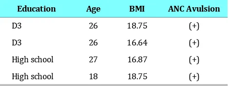
Table 1. Data of patients with levator ani avulsion
Education Age BMI ANC Avulsion
D3 26 18.75 (+)
D3 26 16.64 (+)
High school 27 16.87 (+)
High school 18 18.75 (+)
Table caption: BMI=Body Mass Index, ANC=Antenatal care
We had 4 women who had levator ani avulsion. Diagnosis of levator trauma (avulsion) on to-mographic ultrasound was correlated with prede-livery demographic variables and ultrasound pa-rameters.
Mean maternal age was 24.3 years (range, 18-27). Mean BMI was 17,75 (range 16.64-18.75). From 182 patients, we checked on antenatal care, we found four patients with levator ani avulsion. We can explain the relationship between education and economic conditions that form a positive feed-back. Socio-demographic factors will affect a per-son’s nutritional status, meaning that if economic conditions get better the nutritional status will be better also. This will affect the body mass index to increase also.
DISCUSSION
obstetric risk factors include age of mother at first birth. A number of levator ani trauma mechanism was proposed, which include genetic predisposi-tion associated with the properties of the collagen network, the mechanism strain of the pelvic floor, pelvic floor collagen tissue adaptation, and ultra-structural changes in vaginal tissue.11 If the pelvic
floor has weakened, e.g. during vaginal delivery, the hiatus may not fully close anymore at contrac-tion of the levator muscle. The counterforce will not come into effect, and POP may occur.11,12
Levator ani avulsion can be influenced by vari-ous factors. Race will have more influence on ge-netic conditions (gege-netic predisposition) and the factor of molecular biology. Genetic predisposition factors will affect the ability of the network adap-tation and ultra structural changes. The relation-ship of tissue factor in the incidence of avulsion of the levator ani form a positive feedback or rein-forcing loop. This can be interpreted if the adapt-ability of the network increases (getting better), the incidence of levator ani trauma will decrease. Furthermore, if the levator ani increased the inci-dence of a person’s trauma then network adapt-ability will degrade.8,10
By analogy, the above positive feedback can ex-plain the relationship between education and eco-nomic conditions that form a positive feedback. So-cio-demographic factors will affect a person’s nu-tritional status, meaning that if economic con-ditions get better the nutritional status will be bet-ter also. This will affect the body mass index to in-crease also. Furthermore, body mass index inin-creased the incidence of avulsion of the levator ani.3,6
Old primi factor and duration of second stage of labor will affect the mechanisms of tissue strain on the pelvic floor muscles including levator ani. As a result, the mechanism of strain factor and the in-cidence of levator ani trauma will form a negative feedback. The higher the strain mechanism, the higher the levator ani trauma incidence. However, if the levator ani trauma increase as a result it will reduce the strain mechanism (the ability of the strain is reduced due to trauma). The relationship between second stage of labor and vaginal lacera-tions can be explained as follows: the higher the second stage of labor the higher the potential for vaginal lacerations. The same explanation used to explain the relationship between the variables birth weight, episiotomy, and laceration of the va-gina and the incidence avulsion of the levator ani.
Old primi factors, stage II of labor, vaginal lacera-tions, episiotomy, and weight (baby) at birth, in-cluding the obstetric risk factors.7,8
It has been suggested that the hormonal effects of pregnancy may have an effect on levator ani muscle properties.13 Pregnancy is known to have
negative effects on the integrity of the pelvic floor due to the influence of specific hormones that in-crease during pregnancy. Elevated levels of two hormones: progesterone and relaxin, especially will cause weakness of collagen tissue throughout the body. Influence of the hormones that cause di-lation of blood vessels have also caused a greater mobility of joints, including joints in the pelvis, which causes a slight increase in diameter of the birth canal. These hormones also affect the colla-gen tissue of the pelvis, which causes weakness of ancillary structures from the vagina, bladder and rectum.11-16
The physiological changes occurring during pregnancy and the processes of childbirth have a detrimental effect on the structure and function of the muscles, nerves and fascial tissues (connective tissue) that make up the pelvic floor complex. As the fetus grows, the weight of the fetus and the gravid uterus (pregnant uterus) produce anatomi-cal changes to the bladder and urethra. Studies us-ing ultrasound imagus-ing techniques have shown that the angle between the bladder neck and the urethra increases, producing an increased opening of the bladder neck. There is also an increased mo-bility of the bladder due to the hormonal changes of pregnancy which also affect the pelvic floor com-plex.17 Furthermore, it remains unclear whether
pregnancy itself or factors associated with labor and delivery contribute to the injury of the pelvic floor.18
A long time has passed since Kegel first recom-mended pelvic floor muscle training to prevent and treat pelvic floor dysfunctions such as urinary in-continence (UI) and pelvic organ prolapse in women after child birth. Since then, randomized controlled trials have shown that antenatal pelvic floor muscle training also can prevent and treat UI both during pregnancy and in the immediate post-partum period. However, only a small proportion of pregnant women actually exercise the pelvic floor muscle regularly.19 This may be because
care providers claim that there is anecdotal evi-dence that elite athletes have rigid, inextensible pelvic floors that prolong the second stage of labor and that specific pelvic floor muscle training may make the pelvic floor muscle too strong and less elastic.21 Antenatal pelvic floor muscle training
be-fore and during pregnancy does not seem to affect labor and birth negatively.22 One study assessed
pelvic floor muscle strength and found that it in-creased significantly after the 8-week training pe-riod.21
However, regular pelvic floor muscle training has been shown to increase pelvic floor muscle strength and muscle volume, to lift the levator plate into a higher pelvic position and to narrow the hiatus.23,24 All these factors potentially may
prolong the total length of labor. It can be hypothe-sized that well-trained muscles may contribute to a reduced risk of injury during labor and may heal faster than untrained muscles.21
Previous studies investigating the use of estro-gen as a therapy for prolapse have shown no effect on the stage of prolapse. In contrast, estrogen has been found to be therapeutic in the treatment of mild stress urinary incontinence and symptoms as-sociated with urogenital atrophy. Estrogen recep-tors have been identified throughout the nuclei of the connective tissue and smooth muscle cells of the bladder trigone, urethra, vaginal mucosa, leva-tor ani muscle stroma, the arcus tendineus, and the uterosacral ligament. If estrogen does positively impact the pelvic floor, it most likely acts in a pre-ventative fashion to optimize the function of the pelvic floor muscle and connective tissue.7
An attempt to integrate various risk factors and to consider mechanisms of levator ani trauma. All efforts were done to eventually establish a scoring system that can be used to predict the occurrence of levator ani muscle damage. When scoring showed the risk of damage to the levator ani is low, then the patient can be convinced to choose a vagi-nal delivery without any sense of fear of pelvic floor dysfunction.
CONCLUSION
From this case series, we can learn that there are many complex factors that can cause avulsion of the levator ani. With a greater understanding on the function of the pelvic floor muscles and risk factors for trauma and damage as a result of
preg-nancy and birth, healthcare professionals will have better ability to meet the needs of women in the childbearing year.
This study utilized a scoring system that can be used to predict the occurrence of levator ani mus-cle damage. Thus, it is expected that the decline in quality of life of women due to dysfunction of the pelvic floor after vaginal delivery can be prevented. Physicians were expected to become more aware of the women risks during pregnancy and able to provide a comprehensive choice of solutions. In the end, the decline in quality of life for women, espe-cially after a birth, should be able to be prevented.
REFERENCES
1. Dietz HP. Levator trauma in Labor. A Challenge for Obste-tricians, Surgeons and Sonologists. Ultrasound Obstet Gyne-col 2007; 29:368-71.
2. DeLancey JO, Morgan DM, Fenner DE, Keamey R, Guire K, Miller JM, Hussain H, Umek W, Hsu Y, Ashton-Miller JA. Comparison of levator ani muscle defects and function in women with and without pelvic organ prolapse. Obstet Gynecol 2007; 109:295-302.
3. Dietz HP, Shek C. Levator avulsion and grading of pelvic floor muscle strength. Int Urogynecol J Pelvic Floor Dysfunct 2008; 19:633-6.
4. Buchsbaum GM, Duecy EE, Kerr LA, Huang LS, Perevich M, Guzick DS. Pelvic organ prolapse in nulliparous women and their parous sisters. Obstet Gynecol 2006; 108:1388-93. 5. Nikolova G, Lee H, Berkovitz S, Nelson S, Sinsheimer J, Vilain
E et al. Sequence variant in the laminin gamma 1 (LAMC1) gene associated with familial pelvic organ prolapse. Hum Genetics 2007; 120:847-56.
6. Twiss C, Triaca V, Rodriguez LV. Familial transmission of urogenital prolapse and incontinence. Curr Opin Obstet Gynecol 2007; 19:464-8.
7. Moalli PA, Ivy SJ, Meyn LA, Zyczynski HM. Risk Factors As-sociated with Pelvic Floor Disorder in Women Undergoing Surgical Repair. Obstet Gynecol 2003; 101:869-74 8. Kearney R, Miller JM, Ashton-Miller JA, DeLancey JOL.
Ob-stetric Factors associated with Levator Ani Muscle Injury after Vaginal Birth. Obstet Gynecol 2006; 107:144-9. 9. Viktrup L, Rortweit G, Lose G. Risk of Stress Urinary
Incon-tinence Twelve Years after the First Pregnancy and Deliv-ery. Obstet Gynecol 2006; 108:248-54.
10. Dietz HP. Quantification of Major Morphological Abnormali-ties of the Levator Ani. Ultrasound Obstet Gynecol 2007; 29:329-34
11. Kerkhof MH,Hendriks L, Brolmann HA. Changes in connec-tive tissue in patients with pelvic organ prolapse a review of the current literature. Int Urogynecol J Pelvic Floor Dys-funct 2009; 20:461-74.
12. Shek KL, Dietz HP. Can levator avulsion be predicted ante-natally? Am J Obstet Gynecol 2010; 202:586.
14. Jelovsek JE, Maher C, Barber MD. Pelvic organ prolapse. Lancet 2007; 369: 1027-38.
15. Schwetner-Tiepelmann N, Thakar R, Sultan AH, Turn R. Ob-stetric levator ani muscle injuries - Current status. Ultra-sound Obstet Gynecol 2011.
16. Handa VL, Blomquist JL, McDermott KC, Friedman S, Munoz A. Pelvic floor disorders after vaginal birth: effect of episio-tomy, perineal laceration, and operative birth. Obstet Gyne-col 2012; 119:233-9.
17. Herbert J. Pregnancy and childbirth: the effect on pelvic floor muscles. Nurs Times 2009; 105:38-41.
18. O’Boyle AL, O’Boyle JD, Calhoun B, Davis GD. Pelvic organ support in pregnancy and postpartum. Int Urogynecol J Pel-vic Floor Dysfunct 2005; 16:69-72.
19. Bo K, Haakstad L, Voldner N. Do pregnant women exercise their pelvic floor muscles? Int Urogynecol J Pelvic Floor Dys-funct 2007; 18:733-6.
20. McLennan MT, Melick CF, Alten B, Young J, Hoehn MR. Pa-tients’ knowledge of potential pelvic floor changes associ-ated with pregnancy
21. Bo K, Fleten C, Nystad W. Effect of antenatal pelvic floor muscle training on labor and birth. Obstet Gynecol 2009; 113:1279-84.
22. Agur W, Steggles P, Waterfield M, Freeman R. Does ante-natal pelvic floor training affect the outcome of labour? A randomised controlled trial. Int Urogynecol J Pelvic Floor Dysfunct 2008; 19:85-8.
23. Balmforth JR, Mantle J, Bidmead J, Cardozo L. A prospective observational trial of pelvic floor muscle training for female stress urinary incontinence. BJU Int 2006; 98:811-7.
24. Dumoulin C, Peng Q, Stodkilde-Jorgensen Shishido K, Con-stantinou C. Changes in levator ani anatomical configuration following physiotherapy in women with stress urinary in-continence. Urology 2007; 178:970-7.