The role of estrogen in testis and the male reproductive tract: a review
and species comparison
R. A. Hess1, K. Carnes
Department of Veterinary Biosciences, Reproductive Biology and Toxicology, University of Illinois, Urbana, IL 61802, USA
Abstract
Testosterone and estrogen are hormones important to both sexes. In the adult testis, estrogen is synthesized by Leydig cells and germ cells, producing a relatively high concentration in rete testis fluid and in semen of several species. Estrogen receptors (ER) are present in the testis, efferent ductules and epididymis of most species; however, ERα is reported absent in the testis of a few, including man. ERα is abundant in the efferent ductule epithelium of every species examined to date. Is primary function is the regulated expression of proteins involved in fluid reabsorption. Disruption of ERα, either in the knockout (ERαKO) or by treatment with a pure antiestrogen, results in dilution of cauda epididymal sperm, disruption of sperm morphology, inhibition of sodium transport and subsequent water reabsorption, increased secretion of Cl-, and eventual decreased fertility. Loss of aromatase activity in the ArKO mouse does not result in a ERαKO of antiestrogen phenotype, suggesting that epithelial ERα in the efferent ductules may exhibit ligand-independent activity. In addition to the primary regulation of luminal fluid and ion transport, estrogen is also responsible for maintaining a differentiated epithelial morphology through a mechanism remaining to be discovered. Thus, estrogen or its receptor is important for male reproductive tract function in numerous species.
Key words: estrogen, aromatase, estrogen receptor, testis, efferent ductules, epididymis, prostate, sperm, fertility
Introduction
Estrogen has been found in the semen and fluids of the male reproductive tract of many species (Adamopoulos et al., 1984; Bujan et al., 1993; Claus et al., 1992; Claus et al., 1985; Eiler and Graves, 1977; Free and Jaffe, 1979; Ganjam and Amann, 1976; Setchell et al., 1983; Waites and
Einer-_______________________________________________
1
Corresponding author: [email protected] Received: June 5, 2004
Accepted: July 13, 2004
Jensen, 1974). At first it was thought that this male source of estrogen was produced primarily by the accessory sex glands and that estrogen’s function should be relegated to influencing the female reproductive tract after ejaculation, a role that it may indeed play to some degree (Willenburg et al., 2003). In the 1930’s it was reported that developing testes were responsive to the “female” hormone ( also reviewed by Weniger, 1990; Wolff and Ginglinger, 1935). It was also known in the 1930’s and 40’s that developmental exposure to high doses of estrogens could induce malformations in the male reproductive tract (Arai et al., 1983; Burrows, 1935; Greene et al., 1940; McLachlan, 1979). However, as late as the early 1990’s, many scientists still considered estrogen receptor presence in the adult male reproductive tract to be only a residual of embryological differentiation (Greco et al., 1993). Previous reviews have already covered important aspects of estrogen’s influence on male reproductive development (Hess, 2003; Hess et al., 2001b; Iguchi et al., 2001; O'Donnell et al., 2001; Sharpe, 1998; Sharpe, 2003); therefore, here we will focus on a comparison of estrogen synthesis, receptor localization and potential function in a variety of adult male species.
Estrogen Synthesis and Inactivation
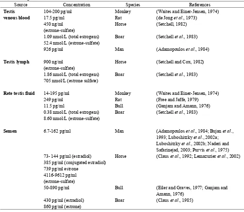
species, with the rat showing the highest, at 249 pg/ml (Free and Jaffe, 1979). In semen, conjugated estrogens are often found at extreme levels in the horse, bull and boar (Claus et
al., 1992; Claus et al., 1985; Eiler and Graves, 1977; Ganjam and Amann, 1976; Lemazurier et al., 2002).
Table 1. Estrogen concentrations in the male
Source Concentration Species References
Testis 104-200 pg/ml Monkey (Waites and Einer-Jensen, 1974)
venous blood 17.5 pg/ml Rat (de Jong et al., 1973)
450 ng/ml (estrone-sulfate)
Horse (Setchell, 1982)
1.09 nmol/L (total estrogens) 52.4 nmol/L (estrone-sulfate)
Boar (Setchell et al., 1983)
926 pg/ml Man (Adamopoulos et al., 1984)
Testis lymph 900 ng/ml
(estrone-sulfate)
Horse (Setchell and Cox, 1982)
1.86 nmol/L (total estrogens) 705 nmol/L (estrone sulfate)
Boar (Setchell et al., 1983)
Rete testis fluid 14-195 pg/ml Monkey (Waites and Einer-Jensen, 1974)
249 pg/ml Rat (Free and Jaffe, 1979)
11.5 pg/ml Bull (Ganjam and Amann, 1976)
0.38 nmol/L (total estrogens) 8.60 nmol/L (estrone-sulfate)
Boar (Setchell et al., 1983)
Semen 6.7-162 pg/ml Man (Adamopoulos et al., 1984; Bujan et al.,
1993; Luboshitzky et al., 2002a; Luboshitzky et al., 2002b; Naderi and Safarinejad, 2003; Purvis et al., 1975) 73- 144 pg/ml (estradiol)
385 pg/ml (conjugated estradiol) 739 pg/ml estrone
4116-9612 pg/ml (estrone-sulfate)
Horse (Claus et al., 1992; Lemazurier et al., 2002)
50-890 pg/ml Bull (Eiler and Graves, 1977; Ganjam and
Amann, 1976) 430 pg/ml (estradiol)
860 pg/ml (estrone)
Boar (Claus et al., 1985)
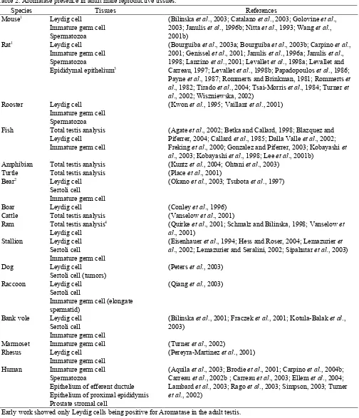
Estrogen synthesis in the male reproductive tract was first thought to occur in Sertoli cells during development, but then only in Leydig cells of the adult testis in most species (Carreau et al., 2003; O'Donnell et al., 2001; Payne et al., 1987; Rommerts and Brinkman, 1981; Rommerts et al., 1982; Sharpe et al., 2003; van der Molen et al., 1981). Table 2 shows the reported locations for estrogen synthesis in the adult male reproductive system. There is a consistent presence of aromatase in Leydig cells, but several species also reportedly show activity in Sertoli cells of the adult testis. In the dog,
Table 2. Aromatase presence in adult male reproductive tissues.
Species Tissues References
Mouse1 Leydig cell Immature germ cell Spermatozoa
(Bilinska et al., 2003; Catalano et al., 2003; Golovine et al., 2003; Janulis et al., 1996b; Nitta et al., 1993; Wang et al., 2001b)
Rat1 Leydig cell
Immature germ cell Spermatozoa
Epididymal epithelium3
(Bourguiba et al., 2003a; Bourguiba et al., 2003b; Carpino et al., 2001; Genissel et al., 2001; Janulis et al., 1996a; Janulis et al., 1998; Lanzino et al., 2001; Levallet et al., 1998a; Levallet and Carreau, 1997; Levallet et al., 1998b; Papadopoulos et al., 1986; Payne et al., 1987; Rommerts and Brinkman, 1981; Rommerts et al., 1982; Tirado et al., 2004; Tsai-Morris et al., 1984; Turner et al., 2002; Wiszniewska, 2002)
Rooster Leydig cell Immature germ cell Spermatozoa
(Kwon et al., 1995; Vaillant et al., 2001)
Fish Total testis analysis Leydig cell Immature germ cell
(Agate et al., 2002; Betka and Callard, 1998; Blazquez and Piferrer, 2004; Callard et al., 1985; Dalla Valle et al., 2002; Freking et al., 2000; Gonzalez and Piferrer, 2003; Kobayashi et al., 2003; Kobayashi et al., 1998; Lee et al., 2001b)
Amphibian Total testis analysis (Kuntz et al., 2004; Ohtani et al., 2003) Turtle Total testis analysis (Place et al., 2001)
Bear2 Leydig cell Sertoli cell
Immature germ cell
(Okano et al., 2003; Tsubota et al., 1997)
Boar Leydig cell (Conley et al., 1996)
Cattle Total testis analysis (Vanselow et al., 2001) Ram Total testis analysis4
Leydig cell
(Quirke et al., 2001; Schmalz and Bilinska, 1998; Vanselow et al., 2001)
Stallion Leydig cell Sertoli cell
Immature germ cell
(Eisenhauer et al., 1994; Hess and Roser, 2004; Lemazurier et al., 2002; Lemazurier and Seralini, 2002; Sipahutar et al., 2003)
Dog Leydig cell
Sertoli cell (tumors)
(Peters et al., 2003)
Raccoon Leydig cell Sertoli cell
Immature germ cell (elongate spermatid)
(Qiang et al., 2003)
Bank vole Leydig cell Sertoli cell
Immature germ cell
(Bilinska et al., 2001; Fraczek et al., 2001; Kotula-Balak et al., 2003)
Marmoset Immature germ cell (Turner et al., 2002) Rhesus Leydig cell
Immature germ cell
(Pereyra-Martinez et al., 2001)
Human Immature germ cell Spermatozoa
Epithelium of efferent ductule Epithelium of proximal epididymis Prostate stromal cell
(Aquila et al., 2003; Brodie et al., 2001; Carpino et al., 2004b; Carreau et al., 2002b ; Carreau et al., 2003; Ellem et al., 2004; Lambard et al., 2003; Rago et al., 2003; Simpson, 2003; Turner
et al., 2002)
1 Early work showed only Leydig cells being positive for Aromatase in the adult testis. 2
Location depended upon the season (Tsubota et al., 1997). 3 Only in primary culture cells (Wiszniewska, 2002).
The first reports to demonstrate aromatase in testicular germ cells and sperm (Fig. 1) were published through a collaborative effort at the University of Illinois (Janulis et al., 1996a; Janulis et al., 1998; Janulis et al., 1996b; Kwon et al., 1995; Nitta et al., 1993). Its presence in germ cells was found in diverse species ranging from mice to chicken testes (Fig. 1). The enzyme was localized in the Golgi of round spermatids and throughout the cytoplasm of elongating and late spermatids. The enzyme is found in the cytoplasmic droplet of epididymal sperm (Fig. 2), but its presence and activity are higher in sperm isolated from the efferent ductules and head of the epididymis than from the cauda region (Janulis et al., 1996a; Rago et al., 2003). Aromatase in germ cells and spermatozoa represent approximately 62% of the total testicular amount (Carreau
et al., 1999; Levallet et al., 1998a; Levallet and Carreau, 1997). Its biological activity in developing germ cells has been found to equal or exceeded that found in interstitial
cells. More recently, Carreau and others have confirmed aromatase presence in testicular germ cells and sperm and have demonstrated aromatase expression and activity in human sperm (Aquila et al., 2003; Aquila et al., 2002; Carani et al., 2002; Carreau, 2000; Carreau, 2001; Carreau, 2002; Carreau, 2003; Carreau et al., 1998; Carreau et al., 2002a; Carreau et al., 2001; Carreau et al., 2002b; Carreau
et al., 2004; Carreau et al., 1999; Carreau et al., 2003; Carreau and Levallet, 1997; Lambard et al., 2003; Lambard
et al., 2004; Rago et al., 2003). Only a few species, such as the horse (Eisenhauer et al., 1994; Hess and Roser, 2004; Lemazurier et al., 2002; Lemazurier and Seralini, 2002; Sipahutar et al., 2003), have not shown testicular germ cells to be aromatase-positive (Table 2). It is unknown if the lack of staining was due to differences in antibodies or if species simply differ in the sources of estrogen found in the reproductive tract.
Figure. 1A. Aromatase in the mouse testis show immunohistochemical staining of Leydig cells (L), elongated spermatids (E), and released sperm (S). 1B. Aromatase in the mouse epididymis showing staining of the cytoplasmic droplet on sperm tails (CD). 1C. Rooster testis showing aromatase in Leydig cells (L), round spermatids, and elongated spermatids (E).
These recent discoveries of germ cell production of estrogen in the male reproductive tract led to new hypotheses regarding estrogen receptor presence in the tract and its potential function. The Leydig cell is no longer considered the only source of estrogen for the reproductive tract and it appears that Leydig cell derived estradiol would more likely target the lymphatics and peripheral circulation, rather than the lumens of rete testis and epididymis. Leydig cells lie adjacent to endothelial cells of the lymphatic system, a region reported to have very high concentrations of estrogens (Setchell, 1982; Setchell et al., 1983). However, blood estrogen concentrations are low in the male, therefore, we presume that estrogens from Leydig cell synthesis would provide limited endocrine activity in the reproductive tract. In the efferent ductules, blood-borne estrogens would likely have even less effect, as these ductules are responsible for reabsorption of over 90% of the luminal fluids (Clulow et al., 1998) and thus display an overwhelming luminal to basal orientation, which could limit the movement of substances from basement membrane into the cell cytoplasm. Although this hypothesis has not been tested directly, there are studies suggesting that this region of the male tract does not respond to exogenous androgens following castration (Fawcett and Hoffer, 1979). More recent studies, however, suggest that after castration the efferent ductules do respond to estrogens and androgens (Oliveira et al., 2004). Nevertheless, current data demonstrate that in most species luminal estrogen, produced by testicular germ cells and luminal sperm, is more than sufficient to target estrogen receptors found in epithelial cells lining the male reproductive tract (Hess, 2002; Hess, 2003; Hess et al., 2002).
Estrogens are inactivated through sulfoconjugation, catalyzed by the enzyme estrogen sulfotransferase, which is abundantly expressed in liver (Song, 2001; Song and Melner, 2000). Interestingly in the male, estrogen sulfotransferase has been found to show the highest concentration and specific organ activity in the testis (Hobkirk and Glasier, 1992; Song, 2001; Song et al., 1995). This enzyme has been studied in the male of only a few species, but was found in testis of pigs, mice, rat, guinea pig and man (Hobkirk and Glasier, 1992; Hobkirk et al., 1989; Miki et al., 2002; Song, 2001; Song et al., 1995). In the testis, its presence is exclusive to the Leydig cell, but along the tract it is found in the epididymal epithelium and epithelium and smooth muscle of the vas deferens of mice (Tong and Song, 2002). It has not been found in prostate or seminal vesicles. The reproductive tracts of other species have not been investigated. Estrogen sulfotransferase is regulated in the testis and epididymis through pituitary gonadotrophins (LH) and androgens (Tong and Song, 2002). The CD-1 mouse testis was shown in 1995 to have the highest organ specific activity (Song et al., 1995) and then in 2001 the testis of this mouse strain was shown to be 16 fold less sensitive to estrogen than the B6 strain of mice
(Spearow et al., 2001). Spearow further showed that the CD-1 testis expresses 3.5 times more estrogen sulfotransferase than the B6 mouse testis (Spearow et al., 2001). Testes of the estrogen sulfotransferase knockout mice are reported to be damaged, with Leydig cell hyperplasia and hypertrophy and decreases in the weights of testis and epididymis (Qian et al., 2001). Sperm motility is also reduced, as well as fertility. Exogenous estrogen treatment of the estrogen sulfotransferase knockout mice induces further decline in sperm quality (Tong and Song, 2002).
Estrogen Receptors in the Male Reproductive Tract
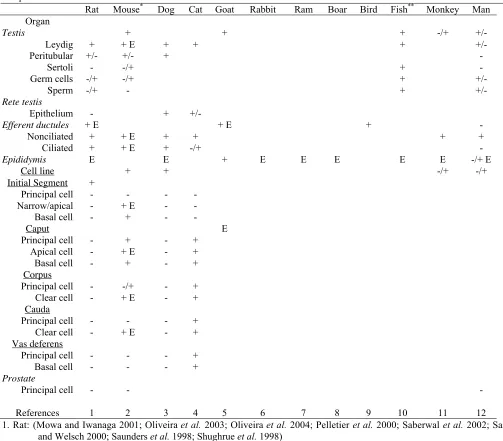
Estrogen receptor-like proteins were found in epididymal tissues over 30 years ago (Danzo et al., 1975). However, early investigations into estrogen receptor presence and function in the male reproductive tract lead to the conclusion that estrogen was more important during development than in the adult (Danzo, 1986). Estrogen binding in epididymal tissues has been noted in numerous species, including the dog (Younes et al., 1979; Younes and Pierrepoint, 1981), human (Murphy et al., 1980), turtle (Dufaure et al., 1983), monkey (Kamal et al., 1985; West and Brenner, 1990), ram (Tekpetey and Amann, 1988), guinea pig (Danzo et al., 1981), and the rat (Kuiper et al., 1997). In the mouse, estrogen binding was found throughout the testis and epididymis (Hess et al., 1997b; Schleicher et al., 1984). The strongest binding was found in the efferent ductule epithelium and initial segment epididymis, with lesser binding in the distal tract (Schleicher et al., 1984). However, binding assays do not differentiate between ERα and ERβ; therefore, other methods, such as immunocytochemistry, in situ hybridization and Northern blot analysis, have been used to separate the two ER subtypes. Unfortunately, these techniques do not provide identical results and disagreements are found in ER presence in the male (Hess et al., 2002).
Using immunocytochemistry, ER has consistently been localized in the epithelium of efferent ductules (Ergun
Table 3. Localization of ERα and estrogen binding (E) in the testis and male reproductive tract epithelium: a species comparison
Rat Mouse* Dog Cat Goat Rabbit Ram Boar Bird Fish** Monkey Man Organ
Testis + + + -/+
+/-Leydig + + E + + +
+/-Peritubular +/- +/- +
-Sertoli - -/+ +
-Germ cells -/+ -/+ +
+/-Sperm -/+ - +
+/-Rete testis
Epithelium - +
+/-Efferent ductules + E + E +
-Nonciliated + + E + + + +
Ciliated + + E + -/+
-Epididymis E E + E E E E E -/+ E
Cell line + + -/+ -/+
Initial Segment +
Principal cell - - -
-Narrow/apical - + E -
-Basal cell - + -
-Caput E
Principal cell - + - +
Apical cell - + E - +
Basal cell - + - +
Corpus
Principal cell - -/+ - +
Clear cell - + E - +
Cauda
Principal cell - - - +
Clear cell - + E - +
Vas deferens
Principal cell - - - +
Basal cell - - - +
Prostate
Principal cell - -
-References 1 2 3 4 5 6 7 8 9 10 11 12
1. Rat: (Mowa and Iwanaga 2001; Oliveira et al. 2003; Oliveira et al. 2004; Pelletier et al. 2000; Saberwal et al. 2002; Sar and Welsch 2000; Saunders et al. 1998; Shughrue et al. 1998)
2. Mouse and vole*: (Atanassova et al. 2001; Bilinska et al. 2001; Prins et al. 2001; Risbridger et al. 2001; Shibayama et al.
2001; Sipila et al. 2004; Takao et al. 2003; Zhou et al. 2002) 3. Dog: (Nie et al. 2002; Telgmann et al. 2001)
4. Cat: (Nie et al. 2002) 5. Goat: (Mansour et al. 2001) 6. Rabbit: Not determined 7. Ram: Not determined 8. Boar: Not determined in adult 9. Bird: (Janssen et al. 1998)
10. Fish, newt** and amphioxus**: (Arenas et al. 2001; Bouma and Nagler 2001; Fang et al. 2003; He et al. 2003; Socorro et al. 2000; Wu et al. 2001)
11. Monkey: (Heikinheimo et al. 1995; Pelletier 2000; Saunders et al. 2001)
Table 3. Localization of ERβ in the testis and male reproductive tract epithelium: a species comparison (continued)
Rat Mouse* Dog Cat Goat Rabbit Ram Boar Bird Fish** Monkey Man
Organ
Testis + + + +
Leydig + + - + +
+/-Sertoli + + - - + +
+/-Peritubular + +/- + + + + +
Germ cells + + + + + -/+
+/-Germ cell tumor +
Rete testis
Epithelium + + + +
Efferent ductules
+
Nonciliated + + + + + +
Ciliated + + +/- + + +
Epididymis
-Cell line +
Initial Segment
Principal cell + + + + + +
Narrow/apical + + + + + +
Basal cell + + + + + +
Caput
Principal cell + +/- + + + + +
Apical cell + + + + + + +
Basal cell + + + + + + +
Corpus
Principal cell + + + + + + +
Clear cell + + + + + + +
Cauda
Principal cell + + + + + + +
Clear cell + + + + + + +
Vas deferens +
Principal cell + + + +
Basal cell + + + + + +
Prostate + +
Principal cell + +
References 1 2 3 4 5 6 7 8 9 10 11 12
1. Rat: (Asano et al. 2003; Atanassova et al. 2001; Makela et al. 2000; Oliveira et al. 2003; Oliveira et al. 2004; Pelletier et al. 2000; Prins et al. 1998; Sar and Welsch 2000; Saunders 1998; Shughrue et al. 1998; Tirado et al. 2004; van Pelt
et al. 1999; Weihua et al. 2001)
2. Mouse and vole*: (Bilinska et al. 2001; Choi et al. 2001; Jefferson et al. 2000; Kuiper et al. 1998; Prins et al. 2001; Risbridger et al. 2001; Rosenfeld et al. 1998; Saunders 1998; Schmalz et al. 1999; Sipila et al. 2004; Takao et al.
2003; Weihua et al. 2001; Zhou et al. 2002) 3. Dog: (Nie et al. 2002)
10. Fish, newt** and amphioxus**: (Arenas et al. 2001; Fang et al. 2003; He et al. 2003; Socorro et al. 2000; Wu et al. 2001) 11. Monkey: (Heikinheimo et al. 1995; McKinnell et al. 2001; Pelletier 2000; Pelletier et al. 1999; Saunders 1998; Saunders
et al. 2001)
12. Man: (Aquila et al. 2004; Denger et al. 2001; Lambard et al. 2004; Makinen et al. 2001; Pais et al. 2003; Pelletier 2000; Pelletier and El-Alfy 2000; Saunders 1998; Saunders et al. 2002; Saunders et al. 2001; Scobie, et al. 2002; Taylor and Al-Azzawi 2000)
The most consistent data across species has been ERα presence in the Leydig or Interstitial cells (Fig. 3), even in the fish testis. There are conflicting reports of ERα in germ cells and sperm (Aquila et al., 2004; Lambard et al., 2004; Nie et al., 2002; Wu et al., 2001; Zhou et al., 2002). Efferent ductules are positive for ERα in all species examined (Fig.4), although one study showed no immunostaining in man (Pelletier and El-Alfy, 2000). Analysis of mRNA from the efferent ductules has indicated that the receptor is expressed 3.5 fold greater than in female tissue (Hess et al., 1997b). The epididymis has generally been found to be ERα negative, although select species, such as the cat and mouse, have shown strong staining for this receptor in specific regions and select cell types (Nie et al., 2002; Zhou et al., 2002). Narrow, apical and clear cells of the rodent epididymis show intense binding affinity for estrogens (Schleicher et al., 1984) and also show intense staining by immunohistochemistry for ERα (Oliveira et al., 2003; Oliveira et al., 2004; Pelletier et al., 2000; Saunders
et al., 1998; Zhou et al., 2002). The prostate epithelium always appears ERα negative, while stromal cells are positive.
The discovery of a second form of ER (ERβ) complicates the interpretation of earlier data from estrogen binding studies, as it is unknown in those studies to which ER binding has occurred. ERβ was originally discovered because of it high expression in prostate (Kuiper et al., 1996), but it has now been found in all tissues of the male reproductive tract, in both epithelium and stromal tissues (Table 4). However, a function for ERβ in the male reproductive tract awaits further investigation, as the ERβ knockout mouse has been shown to be fertile and appears to have a normal testis and epididymis (Krege et al., 1998). ERβ is more widely distributed in the male tract than ERα (Hess et al., 2002) and shows strong reactivity in efferent ductules, similar to ERα. The male tract is an example where both receptors are expressed in high concentrations within the same cell (Nie et al., 2002; Zhou et al., 2002). ERβ appears to be weaker in initial segment epididymis but stronger in the corpus, cauda and vas deferens.
In the testis, ERβ is the more abundant receptor and is typically found in nearly every cell type of the interstitium and the seminiferous tubule (Fig. 3), except for the elongated spermatids (Bilinska et al., 2000; Jefferson et al., 2000; Makinen et al., 2001; McKinnell et al., 2001; Nie
et al., 2002; Pelletier, 2000; Rosenfeld et al., 1998;
Saunders et al., 1998; Saunders et al., 1997; Saunders et al., 2001; Takeyama et al., 2001; Taylor and Al-Azzawi, 2000; van Pelt et al., 1999; Zhou et al., 2002).
In contrast, ERα is found only in the interstitium of the testis in most species examined (Table 4). The ERβ knockout mouse (Couse et al., 1999; Krege et al., 1998) shows no testicular phenotype and double ERαβ knockout mice appear identical to the ERα knockout mice (Couse et al., 1999; Dupont et al., 2000; Eddy et al., 1996; Lubahn et al., 1993; Mahato et al., 2001).
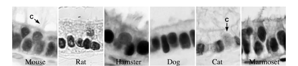
Figure. 4. ERα in the efferent ductule epithelium of several species: mouse, rat, hamster, dog, cat and marmoset monkey. Nonciliated principal cells are strongly positive in all species, but ciliated cells (C) are less positive in some.
Future studies must attempt to resolve conflicting reports found in the literature regarding the presence or absence of ERs in the male reproductive tract of different species. It is difficult to reconcile, for example, the generally accepted lack of ERα expression in germ cells with new reports of ERα expression in human sperm. It will also be important to determine why the cat and mouse express ERα in epididymal tissue, while other species generally show no immunostaining in this region. How could such a divergence in expression evolve? On the other hand, ERβ is nearly ubiquitous in its presence, both in the epithelium and stroma throughout the male reproductive tract. It is possible that in some species ERβ compensates for the lack of ERα, while in the cat and mouse, the duel presence of both receptors may be necessary for balancing unique epididymal functions of fluid reabsorption and sperm maturation.
Estrogen Function in Testis
Estrogen appears to have only a minor role in adult testicular function (see review by O'Donnell et al., 2001). However, Hardy and colleagues (Akingbemi et al., 2003) have demonstrated in mouse cells that antiestrogen treatment inhibitsLeydig cell activity in vitro, but estradiol alone was unable to stimulate Leydig cell steroidogenesis. In the developing testis, estrogen has significant activity in establishing Sertoli cell function (O'Donnell et al., 2001) and potentially even in establishing Sertoli-germ cell adhesion (MacCalman and Blaschuk, 1994; MacCalman et al., 1997). However, in the total absence of estrogen synthesis, the aromatase knockout (ArKO) male shows normal spermatogenesis at the beginning of puberty and only with aging does the testis begin to develop lesions associated with round spermatids (O'Donnell et al., 2001; Robertson et al., 2002). This is not entirely surprising in light of the fact that ERα is not present in the seminiferous epithelium of the mouse (Nie et al., 2002; Zhou et al., 2002) and although ERβ is found in Sertoli cells and nearly all germ cells (Nie et al., 2002; Saunders et al., 2002; Saunders
et al., 2001; Scobie et al., 2002; Zhou et al., 2002), the ERβ knockout (ERβKO) male testis appears normal and the males are fertile (Couse et al., 1999; Dupont et al., 2000; Krege et al., 1998).
There are no data showing that ERα is important in initiating or maintaining spermatogenesis. Transplantation of germ cells from the ERαKO mouse testis into a normal testis (made devoid of germ cells) produces normal spermatozoa capable of fertilization and results in live offspring (Mahato et al., 2001), suggesting that testicular ERα has no influence on spermatogenesis. However, loss of estrogen synthesis in the ArKO mouse (O'Donnell et al., 2001; Robertson et al., 2001) results in decreased fertility with aging. Another study in the mouse also suggests that estrogen may have testicular function, acting through the Leydig cell (Akingbemi et al., 2003). It was suggested that testosterone concentrations are elevated in the ERαKO male (Eddy et al., 1996), due to the disruption in feedback regulation at the hypothalamus, and the more recent study indeed shows that Leydig cells isolated from the ERαKO testis have increased production of testosterone and when treated with the pure ER inhibitor ICI 182,780 show increased steroidogenesis (Akingbemi et al., 2003). Therefore, ER in the testis, although not necessarily essential for spermatogenesis, appear to have a subtle function in Leydig cells.
Although estrogen may not be essential for spermatogenesis, there is indirect evidence of estrogen’s influence on spermatogenesis. Ebling and colleagues (Ebling et al., 2000) found that estradiol implants in the hpg
(Robertson et al., 1999). When the ArKO male is maintained on a soy-free diet, these effects are accelerated and enhanced (O'Donnell et al., 2001; Robertson et al., 2002). Thus, soy based phytoestrogens likely protected the testis somewhat in the ArKO mouse, suggesting that small amounts of estrogen do have testicular effects independent of FSH or LH.
This potential role for estrogen in the testis will most likely be found in the germ cells, as they express ERβ abundantly (Nie et al., 2002; Saunders et al., 2002; Saunders et al., 2001; Zhou et al., 2002) and genistein has a higher affinity for ERβ than for ERα (Kuiper et al., 1998b). Finally, although the Sertoli cell does not express ERα, it is interesting that in the ERαKO testis there is significantly less seminiferous tubular secretion than in the wild-type testis (Hess et al., 1997a). The same effect was suggested for the ArKO testis, as seminiferous tubule luminal volume and tubular length was decreased (Robertson et al., 2002).
Another compelling study that would suggest ERβ having a role in spermatogenesis comes from long-term treatment of the rat and mouse with ICI 182,780 (Cho et al., 2003; Oliveira et al., 2002). Similar to the results seen in the ArKO mouse (O'Donnell et al., 2001; Robertson et al., 2002), at first there was no effect on the testis, as spermatogenesis progressed normally. But with time, the testis shows severe atrophy in the rat (Oliveira et al., 2002) and hypospermatogenesis and abnormal germ cell development in the mouse (Cho et al., 2003). In the rat, seminiferous tubular atrophy was caused by back-pressure induced by fluid accumulation within the rete testis, similar to the reported effects seen in the ERαKO mouse (Hess et a l., 1997a). However, in the mouse there was no seminiferous tubular dilation or increase in testis weight (Cho et al., 2003); therefore, the effects on spermatogenesis could not have been induced by fluid accumulation, but were more likely due to direct effects of the antiestrogen on ERβ found in the germ cells (Zhou et al., 2002). It is also possible that indirect effects due to increases in testosterone concentration or alterations in paracrine factors associated with Leydig cell effects (Akingbemi et al., 2003). Thus overall, estrogen appears to have a function in the adult testis, not only in the Leydig cell but also possibly within the germinal epithelium. However, disruption of this function appears to require an extended period of inhibition.
Estrogen Function in Efferent Ductules
In all species studied to date, efferent ductules are a major site for estrogen function in the male reproductive tract. The ductules connect rete testis to epididymis (Hess, 2002). One-third or more of the head of the epididymis in man and other mammals contains these ducts and it was once thought that they simply transported sperm from testis
to the epididymis. However, it is now known that efferent ductules have an important function in the reabsorption of over 90% of the rete testis fluid and thereby concentrate sperm prior to entering the epididymal lumen (Clulow et al., 1998). Nonciliated cells of the epithelium are reabsorptive, similar to proximal tubules of the kidney, having a brush border of microvilli connecting in the apical cytoplasm to a profusion of apical canaliculi, vesicles, tubules and membrane-bound bodies, which constitutes an elaborate endocytotic/lysosomal system (Hermo et al., 1994). In the basal region, rough endoplasmic reticulum, mitochondria and lipid droplets are common (Ilio and Hess, 1994).
The efferent ductules express an abundance of both androgen and estrogen receptors (Hess et al., 2002; Nie et al., 2002; Oliveira et al., 2003; Oliveira et al., 2004; Zhou et al., 2002). Therefore it was not surprising to discover that the ERαKO mouse and the antiestrogen-treated rodents are infertile or show greatly reduced fertility (Cho et al., 2003; Eddy et al., 1996; Lubahn et al., 1993; Oliveira et al., 2002). Numerous prior reviews have covered this transgenic mouse (Carani et al., 2002; Couse and Korach, 1999a; Couse and Korach, 1999b; Couse and Korach, 2001; Couse et al., 2001; Hess, 2000a; Hess, 2000b; Hess, 2003; Hess et al., 2001a; Hess et al., 2002; Hess et al., 2001b; O'Donnell et al., 2001). Although the ERαKO testis appeared normal before puberty, after the onset of spermatogenesis, the testis began to degenerate and eventually became atrophic (Eddy
et al., 1996). By 150 days, cauda sperm from the ERαKO male were abnormal and sperm concentrations were significantly reduced (Eddy et al., 1996), suggesting that the reproductive tract was also abnormal. A later study by the Eddy’s lab showed that ERαKO germ cells transplanted into a normal testis (treated with busulphan to remove native germ cells) were capable of fertilization (Mahato et al., 2000). That study clearly pointed to extra-testicular regions, such as the efferent ductules and epididymis, being the major source of pathological alterations in ERαKO males (Eddy et al., 1996; Hess et al., 1997a).
The rete testes in the ERαKO mouse and the antiestrogen ICI 182,780 treated male mouse and rat are dilated and protrude into the testis (Eddy et al., 1996; Hess
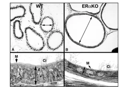
et al., 1997a; Lee et al., 2000; Oliveira et al., 2001). Based upon these data, we hypothesized that the efferent ductules were either a) occluded due to excessive reabsorption, or b) dilated due to an inhibition of fluid reabsorption. After careful examination, we found the second hypothesis to be true (Fig. 5), as the efferent ductule lumen was dilated markedly when ERα was inhibited (Cho et al., 2003; Hess
Figure. 5A. Wild type mouse (WT) showing normally dilated proximal efferent ductules. 5B. In the ERαKO mouse, the proximal efferent ductule lumen is extremely dilated compared to WT. 5C. WT efferent ductule epithelium by light microscopy showing normal columnar height. Nonciliated cells contain lysosomes (L) and endosomes (E) and have a prominent microvillus border (M) lining the lumen. Cilia (Ci) protrude into the lumen from the ciliated cell. 5D. ERαKO efferent ductule epithelium by light microscopy showing decreased epithelial height. Nonciliated cells contain few cytoplasmic organelles and the microvillus border (M) lining the lumen is greatly reduced. Cilia (Ci) protrude into the lumen from the ciliated cell.
in the lumen overloaded the funnel-like ductal system that is found in the rodent. As predicted, the accumulation of fluid caused a transient increase in testis weight in ERαKO males between 32-81 days of age and then a steady decrease in weight out to 185 days of age, when total atrophy was observed (Hess et al., 1997a). These data suggested that long-term atrophy of testes in the knockout mouse was caused by backpressure of the accumulating luminal fluids, a well-recognized pathogenesis found after exposure to various toxicants (Hess et al., 1997a; Hess et al., 2000). However, atrophy was only partially induced by the antiestrogen treatment in the adult mice (Cho et al., 2003), but was induced by long-term treatment with ICI 182,780 in the rat (Oliveira et al., 2001; Oliveira et al., 2002). These data have led us to hypothesize that ERβ that is present in the seminiferous epithelium, which would be blocked in the ICI 182,780 treated males, does have a role in normal spermatogenesis, but is disrupted only after inhibition for an extended period of time.
Figure. 6A. Wild type mouse (WT) efferent ductule epithelium at higher magnification by electron microscopy. The nonciliated principal cells are columnar and the apical cytoplasm is filled with lysosomes (L) and the endocytotic apparatus (E). The microvillus brush border (M) shows extensive individual protrusions. N, nucleus. 6B. ERαKO efferent ductule epithelium at higher magnification by electron microscopy. The nonciliated principal cells are short and the apical cytoplasm lacks the typical lysosomes and endocytotic apparatus. The microvillus brush border (M) consists of short irregular protrusions. The nuclei (N) are somewhat distorted and flattened.
The ERαKO mouse provided the first strong evidence that estrogen, or more specifically, a functional ERα, is involved in the regulation of fluid transport in the male reproductive tract, and responsible for increasing the concentration of sperm as they enter the epididymis. Subsequent studies have shown that the major Na+ transporter in the efferent ductule epithelium (NHE3) is down regulated in the ERαKO male reproductive tract). Both the mRNA and NHE3 protein are decreased substantially in ERαKO and ICI 182,780 treated efferent ductule tissue (Oliveira et al., 2002; Zhou et al., 2001). Because the ERαKO mouse lacks a functional ERα throughout development, the antiestrogen treatment studies are the only ones that effectively demonstrate that ERα is essential for adult function of the efferent ductule epithelium (Cho et al., 2003; Lee et al., 2001a; Lee et al., 2000; Oliveira et al., 2003; Oliveira et al., 2002; Zhou et al., 2001).
ICI 182,780 treatment of the adult male rat (Oliveira et al., 2001; Oliveira et al., 2002) demonstrated that there were species differences in response, with the rat showing greater variability than the mouse (Cho et al. 2003). It is interesting that the rat testes became totally atrophic, similar to the ERαKO mouse, while the ICI treated mice testes showed only limited atrophic seminiferous tubules and partial disruption of spermatogenesis. Other species are currently under investigation and it will be interesting to determine whether different species and even strains of rodents show varying sensitivity to the pure
antiestrogen. As new ER inhibitors are developed it will be possible to determine the separate contributions of the two receptors in male reproduction. Because both receptors are present in the same cell types of the male reproductive tract, it is possible that ERβ functions to dampen ERα in a manner similar to that found in other tissues (Gustafsson, 2003; Lindberg et al., 2003; Strom et al., 2004).
The aromatase knockout mouse (ArKO) does not exhibit the ERαKO and ICI 182,780 treatment phenotypes (Fisher et al., 1998; Robertson et al., 2002; Robertson et al., 2001). This raises several questions regarding the physiology of estrogen in the testis and efferent ductules, but the most likely answer lies in the fact that ERα is constitutively expressed in the rodent species (Oliveira et al., 2004), although regulated by testosterone (it is not clear that the receptor in this study was ERα) in the goat (Goyal
et al., 1998). The ArKO mouse, which lack estrogen, most likely still expresses ERα abundantly in the efferent ductules. If so, this will be an excellent example of ligand-independent activity of ERα, which could maintain NHE3 expression and subsequent ion transport and fluid reabsorption. Evidence has been accumulating that ERα can be activated in the absence of ligand by several mechanisms; the most well established being EGF induced tyrosine phosphorylation of ERα (Coleman and Smith, 2001; Marquez et al., 2001). Activation of MAP kinase induces ERα translocation to the nucleus (Lu et al., 2002; Osborne et al., 2001) and recently it was shown that acetylation of ERα by p300 cofactors also provides a ligand-independent mechanism for ERα signaling (Wang et al., 2001a). It is possible that fluid reabsorption in the efferent ductules commands extreme important for maintenance of fertility such that down regulation of ion transporter expression in this epithelium requires the loss of more than one receptor to cause a reduction in fluid and ion transport. Thus, it appears that estrogen ‘receptor’ action in this epithelium is more important than the presence of hormone itself.
Estrogen Function in Epididymis and Vas Deferens
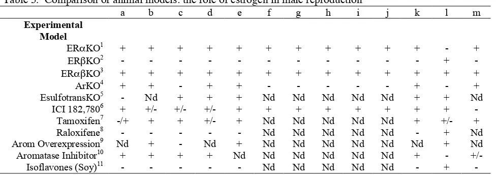
Table 5. Comparison of animal models: the role of estrogen in male reproduction
a b c d e f g h i j k l m
Experimental Model
ERαKO1 + + + + + + + + + + + - +
ERβKO2 - - - +
-ERαβKO3 + + + + + + + + + + + + +
ArKO4 + + - + + - - - + - +
EsulfotransKO5 - Nd + + + Nd Nd Nd Nd Nd + + Nd
ICI 182,7806 + +/- +/- +/- + + + + + + + +
-Tamoxifen7 -/+ + + +/- + Nd Nd Nd Nd Nd + +/- +
Raloxifene8 - - - Nd Nd Nd Nd Nd - + Nd
Arom Overexpression9 Nd + - Nd + Nd Nd Nd Nd Nd Nd + Nd
Aromatase Inhibitor10 + + + + Nd Nd Nd Nd Nd Nd + -
+/-Isoflavones (Soy)11 - - - Nd Nd Nd Nd Nd - +
-a- Infertility or decreased fertility or delayed infertility; b- Increased or decreased LH and/or testosterone; c- Change in testis weight or testicular atrophy d- Seminiferous tubular disruption
e- Leydig cell effects
f- Efferent ductule luminal dilation
g- Decreased efferent ductule epithelial height
h- Decreased efferent ductule endocytosis and/or microvilli
i- Decreased expression of sodium/hydrogen exchanger 3 and carbonic anhydrase II j- Increased expression of efferent ductule ion transporters
k- Effects on sperm, including cauda sperm counts and/or motility l- Effects on prostate or prostate cancer cells
m- Effects on sexual behavior n- Nd- Not determined 1
ERαKO: (Akingbemi et al., 2003; Dupont et al., 2000; Eddy et al., 1996; Hess et al., 1997a; Hess et al., 2000; Lee et al., 2001a; Lee et al., 2000; Lubahn et al., 1993; Lubahn et al., 1989; Mahato et al., 2000; Mahato et al., 2001; Nakai et al., 2001; Ogawa et al., 2000; Prins et al., 2001; Zhou et al., 2001)
2
ERβKO: (Dupont et al., 2000; Gustafsson and Warner, 2000; Krege et al., 1998; Risbridger et al., 2001; Weihua et al., 2001) 3
ERαβKO: (Couse et al., 1999; Dupont et al., 2000) 4
ArKO: (Fisher et al., 1998; Robertson et al., 2002; Robertson et al., 2001) 5
Estrogen sulfotransferase knockout: (Qian et al., 2001) 6
ICI 182,780: Mouse; (Cho et al., 2003; Hess et al., 1997a; Lee et al., 2000); Rat; (Oliveira et al., 2001; Oliveira et al., 2002); Prostate; (Ho, 2004; Huynh et al., 2001; Turner et al., 2001); Human Sperm; (Aquila et al., 2004)
7
Tamoxifen: (Adamopoulos et al., 1997; Belmonte et al., 1998; Brigante et al., 1985; Buvat et al., 1983; Chou et al., 1992; Corrada
et al., 2004; Danner et al., 1983; Dony et al., 1985; Du Mond et al., 2001; Gill-Sharma et al., 2001; Gill-Sharma et al., 2003; Gill-Sharma et al., 1993; Gopalkrishnan et al., 1998; Kotoulas et al., 1994; Li, 1991; Minucci et al., 1997; Nam et al., 2003; Noci et al., 1985; Padmalatha Rai and Vijayalaxmi, 2001; Parte et al., 2000; Robinzon et al., 1990; Rozenboim
et al., 1989; Rozenboim et al., 1986; Saberwal et al., 2002; Schill and Landthaler, 1981; Sethi-Saberwal et al., 2003) 8
Raloxifene: (Hoyt et al., 1998; Neubauer et al., 1993; Neubauer et al., 1995) 9
Arom Overexpression: (Fowler et al., 2000; Gill et al., 2001; Hiramatsu et al., 1997; Luthra et al., 2003; Simpson, 2003) 10
Aromatase Inhibitor: (Hayes et al., 2001; Hayes et al., 2000; Leder et al., 2004; Luthra et al., 2003; Mauras et al., 2000; Omura et al., 2001; Panno et al., 1995; Shetty et al., 1998; Smith et al., 2002; Trunet et al., 1993; Turner et al., 2000; Ulisse et al., 1994)
11
Isoflavones (soy): (Faqi et al., 2004; Mitchell et al., 2001; Morrissey and Watson, 2003; Robertson et al., 2002)
passage of immature sperm into the cauda epididymis, resulting in total sterility. The study did not determine effects on serum hormone concentrations, which leaves open the possibility that estrogen was not acting directly, but instead interfering with gonadotropin secretions and the
Other studies have shown that estrogen can influence contractions of the reproductive tract (Elmallah et al., 1995; Markus et al., 1980; Velasco et al., 1997). This potential mechanism for estrogen action in the epididymis should be further studies, as environmental estrogens, when given developmentally, also inhibit sperm transit time in the male reproductive tract (Gray et al., 1995).
Other studies have shown that estrogen, even in the presence of maintenance levels of testosterone, produces harmful effects on the epididymis and reduces fertilizing
ability of epididymal sperm (Lubicz-Nawrocki, 1974). Although other specific effects have been noted after estrogen treatment, it is not clear whether or not the effects on the epididymis were direct or indirect. In general, the effects of castration on the epididymis are reversible by testosterone administration and estrogen is antagonistic (Jones et al., 1980; Ma et al., 1998). Therefore, the question of estrogen’s importance in regulation of the epididymis and vas deferens remains unanswered.
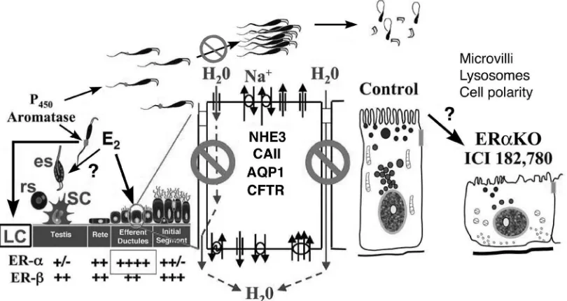
Figure. 7. This summarizes the presence of P450aromatase, estrogen receptors (ER) and targets for estrogen function and dysfunction in the
male reproductive tract. In the adult testis of many species, Leydig (LC) and germ cells (round spermatids-rs; elongated spermatids-es) and sperm express aromatase. Sertoli cells (SC) in the adult do not synthesize estrogen to any great extent. Estrogen (E2) synthesized by these sources target the abundance of ERα and ERβ found down stream in the efferent ductules. Estrogen does influence Leydig cell function but questions remain regarding its effect on the germ cells. In the mouse there are many epithelial cell types that contain ERα along the reproductive tract, but in other species only the efferent ductules express this receptor, while ERβ is nearly ubiquitous in epithelial cells of testis and epididymis of all species examined. Estrogen’s primary function in the male tract is the regulation of fluid reabsorption in the efferent ductules via ERα, which increases the concentration of sperm prior to entering the epididymis. Disruption of ERα results in decreased Na+ transport and thus decreased water (H2O) and fluid reabsorption. This inhibition is mediated by a decrease in the expression
of NHE3 mRNA and protein and also decreases in carbonic anhydrase II (CAII) and aquaporin I (AQP-1) proteins. There is also an increase in cystic fibrosis transmembrane conductance regulator (CFTR) protein and mRNA, which adds to the NHE3 effect by secreting Cl- into the lumen (Lee et al., 2001a). This inhibition (indicated by ) of fluid reabsorption results in the dilution of cauda epididymal sperm, disruption of sperm morphology, and eventual decreased fertility. In addition to this primary regulation, estrogen is also responsible for maintaining a differentiated epithelial morphology, which includes the expression of microvilli, lysosomes through an unknown mechanism that is apparently associated with cell polarity.
Summary and Conclusions
Estrogen is found in abundance in the testis, rete testis fluid and semen of many species. Its importance in the regulation of the male reproductive tract is now evident (Fig. 7), with convincing data showing direct effects on the function of Leydig cells and the efferent ductule epithelium.
Estrogen’s primary function in the male tract appears to be the regulation of fluid reabsorption in the efferent ductules via ERα. Disruption of the receptor results in dilution of cauda epididymal sperm, disruption of sperm morphology, inhibition of sodium transport and subsequent water reabsorption, increased secretion of Cl-, and eventual decreased fertility. The mechanism by which estrogen regulates epithelial morphology, such as microvillus growth and expression of endosomes and lysosomes, remains to be determined. Based upon the data reviewed, we must conclude that estrogen or its receptor is important for male reproductive tract function in numerous species.
Acknowledgments
We would like to acknowledge past and present trainees and visiting scientists whose work has helped to shape our understanding of estrogen function in the male: Hiro Nitta, Ken Ilio, Yu-Chyu Chen, Dan Gist, Masaaki Nakai, Sarah Janssen, Lynn Janulis, Hyun Wook Cho, Ki-ho Lee, Seok Kwon, Rong Nie, Qing Zhou, Cleida Oliveira, Paul Klopfenstein, James Ford, Jr., Tameka Phillips, Carla Morrow and Avenel Joseph. Supported by NIH grants HD35126, ES07326 and the Conrad program.
References
Adamopoulos, D, Lawrence, DM, Vassilopoulos, P, Kapolla, N, Kontogeorgos, L and McGarrigle, HH. 1984. Hormone levels in the reproductive system of normospermic men and patients with oligospermia and varicocele. J Clin Endocrinol Metab. 59:447-452.
Adamopoulos, DA, Nicopoulou, S, Kapolla, N, Karamertzanis, M and Andreou, E. 1997. The combination of testosterone undecanoate with tamoxifen citrate enhances the effects of each agent given independently on seminal parameters in men with idiopathic oligozoospermia. Fertil Steril. 67:756-762.
Agate, RJ, Perlman, WR and Arnold, AP. 2002. Cloning and expression of zebra finch (Taeniopygia guttata) steroidogenic factor 1: overlap with hypothalamic but not with telencephalic aromatase. Biol Reprod. 66:1127-1133. Akingbemi, BT, Ge, R, Rosenfeld, CS, Newton, LG, Hardy, DO, Catterall, JF, Lubahn, DB, Korach, KS and Hardy, MP. 2003. Estrogen receptor-alpha gene deficiency enhances androgen biosynthesis in the mouse Leydig cell.
Endocrinology. 144:84-93.
Al-Awqati, Q, Vijayakumar, S and Takito, J. 2003. Terminal differentiation of epithelia. Biol Chem. 384:1255-1258.
Aquila, S, Sisci, D, Gentile, M, Carpino, A, Middea, E, Catalano, S, Rago, V and Ando, S. 2003. Towards a physiological role for cytochrome P450 aromatase in ejaculated human sperm. Hum Reprod. 18:1650-1659.
Aquila, S, Sisci, D, Gentile, M, Middea, E, Catalano, S, Carpino, A, Rago, V and Ando, S. 2004. Estrogen Receptor (ER)alpha and ERbeta Are Both Expressed in Human Ejaculated Spermatozoa: Evidence of Their Direct Interaction with Phosphatidylinositol-3-OH Kinase/Akt Pathway. J Clin Endocrinol Metab. 89:1443-1451.
Aquila, S, Sisci, D, Gentile, M, Middea, E, Siciliano, L and Ando, S. 2002. Human ejaculated spermatozoa contain active P450 aromatase. J Clin Endocrinol Metab. 87:3385-3390.
Arai, Y, Mori, T, Suzuki, Y and Bern, H. 1983. Long-term effects of perinatal exposure to sex steroids and diethylstilbestrol on the reproductive system of male mammals. Int Rev Cytol. 84:235-265.
Arenas, MI, Royuela, M, Lobo, MV, Alfaro, JM, Fraile, B and Paniagua, R. 2001. Androgen receptor (AR), estrogen alpha (ER-alpha) and estrogen receptor-beta (ER-receptor-beta) expression in the testis of the newt, Triturus marmoratus marmoratus during the annual cycle. J Anat. 199:465-472.
Asano, K, Maruyama, S, Usui, T and Fujimoto, N. 2003. Regulation of estrogen receptor alpha and beta expression by testosterone in the rat prostate gland. Endocr J. 50:281-287.
Atanassova, N, McKinnell, C, Williams, K, Turner, KJ, Fisher, JS, Saunders, PT, Millar, MR and Sharpe, RM. 2001. Age-, cell- and region-specific immunoexpression of estrogen receptor alpha (but not estrogen receptor beta) during postnatal development of the epididymis and vas deferens of the rat and disruption of this pattern by neonatal treatment with diethylstilbestrol. Endocrinology. 142:874-886.
Belmonte, S, Maturano, M, Bertini, MF, Pusiol, E, Sartor, T and Sosa, MA. 1998. Changes in the content of rat epididymal fluid induced by prolonged treatment with tamoxifen. Andrologia. 30:345-350.
Betka, M and Callard, GV. 1998. Negative feedback control of the spermatogenic progression by testicular oestrogen synthesis: insights from the shark testis model.
Apmis. 106:252-257; discussion 257-258.
Bilinska, B, Kotula-Balak, M, Gancarczyk, M, Sadowska, J, Tabarowski, Z and Wojtusiak, A. 2003. Androgen aromatization in cryptorchid mouse testis. Acta Histochem. 105:57-65.
Bilinska, B, Schmalz-Fraczek, B, Kotula, M and Carreau, S. 2001. Photoperiod-dependent capability of androgen aromatization and the role of estrogens in the bank vole testis visualized by means of immunohistochemistry.
Mol Cell Endocrinol. 178:189-198.
Blazquez, M and Piferrer, F. 2004. Cloning, sequence analysis, tissue distribution, and sex-specific expression of the neural form of P450 aromatase in juvenile sea bass (Dicentrarchus labrax). Mol Cell Endocrinol. 219:83-94. Bouma, J and Nagler, JJ. 2001. Estrogen receptor-alpha protein localization in the testis of the rainbow trout (Oncorhynchus mykiss) during different stages of the reproductive cycle. Biol Reprod. 65:60-65.
Bourguiba, S, Genissel, C, Lambard, S, Bouraima, H and Carreau, S. 2003a. Regulation of aromatase gene expression in Leydig cells and germ cells. J Steroid Biochem Mol Biol. 86:335-343.
Bourguiba, S, Lambard, S and Carreau, S. 2003b. Steroids control the aromatase gene expression in purified germ cells from the adult male rat. J Mol Endocrinol. 31:83-94.
Brand, H, Kos, M, Denger, S, Flouriot, G, Gromoll, J, Gannon, F and Reid, G. 2002. A novel promoter is involved in the expression of estrogen receptor alpha in human testis and epididymis. Endocrinology. 143:3397-3404.
Brigante, C, Motta, G, Fusi, F, Coletta, MP and Busacca, M. 1985. Treatment of idiopathic oligozoospermia with tamoxifen. Acta Eur Fertil. 16:361-364.
Brodie, A, Inkster, S and Yue, W. 2001. Aromatase expression in the human male. Mol Cell Endocrinol. 178:23-28.
Bujan, L, Mieusset, R, Audran, F, Lumbroso, S and Sultan, C. 1993. Increased oestradiol level in seminal plasma in infertile men. Hum Reprod. 8:74-77.
Burrows, H. 1935. Pathological conditions induced by oestrogenic compounds in the coagulating gland and prostate of the mouse. Am J Cancer. 23:490-512.
Buvat, J, Ardaens, K, Lemaire, A, Gauthier, A, Gasnault, JP and Buvat-Herbaut, M. 1983. Increased sperm count in 25 cases of idiopathic normogonadotropic oligospermia following treatment with tamoxifen. Fertil Steril. 39:700-703.
Callard, GV, Pudney, JA, Mak, P and Canick, JA. 1985. Stage-dependent changes in steroidogenic enzymes and estrogen receptors during spermatogenesis in the testis of the dogfish, Squalus acanthias. Endocrinology. 117:1328-1335.
Carani, C, Fabbi, M, Zirilli, L and Sgarbi, I. 2002. [Estrogen resistance and aromatase deficiency in humans]. J Soc Biol. 196:245-248.
Carpino, A, Bilinska, B, Siciliano, L, Maggiolini, M and Rago, V. 2004a. Immunolocalization of estrogen receptor beta in the epididymis of mature and immature pigs. Folia Histochem Cytobiol. 42:13-17.
Carpino, A, Pezzi, V, Rago, V, Bilinska, B and Ando, S. 2001. Immunolocalization of cytochrome P450 aromatase in rat testis during postnatal development. Tissue Cell. 33:349-353.
Carpino, A, Romeo, F and Rago, V. 2004b. Aromatase immunolocalization in human ductuli efferentes and proximal ductus epididymis. J Anat. 204:217-220.
Carreau, S. 2000. Estrogens and male reproduction. Folia Histochemica et Cytobiologica. 38:47-52.
Carreau, S. 2001. Germ cells: a new source of estrogens in the male gonad. Mol Cell Endocrinol. 178:65-72.
Carreau, S. 2002. The testicular aromatase: from gene to physiological role. Reprod Biol. 2:5-12.
Carreau, S. 2003. Estrogens - male hormones? Folia Histochem et Cytobiol. 41:107-111.
Carreau, S, Balinski, B and Levallet, J. 1998. Male germ cells: a new source of estrogens in the mammalian testis.
Annales D Endocrinologie. 59:79-92.
Carreau, S, Bourguiba, S, Lambard, S and Galeraud-Denis, I. 2002a. [Testicular aromatase]. J Soc Biol. 196:241-244.
Carreau, S, Bourguiba, S, Lambard, S, Galeraud-Denis, I, Genissel, C, Bilinska, B, Benahmed, M and Levallet, J. 2001. Aromatase expression in male germ cells. J Steroid Biochem Mol Biol. 79:203-208.
Carreau, S, Bourguiba, S, Lambard, S, Galeraud-Denis, I, Genissel, C and Levallet, J. 2002b. Reproductive system: aromatase and estrogens. Mol Cell Endocrinol. 193:137-143.
Carreau, S, Bourguiba, S, Lambard, S, Silandre, D and Delalande, C. 2004. The promoter(s) of the aromatase gene in male testicular cells. Reprod Biol. 4:23-34.
Carreau, S, Genissel, C, Bilinska, B and Levallet, J. 1999. Sources of oestrogen in the testis and reproductive tract of the male. Int J Androl. 22:211-223.
Carreau, S, Lambard, S, Delalande, C, Denis-Galeraud, I, Bilinska, B and Bourguiba, S. 2003. Aromatase Carpino, A, Young, M, McPhaul, MJ and Ando, S. 2003. Triiodothyronine Decreases the Activity of the Proximal Promoter (PII) of the Aromatase Gene in the Mouse Sertoli Cell Line, TM4. Mol Endocrinol. 17:923-934.
Cho, HW, Nie, R, Carnes, K, Zhou, Q, Sharief, NA and Hess, RA. 2003. The antiestrogen ICI 182,780 induces early effects on the adult male mouse reproductive tract and long-term decreased fertility without testicular atrophy. Reprod Biol Endocrinol. 1:57.
Chou, YC, Iguchi, T and Bern, HA. 1992. Effects of antiestrogens on adult and neonatal mouse reproductive organs. Reprod Toxicol. 6:439-446.
Claus, R, Dimmick, MA, Gimenez, T and Hudson, LW. 1992. Estrogens and prostaglandin F2a in the semen and blood plasma of stallions. Theriogenology. 38:687-693. Claus, R, Schopper, D and Hoang-Vu, C. 1985. Contribution of individual compartments of the genital tract to oestrogen and testosterone concentrations in ejaculates of the boar. Acta Endocrinol. 109:281-288.
Clulow, J, Jones, RC, Hansen, LA and Man, SY. 1998. Fluid and electrolyte reabsorption in the ductuli efferentes testis. J Reprod Fertil Suppl. 53:1-14.
Coleman, KM and Smith, CL. 2001. Intracellular signaling pathways: nongenomic actions of estrogens and ligand-independent activation of estrogen receptors. Front Biosci. 6:D1379-1391.
Conley, AJ, Corbin, CJ, Hinshelwood, MM, Liu, Z, Simpson, ER, Ford, JJ and Harada, N. 1996. Functional aromatase expression in porcine adrenal gland and testis.
Biol Reprod. 54:497-505.
Corrada, Y, Arias, D, Rodriguez, R, Spaini, E, Fava, F and Gobello, C. 2004. Effect of tamoxifen citrate on reproductive parameters of male dogs. Theriogenology. 61:1327-1341.
Couse, JF, Hewitt, SC, Bunch, DO, Sar, M, Walker, VR, Davis, BJ and Korach, KS. 1999. Postnatal sex reversal of the ovaries in mice lacking estrogen receptors alpha and beta. Science. 286:2328-2331.
Couse, JF and Korach, KS. 1999a. Estrogen receptor null mice: what have we learned and where will they lead us?
Endocr Rev. 20:358-417.
Couse, JF and Korach, KS. 1999b. Reproductive phenotypes in the estrogen receptor-alpha knockout mouse.
Ann Endocrinol (Paris). 60:143-148.
Couse, JF and Korach, KS. 2001. Contrasting phenotypes in reproductive tissues of female estrogen receptor null mice. Ann N Y Acad Sci. 948:1-8.
Couse, JF, Mahata, D, Eddy, EM and Korach, KS. 2001. Molecular mechanism of estrogen action in the male: insights from the estrogen receptor null mice. Reprod Fertil Develop. 13:211-219.
Dalla Valle, L, Lunardi, L, Colombo, L and Belvedere, P. 2002. European sea bass (Dicentrarchus labrax L.) cytochrome P450arom: cDNA cloning, expression and genomic organization. J Steroid Biochem Mol Biol. 80:25-34.
Danner, C, Frick, J and Maier, F. 1983. Results of treatment with tamoxifen in oligozoospermic men.
Andrologia. 15 Spec No:584-587.
Danzo, BJ. 1986. A protease acting on the estrogen receptor may modify its action in the adult rabbit epididymis. J Steroid Biochem. 25:511-519.
Danzo, BJ, Eller, BC, Judy, LA, Trautman, JR and Orgebin-Crist, MC. 1975. Estradiol binding in cytosol
from epididymides of immature rabbits. Mol Cell Endocrinol. 2:91-105.
Danzo, BJ, St. Raymond, PA and Davies, J. 1981. Hormonally responsive areas of the reproductive system of the male guinea pig. III. Presence of cytoplasmic estrogen receptors. Biol Reprod. 25:1159-1168.
de Jong, F, Hey, A and van der Molen, H. 1973. Effect of gonadotrophins on the secretion of oestradiol-17β and testosterone by the rat testis. J Endocrinol. 57:277-284. Denger, S, Reid, G, Brand, H, Kos, M and Gannon, F. 2001. Tissue-specific expression of human ERalpha and ERbeta in the male. Mol Cell Endocrinol. 178:155-160. Dony, JM, Smals, AG, Rolland, R, Fauser, BC and Thomas, CM. 1985. Effect of lower versus higher doses of tamoxifen on pituitary-gonadal function and sperm indices in oligozoospermic men. Andrologia. 17:369-378.
Du Mond, JW, Jr., Singh, KP and Roy, D. 2001. The biphasic stimulation of proliferation of Leydig cells by estrogen exposure. Int J Oncol. 18:623-628.
Dufaure, JP, Mak, P and Callard, IP. 1983. Estradiol binding activity in epididymal cytosol of the turtle,
Chrysemys picta. Gen Comp Endocrinol. 51:61-65.
Dupont, S, Krust, A, Gansmuller, A, Dierich, A, Chambon, P and Mark, M. 2000. Effect of single and compound knockouts of estrogen receptors α (ERα) and β (ERβ) on mouse reproductive phenotypes. Development. 127:4277-4291.
Ebling, FJ, Brooks, AN, Cronin, AS, Ford, H and Kerr, JB. 2000. Estrogenic induction of spermatogenesis in the hypogonadal mouse. Endocrinology. 141:2861-2869.
Eddy, EM, Washburn, TF, Bunch, DO, Goulding, EH, Gladen, BC, Lubahn, DB and Korach, KS. 1996. Targeted disruption of the estrogen receptor gene in male mice causes alteration of spermatogenesis and infertility.
Endocrinol. 137:4796-4805.
Eiler, H and Graves, C. 1977. Oestrogen content of semen and the effect of exogenous oestradiol-17α on the oestrogen and androgen concentration in semen and blood plasma of bulls. J Reprod Fert. 50:17-21.
Eisenhauer, KM, McCue, PM, Nayden, DK, Osawa, Y and Roser, JF. 1994. Localization of aromatase in equine Leydig cells. Domest Anim Endocrinol. 11:291-298.
Ellem, SJ, Schmitt, JF, Pedersen, JS, Frydenberg, M and Risbridger, GP. 2004. Local aromatase expression in human prostate is altered in malignancy. J Clin Endocrinol Metab. 89:2434-2441.
Elmallah, AI, Sharabi, F, Omar, AG and El-Mas, MM. 1995. Prazosin-induced blockade of extraneuronal uptake facilitates dopaminergic modulation of muscle twitches in rat vas deferens. J Pharm Pharmacol. 47:932-936.
Fang, YQ, Weng, YZ, Huang, WQ and Sun, L. 2003. [Localization of the estrogen receptor alpha and beta-subtype in the nervous system, Hatschek's pit and gonads of amphioxus, Branchiostoma belcheri]. Shi Yan Sheng Wu Xue Bao. 36:368-374.
Faqi, AS, Johnson, WD, Morrissey, RL and McCormick, DL. 2004. Reproductive toxicity assessment of chronic dietary exposure to soy isoflavones in male rats. Reprod Toxicol. 18:605-611.
Fawcett, DW and Hoffer, AP. 1979. Failure of exogenous androgen to prevent regression of the initial segments of the rat epididymis after efferent duct ligation or orchidectomy.
Biol Reprod. 20:162-181.
Fisher, CR, Graves, KH, Parlow, AF and Simpson, ER. 1998. Characterization of mice deficient in aromatase (ArKO) because of targeted disruption of the cyp19 gene.
Proc Natl Acad Sci USA. 95:6965-6970.
Fisher, JS, Millar, MR, Majdic, G, Saunders, PT, Fraser, HM and Sharpe, RM. 1997. Immunolocalisation of oestrogen receptor-alpha within the testis and excurrent ducts of the rat and marmoset monkey from perinatal life to adulthood. J Endocrinol. 153:485-495.
Fowler, KA, Gill, K, Kirma, N, Dillehay, DL and Tekmal, RR. 2000. Overexpression of aromatase leads to development of testicular leydig cell tumors : an in vivo model for hormone-mediated TesticularCancer. Am J Pathol. 156:347-353.
Fraczek, B, Bourguiba, S, Carreau, S and Bilinska, B. 2001. Immunolocalization and activity of aromatase in the bank vole testes. Folia Histochem Cytobiol. 39:315-319. Free, MJ and Jaffe, RA. 1979. Collection of rete testis fluid from rats without previous efferent duct ligation. Biol Reprod. 20:269-278.
Freking, F, Nazairians, T and Schlinger, BA. 2000. The expression of the sex steroid-synthesizing enzymes CYP11A1, 3beta-HSD, CYP17, and CYP19 in gonads and adrenals of adult and developing zebra finches. Gen Comp Endocrinol. 119:140-151.
Ganjam, VK and Amann, RP. 1976. Steroids in fluids and sperm entering and leaving the bovine epididymis, epididymal tissue, and accessory sex gland secretions.
Endocrinology. 99:1618-1630.
Genissel, C, Levallet, J and Carreau, S. 2001. Regulation of cytochrome P450 aromatase gene expression in adult rat Leydig cells: comparison with estradiol production. J Endocrinol. 168:95-105.
Gill, K, Kirma, N and Tekmal, RR. 2001. Overexpression of aromatase in transgenic male mice results in the induction of gynecomastia and other biochemical changes in mammary glands. J Steroid Biochem Mol Biol. 77:13-18. Gill-Sharma, MK, Balasinor, N, Parte, P, Aleem, M and Juneja, HS. 2001. Effects of tamoxifen metabolites on fertility of male rat. Contraception. 63:103-109.
Gill-Sharma, MK, D'Souza, S, Parte, P, Balasinor, N, Choudhuri, J, Majramkar, DD, Aleem, M and Juneja,
HS. 2003. Effect of oral tamoxifen on semen characteristics and serum hormone profile in male bonnet monkeys.
Contraception. 67:409-413.
Gill-Sharma, MK, Gopalkrishnan, K, Balasinor, N, Parte, P, Jayaraman, S and Juneja, HS. 1993. Effects of tamoxifen on the fertility of male rats. J Reprod Fertil. 99:395-402.
Golovine, K, Schwerin, M and Vanselow, J. 2003. Three different promoters control expression of the aromatase cytochrome p450 gene (cyp19) in mouse gonads and brain.
Biol Reprod. 68:978-984.
Gonzalez, A and Piferrer, F. 2003. Aromatase activity in the European sea bass (Dicentrarchus labrax L.) brain. Distribution and changes in relation to age, sex, and the annual reproductive cycle. Gen Comp Endocrinol. 132:223-230.
Gonzalez-Unzaga, M, Tellez, J and Calzada, L. 2003. Clinical significance of nuclear matrix-estradiol receptor complex in human sperm. Arch Androl. 49:77-81.
Gopalkrishnan, K, Gill-Sharma, MK, Balasinor, N, Padwal, V, D'Souza, S, Parte, P, Jayaraman, S and Juneja, HS. 1998. Tamoxifen-induced light and electron microscopic changes in the rat testicular morphology and serum hormonal profile of reproductive hormones.
Contraception. 57:261-269.
Goyal, HO, Bartol, FF, Wiley, AA, Khalil, MK, Chiu, J and Vig, MM. 1997a. Immunolocalization of androgen receptor and estrogen receptor in the developing testis and excurrent ducts of goats. Anat Rec. 249:54-62.
Goyal, HO, Bartol, FF, Wiley, AA, Khalil, MK, Williams, CS and Vig, MM. 1998. Regulation of androgen and estrogen receptors in male excurrent ducts of the goat: an immunohistochemical study. Anat Rec. 250:164-171. Goyal, HO, Bartol, FF, Wiley, AA and Neff, CW. 1997b. Immunolocalization of receptors for androgen and estrogen in male caprine reproductive tissues: unique distribution of estrogen receptors in efferent ductule epithelium. Biol Reprod. 56:90-101.
Gray, LE, Jr., Kelce, WR, Monosson, E, Ostby, JS and Birnbaum, LS. 1995. Exposure to TCDD during development permanently alters reproductive function in male Long Evans rats and hamsters: reduced ejaculated and epididymal sperm numbers and sex accessory gland weights in offspring with normal androgenic status. Toxicol Appl Pharmacol. 131:108-118.
Greco, TL, Duello, TM and Gorski, J. 1993. Estrogen receptors, estradiol, and diethylstilbestrol in early development: the mouse as a model for the study of estrogen receptors and estrogen sensitivity in embryonic development of male and female reproductive tracts. Endocr Rev. 14:59-71.