CHAPTER II
LITERATURE REVIEW
When two diseases happen in one patient, this might indicate that both
share the same genetic defect.1, 2History behind MR and epilepsy might help us
understand these two conditions. In ancient times, patients with MR seem to be
neglected by people in the society.3 People think they are not fit enough to fulfill
at least minimum requirement set from the environment, being at least to be
independent. Being capable to work and improving knowledge for human kind
benefit is the highest goal for people nowadays. If they couldn’t fulfill those
criteria, then they might not be accepted by the society to be joining them in a
higher rank of group.3
People with epilepsy also have not always been accepted by society. They
thought that epilepsy was caused by supranatural forces. The idea that MR and
epilepsy are caused by dysfunction in the brain slowly progressed after the
Hippocrates era (460-370 BC). Quran (619-623 M) stated how to treat people
with mental disability in the Sura 4:5.4 Starting later, people gradually have better
understanding due to extensive researchsfocused on pathogenesis of MR and
II.1 MENTAL RETARDATION II.1.1 Definition
Mental retardation (MR) is described to substantial limitations either in
cognitive or non-cognitive functioning. There are three most widely used criteria
for defining MR for physicians. The first one is DSM-IV-TR (Diagnostic and
Statistical Manual of Mental Disorders) published by the American Psychiatric
Association, which is explained as follows: (i) significant sub-average intellectual
functioning, (ii) concurrent deficits or impairments in present adaptive
functioning, and (iii) onset before 18 years old.6-9
Another common used definition comes from the American Association
on Intellectual and Developmental Disabilities (AAIDD). Similarly, AAIDD
defines MR as a disorder that has limitation in intellectual and adaptive behavioral
functioning that is expressed in conceptual, social and practical adaptive skills
before onset of 18 years old.10, 11 The third one is explained by the World Health
Organization (WHO) as International Classification of Disease (ICD-10), that MR
is a condition of incomplete development of the mind, that can be shown by
impairment of skills during the developmental period, skills that have effect on
their intelligence like cognitive, language, motor, and social abilities.12 These
definitions are widely used and very important to distinguish MR patients with
other similar mental illness for many MR researchers.6, 13-16
Diagnosing MR needs a proper and valid intelligence assessment. There
are countless types of IQ (Intelligence Quotients) tests available, with varying
Children). For young children most clinicians are using Stanford-Binet-IV,
Woodcock-Johnson-R and WPPSI (Wechsler Preschool and Primary Scale of
Intelligence).17 For children less than 5 years old before IQ test can be done,
clinicians usually use Development Delay (DD) terms. Not all children with
Developmental Delay develop MR, for example on cases with cerebral palsy,
some of neuromuscular disruptions could lead to learning disturbance, but an IQ
test performed later shows normal results.18
II.1.2 Classification of MR
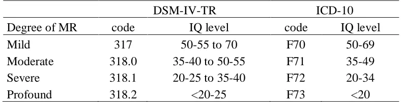
Degree of severity on MR as mild, moderate, severe or profound can be
seen from DSM-IV-TR and ICD-10.7, 12Also described in IQ score.
Table 2.Degree of MR based on IQ scores from DSM-IV-TR and ICD-10.6
DSM-IV-TR ICD-10
Degree of MR code IQ level code IQ level
Mild 317 50-55 to 70 F70 50-69
Moderate 318.0 35-40 to 50-55 F71 35-49
Severe 318.1 20-25 to 35-40 F72 20-34
Profound 318.2 <20-25 F73 <20
Another classification of MR are syndromic and non syndromic cases. MR
is classified as syndromic if it is related with dysmorphic or specific groups of
features. Also if it is related to specific clinical, radiological, metabolic or
biological features. While non syndromic MR is defined as a condition that only
cognitive impairment is the only feature that manifested in that patient.19
II.1.3 Prevalence
It’s generally known that the MR prevalence is around 1-3% of the
MR cases are in the mild group. Most of them remain classified as normal
children until they reach the first or second grade of elementary school. Moderate
MR comprises around 10% of all MR cases. Their language and communication
skills start to have problems during their second grade of elementary school. Even
during their adolescence they have also problems on socializing so that they
become alienated. Next is severe MR, coping around 4% of MR cases. While
profound MR happens on 1-2% MR cases. The more severe MR, the more
causative factors can be identified.20
II.1.4 Etiology
Being able to know and understand the causes of MR has a huge impact
for a lot of people. For the patient themselves, it can be a promising future in
order to get the correct further management, early screening for various
complications and protection from unnecessary tests. For their family and society,
knowing the recurrence risk, possibility to perform prenatal diagnosis, more
information of the diseases are important also.21 Regrettably more than half of the
cases still are unexplained.6, 21, 22
There are many factors causing MR; it could be genetic, non genetic, or
unknown. For genetic factors, chromosomal abnormalities, single gene disorders
(Mendelian disorders/ mitochondrial), multi-factorial, or other genetic causes
disorders may be the causes. Non-genetic factors, could be environmental such as
toxins and infections, or it might be developmental disturbances during prenatal,
A nice overview of the distribution of MR causes, taken from 10.997 MR
patients published by Stevenson et al. in 2003 (adapted from Koolen DA thesis on
2008) is depicted in this picture below.
Figure 1.Causes of mental retardation.6, 22
From that figure, around 11% causes of MR are chromosomal, 8% single gene, 7 % multifactorial, 2% other genetic causes, 16% environmental, and 56% remain unknown.
There are three main periods of time that are very important to be
evaluated in order to know the causes of MR in a patient, which are prenatal,
perinatal and postnatal. During prenatal, genetic play roles for about 60-80%
cases.12
MR is classified as prenatal if the causes are due to chromosomal
aberration, single gene disorder, or environmental factors such as Iodine and/or
folic acid deficiency, severe malnutrition during pregnancy, drug induced
(alcohol, nicotine, cocaine), radiation, infection (rubella, syphilis, toxoplasma,
cytomegalovirus, HIV) during pregnancy etc. It will be classified as perinatal
when it comprises any causative agents during late pregnancy period, while giving
birth and during neonatal period (first four weeks of infancy). A lot of risk factors
could have an effect, such as diseases of the mother (heart failure, kidney failure,
diabetes, etc), prematurity, very low birth weight, asphyxia, complication during Chromosomal
delivery. Another possibility is poor baby condition such as septicemia,
hypoglycemia, icteric (yellow baby), etc. Postnatal category refers to condition
that affect MR patient after 4 weeks old, such as brain infection, head trauma,
chronic toxicity, severe malnutrition and lack of stimulation.12 Nevertheless,
genetic factors play a crucial role since approximately half of MR cases have a
familial history.24-27
II.2 THE DEVELOPMENT OF GENETIC RESEARCH
Chromosomal abnormalities such as trisomies, monosomies, supernumary
marker chromosomes, unbalanced translocations and large deletions and
duplications are the most common genetic cause of MR. They occupy 7% of MR
cases.28 After new molecular techniques became more available and cheaper,
many smaller cytogenetic rearrangements started to be revealed. Using FISH and
MLPA scientist could find around 5% more of pathogenic chromosomal
aberrations.29, 30 But still, those two techniques are not enough to detect
aberrations on a large genomewide scale. The latest technology used is genome
wide array such as CGH (comparative genomic hybridization), then SNP (Single
Nucleotide Polymorphism) arrays. That are hoped to be able to detect
chromosomal aberrations in 5-10% more.29-31
The search for underlying genetic defect of mental retardation and epilepsy
plays a very important role in the field of pediatrics and neurology. Detecting the
molecular basis for the disorder may provide precise genetic counseling.32-38
Genetics diagnosis for MR and epilepsy individual is based on
neuro imaging, or many other advanced techniques are considered as the tool to
confirm the genetics diagnosis. None of themhas the highest priority and have to
perform first. On MR and epilepsy cases, all tools are necessary, because it is a
very complex disorder. In order to get the exact diagnosis, complete and reliable
data are needed.24
The Online Mendelian Inheritance in Man (OMIM) database for genetic
conditions (http://www.ncbi.nlm.nih.gov/omim) contains 1684 entries for mental
retardation and 284 for mental retardation epilepsy (October 2010). This data
shows so many differential diagnose and other diseases that related to MR
phenotype.6 Sensory disturbances found in more than 10% of MR population, four
times higher than in normal population. Seizures also often happen in MR
patients.20
II.3 EPILEPSY II.3.1 Definition
The fact that epilepsy is found in 10-25,5% of the MR population makes
us more interested to get deeper information about it.1, 20, 39 Starting with the
definition, epilepsy is a brain disorder that is characterized by repeated events of
epileptic seizures. It has neurobiologic, cognitive, psychological, and social
consequences. Epilepsy is different with epileptic seizure. An epileptic seizure is a
temporary incident of signs and/or symptoms due to abnormal extreme or
synchronous neuronal activity in the brain.40, 41 First the most important diagnostic
test in epilepsy is a thorough and detailed history of the patients episodes. A
cause and location of the seizure. For epilepsy, the history is usually more vital
than the physical test and EEG.
The International League Against Epilepsy (ILAE) and the International
Bureau for Epilepsy (IBE) have come to compromise definitions for the terms
epileptic seizure and epilepsy. An epileptic seizure is a temporary occurrence of
signs and/or symptoms due to abnormal excessive or synchronous neuronal
activity in the brain. Epilepsy is a condition of the brain characterized by an
enduring predisposition to generate epileptic seizures and by the neurobiologic,
cognitive, psychological, and social consequences of this condition. The definition
of epilepsy requires the manifestation of at least one epileptic seizure.40
II.3.2 Classification
Classification of syndromic epilepsy relied on seizures factors type
(general or localized), etiology (symptomatic or idiopathic), age of onset and
seizures related conditions. While classification that is based on seizures type can
be defined from clinical assessment and electro encephalogram (EEG).
Description based on seizures type shown in the next list.40, 42
I. Partial Seizures (Focal, start in one place)
- Simple (no loss of consciousness/ memory) - Complex (loss of consciousness/ memory)
With or without aura (warning) With or without automatisms - Secondarily generalized (spreads)
II. Generalized
- Absence, typical or atypical (petit mal) - Tonic-Clonic (grand mal)
- Myoclonic
- Atonic
- Tonic
The term of “epilepsy” also encompasses various different syndromes.
Epilepsy syndromes fall into two broad categories: generalized and partial (or
localization-related) syndromes. In generalized epilepsies, the major type of
seizures begins at once in both cerebral hemispheres. Many forms of generalized
epilepsy have a genetic basic. In most of them, the neurologic function is found to
be normal. In partial epilepsies, seizures initiate in one or more localized foci,
although they can spread to the whole brain.40, 42
II.3.3 Prevalence
Among all of the neurological problems, epilepsy is the most common
one. It affects at least 50 million or approximately 1% of people worldwide.
Complex partial seizures are reported for about 40% of all seizure types in adults.
Simple partial seizures account for about 20%, primary generalized tonic-clonic
seizures about 20%, absence around 10% and other seizure types for 10%. In a
pediatric population, absence seizures engage a greater section.2, 43, 44
Studies conducted in India, show the prevalence between 4.6 and 8.5 per
1000, while in Sri Lanka they found 9.02 per 1000 rates. Jallon showed almost
similar prevalence by several studies, Pakistan (9.99), China (4.4), and Japan
(1.5).45China studies illustrated the rate was 1.54 per 1000 and 6.2 per 1000.46, 47
The WHO conclusion from general studies is that estimation of the active epilepsy
in developing country prevalence rates, range from 5 to 10 per 1000 people.48
II.3.4 Etiology
The most regular subject asked in every epilepsy patient is “Why do I have
supernatural motive behind it. Not only in the past, even to these days in countries
like Tanzania,epilepsy still being associated with possession ofgenie or spirits,
witch, poison and is believed to be contagious49. Epilepsy actually has physical
causes. The end result of many factors that could cause destruction of the brain
may cause epilepsy. In order to make epilepsy understandable, based on epilepsy
causes, it is divided into three categories: symptomatic, idiopathic and
cryptogenic.42
Around 70% of epilepsy cases are caused by non-genetic factors like head
trauma, congenital malformation of the brain, lack of oxygen during birth, a brain
tumor, a stroke, a cerebral hemorrhage, alcoholism, brain infections (encephalitis
or meningitis). In the remaining group (30%) the cause of the epilepsy is
genetically inherited.50, 51
It is indeed difficult to interpret etiology of research patients without good
history archives from each MR patient. That is the importance of this research, to
search the genetic possibility as the cause of MR with epilepsy in Indonesia. It is
indeed difficult to interpret etiology of MR epilepsy patients without good history
archives from each patient.
II.3.5 Epilepsy pathogenesis
Seizures happen mostly due to imbalance of excitation and inhibition in a
part of the brain. Or in other words, it is an electrochemical disorder. The etiology
of seizures varies with the type of seizure, it depends whether it starts focally in
one part of the brain or generalized all over the brain. Causes for focal seizures are
dysplasia, mesial temporal sclerosis, etc. General seizures causes vary from
metabolic, medication reactions, idiopathic until genetic.33, 35, 41, 50-53
One of the general epilepsy that has explainable mechanism is absence
epilepsy. Having 3-8 years as age of onset, absence epilepsy also has specific
clinical characteristics. The patients lose their consciousness for a moment in the
middle of their activity and suddenly come back to consciousness and don’t know
what was happening. There are several hypotheses behind it. Some studies
conclude that absence epilepsy happens due to circuit changes between thalamus
and cerebral cortex. They suspect that the abnormal circuit is caused by disruption
of T-type calcium channels or altered function of g-aminobutyric acid (GABA)
receptor.43
II.3.6 Genetic research in epilepsy
New gene hunting techniques may allow recognition of some new genes in
patients without family history of seizures. Those genes known to be related to the
development of epilepsy were initially identified in a few rare families with
multiple affected individuals. This is a quickly evolving field and the current
concept is that the ‘‘common’’ epilepsies (the familial and the
non-encephalopathic epilepsies) are complex diseases resulting from an interaction of
multiple genetic and environmental factors.35, 41
As a complex disorder with multiple sub classifications and etiologies, it is
a very challenging job to find genetic causes for epilepsy cases. Andrade CS et al
(2009) explained in his study about epilepsy due to Malformation of Cortical
involved in neuronal proliferation, migration and cortical lamination during
embryogenesis. For epilepsies with normal macroscopic appearance, most of their
genetic defects caused by alterations in voltage-gated or ligand-gated channels.2,
43, 44
Figure 2.Ion-Channel Dysfunction Associated with Epilepsy
Section A illustrates neuronal-ion-channel in a normal function and the action potential. Section B shows the effect of mutations in SCN1B, which encodes a voltage-gated sodium-channel subunit. While section C describes the consequences if there are mutations in KCNQ2 and KCNQ3, which both encode potassium channels happened. They both are associated with benign familial neonatal convulsions.43
Focal cortical dysplasia is said to be related with mutations in the TSC1
gene.54 Polymicrogyria related with mutations in SPRX2.55Lissencephaly is
caused by mutations in LIS1andARX.56LGI1 and ARX genes are involved in cell
migration during development.57
Genetic epilepsy with febrile seizures plus, mutations found in
SCN1B58andSCN1A.59, 60Severe myoclonic epilepsy of infancy (SMEI) mutations
Autosomal dominant temporal lobe epilepsy with auditory features are
related with mutations in LGI1.50, 62 Other gene mutations have been identified in
potassium channels (KCNQ2 and KCNQ3) associated with benign neonatal
familial convulsions, and in the nicotinic acetylcholine receptor (CHRNA4 and
CHRNB2) in autosomal dominant temporal lobe epilepsy.57
Although many patients meet the clinical criteria for a particular
syndrome, not all of them have the same genetic abnormalities (for example only
30–50% of patients with Dravet’s syndrome possess an abnormality of the
sodium-channel gene). In general, epilepsy syndromes occur sporadically, and
genetic analysis is not always easy. When genetic analysis became more
accessible and cheaper, there were more hopes that it would clarify the
mechanisms of epileptogenesis, seizure generation and transmission.57
II.4 MENTAL RETARDATION AND EPILEPSY
Genetic association between MR and epilepsy has been suggested in both
syndromic and monogenic cases, which are completing each other. For
syndromic, there are early infantile epileptic encephalopathy or known also as
Ohtahara syndrome, severe myoclonic epilepsy in infancy (SMEI) or Dravet
syndrome, Lennox–Gastaut syndrome, and West syndrome (WS). The studies
where MR and epilepsy are present together provides convincing evidence for the
1. Hirose S, Mitsudome A. X-linked mental retardation and epilepsy: pathogenetic significance of ARX mutations. Brain Dev. 2003 Apr;25(3):161-5.
2. Andrade DM. Genetic basis in epilepsies caused by malformations of cortical development and in those with structurally normal brain. Hum Genet. 2009;126(1):173-93.
3. Davison K. Historical aspects of mood disorders: Psychiatry; 2006.
4. Paladin AV. Ethics and neurology in the Islamic world. Continuity and change. Ital J Neurol Sci. 1998 Aug;19(4):255-8.
5. Aragona M. The concept of mental disorder and the DSM-V. 1 ed. 2009 DPMNS, editor2009.
6. Koolen D. Copy Number Variation and Mental Retardation. Nijmegen NL: Radboud Universiteit 2008.
7. APA. Diagnostic and Statistical Manual of Mental Disorders. 4th ed. tre, editor. Washington DC: American Psychiatric Association; 2000.
8. Schroeder SR. Mental retardation and developmental disabilities influenced by environmental neurotoxic insults. Environ Health Perspect. 2000;3:395-9.
9. Staden V. "Liminal Perils: Early Roman Receptions of Greek Medicine," in Tradition, Transmission, Transformation, ed. Livesey FJRaSPRwS, editor. Leiden: Brill; 1996.
10. Luckasson B-DS, Buntinx WHE. Mental retardation: Definition, classification, and system of support. 10th, editor. Washington DC: American Association on Mental Retardation; 2002.
11. Snell ME, Luckasson, R. Characteristics and needs of people with intellectual disability who have higher IQs. : Intellectual and Developmental Disabilities; 2009. 12. WHO. International Statistical Classification of Diseases and Health Related Problems ICD-10. Geneva: World Health Organization; 2001.
13. Biasini FJ HL, Bray NW. Mental Retardation: A Symptom and a Syndrome. Netherton DHS WC, (Eds.), editor. New York: Oxford University Press; 1996.
14. Faradz SM, Buckley M, Lam Po T, Leigh D, Holden JJ. Molecular screening for fragile X syndrome among Indonesian children with developmental disability. Am J Med Genet. 1999 Apr 2;83(4):350-1.
15. Inlow JK, Restifo LL. Molecular and comparative genetics of mental retardation. Genetics. 2004 Feb;166(2):835-81.
16. Yntema H. Molecular genetics of nonspecific X-linked mental retardation. Nijmegen (NL): Radboud Univ.; 2001.
17. McDaniel M. Estimating state IQ: Measurement challenges and preliminary correlates. Intelligence. 2006;34:607-19.
18. Shaffer LG. American College of Medical Genetics guideline on the cytogenetic evaluation of the individual with developmental delay or mental retardation. Genet Med. 2005 Nov-Dec;7(9):650-4.
19. Chelly J, Khelfaoui M, Francis F, Cherif B, Bienvenu T. Genetics and pathophysiology of mental retardation. Eur J Hum Genet. 2006 Jun;14(6):701-13. 20. Sadock BJ K. Mental Retardation. Synopsis of psychiatry. Behavioral
Sciences/Clinical Psychiatry. ed. t, editor. USA: Lippincot Williams & Wilkins; 2007. 21. Knight S. Genetic of Mental Retardation. Disability AOELDaI, editor.
Switzerland: Karger 2010.
22. Stevenson RE, Procopio-Allen AM, Schroer RJ, Collins JS. Genetic syndromes among individuals with mental retardation. Am J Med Genet A. 2003 Nov
23. Durkin M. Prevalence and Correlates of Mental retardation among children in Karachi, Pakistan. Am J Epidemiol. 1998;147:3:281-8.
24. Rimoin DL CJM, Pyeritz R E, Korf BR. Emery and Rimoin’s Principles and Practice of Medical Genetics. edition t, editor. New York2002.
25. De Vries BB, Winter R, Schinzel A, van Ravenswaaij-Arts C. Telomeres: a diagnosis at the end of the chromosomes. J Med Genet. 2003;40(6):385-98.
26. Strachan T RA. Human Molecular Genetics. Edition T, editor. United States: Taylor & Francis Inc.; 2003.
27. Koolen DA, Nillesen WM, Versteeg MH, Merkx GF, Knoers NV, Kets M, et al. Screening for subtelomeric rearrangements in 210 patients with unexplained mental retardation using multiplex ligation dependent probe amplification (MLPA). J Med Genet. 2004 Dec;41(12):892-9.
28. Leonard H, Wen X. The epidemiology of mental retardation: challenges and opportunities in the new millennium. Ment Retard Dev Disabil Res Rev. 2002;8(3):117-34.
29. de Vries BB, Pfundt R, Leisink M, Koolen DA, Vissers LE, Janssen IM, et al. Diagnostic genome profiling in mental retardation. Am J Hum Genet. 2005;77(4):606-16. 30. Vissers LE, Veltman JA, van Kessel AG, Brunner HG. Identification of disease genes by whole genome CGH arrays. Hum Mol Genet. 2005 Oct 15;14 Spec No. 2:R215-23.
31. VanBon B. Emerging genomic disorders in mental retardation. Nijmegen The Netherland: Radboud Universiteit; 2010.
32. Nabbout R, Dulac O. Epileptic syndromes in infancy and childhood. Curr Opin Neurol. 2008 Apr;21(2):161-6.
33. Coppola G, Veggiotti P, Del Giudice EM, Bellini G, Longaretti F, Taglialatela M, et al. Mutational scanning of potassium, sodium and chloride ion channels in malignant migrating partial seizures in infancy. Brain Dev. 2006;28(2):76-9.
34. Claes L, Del-Favero J, Ceulemans B, Lagae L, Van Broeckhoven C, De Jonghe P. De novo mutations in the sodium-channel gene SCN1A cause severe myoclonic epilepsy of infancy. Am J Hum Genet. 2001;68(6):1327-32.
35. Degen R, Holthausen H, Wolf P. The genetics of localization-related symptomatic epilepsy: risk of a family history with seizures in patients who have undergone surgery. J Neurol. 1997 Jul;244(7):439-45.
36. Fujiwara T. Clinical spectrum of mutations in SCN1A gene: severe myoclonic epilepsy in infancy and related epilepsies. Epilepsy Res. 2006 Aug;70 Suppl 1:S223-30. 37. Scheffer IE, Wallace R, Mulley JC, Berkovic SF. Clinical and molecular genetics of myoclonic-astatic epilepsy and severe myoclonic epilepsy in infancy (Dravet
syndrome). Brain Dev. 2001 Nov;23(7):732-5.
38. Chang BS, Lowenstein DH. Epilepsy. N Engl J Med. 2003;349(13):1257-66. 39. McDermott S, Moran R, Platt T, Wood H, Isaac T, Dasari S. Prevalence of epilepsy in adults with mental retardation and related disabilities in primary care. Am J Ment Retard. 2005 Jan;110(1):48-56.
40. Fisher RS, van Emde W, Blume W, Elger C, Genton P, Lee P, et al. Epileptic seizures and epilepsy: definitions proposed by the International League Against Epilepsy (ILAE) and the International Bureau for Epilepsy (IBE). Epilepsia. 2005 Apr;46(4):470-2.
42. Browne TR HG. Epilepsy: definitions and background. In: Handbook of epilepsy, 2nd edition. Philadelphia: Lippincott Williams & Wilkins; 2000.
43. Chang BS LD. Epilepsy: Mechanisms of Disease.2003.
44. Beckung E, Uvebrant P. Motor and sensory impairments in children with intractable epilepsy. Epilepsia. 1993 Sep-Oct;34(5):924-9.
45. Jallon P. Epilepsy in developing countries. Epilepsia. 1997 Oct;38(10):1143-51. 46. Fong GC, Mak W, Cheng TS, Chan KH, Fong JK, Ho SL. A prevalence study of epilepsy in Hong Kong. Hong Kong Med J. 2003 Aug;9(4):252-7.
47. Wang W, Wu J, Dai X, Ma G, Yang B, Wang T, et al. Global campaign against epilepsy: assessment of a demonstration project in rural China: Bulletin of the WHO; 2008.
48. Scott RA LS, Sander JWAS. The treatment of epilepsy in developing countries: where do we go from here? Bulletin of the WHO. 2001:344-51.
49. Jilek-Aall L. Morbus sacer in Africa: some religious aspects of epilepsy in traditional cultures. Epilepsia. 1999 Mar;40(3):382-6.
50. Ottman R, Annegers JF, Risch N, Hauser WA, Susser M. Relations of genetic and environmental factors in the etiology of epilepsy. Ann Neurol. 1996 Apr;39(4):442-9. 51. Ottman R, Hirose S, Jain S, Lerche H, Lopes-Cendes I, Noebels JL, et al. Genetic testing in the epilepsies--report of the ILAE Genetics Commission. Epilepsia.
2010;51(4):655-70.
52. Gunadharma S. Psycho-Social Issues: Public awareness, understanding and attitude towards epilepsy in Bandung, Indonesia. Neurology Asia [Supplement]. 2004;9:133 - 4.
53. The epilepsies: The diagnosis and management of the epilepsies in adults and children in primary and secondary care. [database on the Internet]. National Collaborating Centre for Primary Care Epilepsy. 2004 [cited 26 May 2010]. Available from:
www.nice.org.uk/CG020NICEguideline.
54. Becker AJ, Urbach H, Scheffler B, Baden T, Normann S, Lahl R, et al. Focal cortical dysplasia of Taylor's balloon cell type: mutational analysis of the TSC1 gene indicates a pathogenic relationship to tuberous sclerosis. Ann Neurol. 2002 Jul;52(1):29-37.
55. Roll P, Rudolf G, Pereira S, Royer B, Scheffer IE, Massacrier A, et al. SRPX2 mutations in disorders of language cortex and cognition. Hum Mol Genet. 2006 Apr 1;15(7):1195-207.
56. Kato M, Saitoh S, Kamei A, Shiraishi H, Ueda Y, Akasaka M, et al. A longer polyalanine expansion mutation in the ARX gene causes early infantile epileptic encephalopathy with suppression-burst pattern (Ohtahara syndrome). Am J Hum Genet. 2007 Aug;81(2):361-6.
57. Kelso AR, Cock HR. Advances in epilepsy. Br Med Bull. 2004;72:135-48. 58. Wallace RH, Wang DW, Singh R, Scheffer IE, George AL, Jr., Phillips HA, et al. Febrile seizures and generalized epilepsy associated with a mutation in the Na+-channel beta1 subunit gene SCN1B. Nat Genet. 1998 Aug;19(4):366-70.
59. Baulac S, Gourfinkel-An I, Picard F, Rosenberg-Bourgin M, Prud'homme JF, Baulac M, et al. A second locus for familial generalized epilepsy with febrile seizures plus maps to chromosome 2q21-q33. Am J Hum Genet. 1999;65(4):1078-85.
61. Kamiya K, Kaneda M, Sugawara T, Mazaki E, Okamura N, Montal M, et al. A nonsense mutation of the sodium channel gene SCN2A in a patient with intractable epilepsy and mental decline. J Neurosci. 2004 Mar 17;24(11):2690-8.