COLORECTAL CANCER
- PATHOHISTOLOGICAL STANDARDS
SANDA [ITI]1, MARIJA MILKOVI] PERI[A1, IVA BOBU[ KEL^EC2, TAJANA SILOVSKI3, HRVOJE SILOVSKI4 and TOMISLAV ORE[I]5
1 Department for Pathology Ljudevit Jurak, Unit for Oncological Pathology, Universitiy Hospital Center Sestre milosrdnice, Zagreb, Croatia
2 Department for Cytology, Universitiy Hospital Center Sestre milosrdnice, Zagreb, Croatia 3Department of Radiotherapy, University Hospital for Tumors,
Universitiy Hospital Center Sestre milosrdnice, Zagreb, Croatia
4 Department for Gastrointestinal Surgery, Universitiy Hospital Center Zagreb, Zagreb, Croatia 5Department for Surgical Oncology, University Hospital for Tumors,
Universitiy Hospital Center Sestre milosrdnice, Zagreb, Croatia
Summary
Colorectal carcinoma is the third most common cancer site in Croatia, according to the data published by the The Croatian National Cancer Registry. In the last decade, the knowledge about pathogenesis and molecular background of colorectal carcinoma has increased dramatically. More than 90% of colorectal carcinomas are adenocarcinomas originating from epithelial cells of the colorectal mucosa. Tumor staging is the most important prognostic predictor of clinical outcome for patients with colorectal carcinoma. The TNM classification has nowdays replaced other classification systems (Dukes, Astler-Coller) and serves as golden standard in everyday practise.In 2012, 5th Croatian Congress for Pathology resulted in uniform standard for pathologic reporting for all cancer sites. The future for colorectal cancer prognosis and therapy is to discover new molecular subtypes of colorectal cancer which represents the future of personalized oncology and will guide drug-development strategies.
KEY WORDS: colorectal cancer, TNM classification, personalized oncology
KOLOREKTALNI KARCINOM - PATOHISTOLO[KI STANDARDI
SA@ETAK
Prema podacima Registra za rak karcinom kolorektuma je tre}e naj~e{}e tumorsko sijelo u Hrvatskoj. U zadnjih deset godina imamo brojna nova saznanja o patogenezi i molekularnim karakteristikama karcinoma kolorektuma. Vi{e od 90% kolorektalnih karcinoma su adenokarcinomi po histolo{kom tipu, porijekla epitelnih stanica kolorektalne mukoze. Stadij tumora je najbitniji prognosti~ki ~imbenik za pacijente s tom bole{}u. U dana{nje je vrijeme TNM klasifikacija zamijenila druge klasifikacijske sustave (Dukes, Astler-Coller) i koristi se kao zlatni standard u svakodnevnoj praksi. 2012. na Hrva-tskom kongresu patologa, doneseni su standardi za sva tumorska sjela, koje mora zadovoljavati svaki patohistolo{ki nalaz. Budu}nost prognoze i terapije kolorektalnog karcinoma je otkri}e novih molekularnih podtipova prema kojima bi se odre|ivala personalizirana onkolo{ka terapija te odredile nove strategije lije~enja.
INTRODUCTION
Colorectal carcinoma is the third most com-mon cancer site in Croatia, according to the data published by the The Croatian National Cancer Registry, with the 9% of incidence in men and 8% of incidence in women in 2013, after prostate and lung/bronchus cancers in men and after breast and lung/bronchus cancers in women. It is also the third leading cause of cancer-related death in Croatia after lung/bronchus and prostate cancers in men and after lung/bronchus and breast can-cers in women in the same year (1). With the rapid therapeutic advancement in the era of personal-ized medicine, the role of pathologists in the man-agement of patients with colorectal carcinoma has expanded from traditional morphologists to clini-cal consultants for gastroenterologists, colorectal surgeons, oncologists and medical geneticists. Nowdays, pathologists are not only responsible for providing accurate histopathologic diagnosis, but also for assessing pathologic staging, analyz-ing surgical margins, searchanalyz-ing for prognostic pa-rameters that are not included in the staging such as lymphovascular and perineural invasion, and assessing therapeutic effect in patients who have received neoadjavant therapy.
In the last decade,the knowledge about patho-genesis and molecular backgroundof colorectal carcinoma has increased dramatically. As a result, pathological diagnosis of colorectal carcinoma is now more complexand includes analyzing histo-logic features of the tumours that are suggestive of microsatelitte instability (MSI) andselecting ap-propriate tissue sections for MSI for identification of patients with increased risk for Lynch syndrome (2). Introduction of specific targeted therapies re-quires implementationof results of additional mu-tation analysis tests, such as analysis of the BRAF and KRAS genesmutations (3). Pathologists also play a central role in selecting appropriate tissue sections for these two mutations analysis.
HISTOPATHOLOGIC DIAGNOSIS
According to the 4th Edition of WHO Classifi-cation of Tumours of the Digestive system, several histopathological variants of colorectal carcinoma can be distinguished (4). More than 90% of colorec-tal carcinomas are adenocarcinomas originating
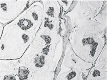
from epithelial cells of the colorectal mucosa (5). Other histopathological variants of colorectal car-cinoma, some of which associaed with specific molecular characteristics include mucinous, sig-net ring cell, medullary, serrated, cribriform comedo-type, micropapillary, neuroendocrine, squamous cell, adenosquamous, spindle cell and undifferentiated carcinomas. Conventional ade-nocarcinoma is characterized by glandular forma-tion, which is the basis for histologic tumor grad-ing. In well differentiated adenocarcinoma >95% of the tumor is gland forming. Moderately differ-entiated adenocarcinoma shows 50-95% gland formation. Poorly differentiated adenocarcinoma is mostly solid with <50% gland formation. The terms “low-grade” and “high-grade” are nowdays in clinical, as well histopathologic, use due to the similar behaviour of well- and moderately differ-entiated carcinomas. Morphological grading of tumours applies only to adenocarcinoma, ”NOS” (Figure 1), because other morphological variants carry their own prognostic significance.
Figure 1. Adenocarcinoma (“NOS”), (original magnification X200)
Most of colorectal carcinomas are initially di-agnosed by endoscopic biopsy or polypectomy. The major aspect of microscopic examination is the evidence of invasion. This is very difficult in every day practiseespecially when the biopsy is superficial or poorly oriented. Material is some-times insufficiant. If the muscularis mucosae can be identified, it is important to determine whether it is disrupted by neoplastic cells. Invasive carci-noma invades through the muscularis mucosae at least into the submucosa. If carcinoma is subjected
to mucosa only, it is classified as carcinoma in situ. Another important feature of invasion is the pres-ence of desmoplasia, a type of fibrous prolifera-tion surrounding tumor cells.
The major difference between colorectal and other gastrointestinal carcinomas is the following: in the colorectum, submucosal invasion is re-quired for the diagnosis of a pT1 tumor and in other parts of the gastrointestinal tract (esopha-gus, stomach and small intestine), mucosal inva-sion is sufficient for the diagnosis of invasive car-cinoma (pT1). For reasons that are not entirely clear but generally thought to be due to the rela-tive paucity of lymphatics, invasion confined to the lamina propria and muscularis mucosae has no risk of nodal or distant metastasis(6). No mat-ter what mat-term is used by pathologists, the identifi-cation of high grade dysplasia or intramucosal carcinoma in a biopsy specimen should not affect the decision-making for patient management. The decision to perform surgical resection should be determined by the gross appearance of the lesion, endoscopic ultrasound findings, and endoscopic resectability.
HISTOLOGIC VARIANTS Mucinous adenocarcinoma
This special type of colorectal carcinoma is used if >50% of the lesionis composed of pools of extracellular mucin that contain malignant epithe-lial cells, either individual or forming clusters,
aci-nar structers or layers. Tumors with a significant mucinous component (>10%) but <50% are cate-gorized as adenocarcinomas with mucinous fea-tures or mucinous differentiation. (Figure 2). The prognosis of mucinous adenocarcinoma in com-parison with conventional adenocarcinoma has been controversial (7,8). Some of them occur in pa-tients with hereditary nonpolyposis colorectal cancer (HNPCC or Lynch syndrome) and thus represent high-level MSI (MSI-H) tumors with ex-pected better clinical outcome (9). In contrast, mu-cinous adenocarcinomas that are microsatellite stable (MSS) are expected to behave more aggres-sively.
Signet ring cell adenocarcinoma
Signet ring cell adenocarcinoma is rare in the colorectum, with frequency <1% of all colorectal carcinomas. It is defined by the presence of >50% of tumor cells with prominent intracytoplasmic mucin, displacing the nucleus to the periphery. Signet ring cells may show an infiltrative growth pattern or are present within the pools of extracel-lular mucin. Most of them are poorly differentiat-ed (high grade) and have worse outcome than conventional adenocarcinoma (10-12). However, some signet ring cell carcinomas may be MSI-H tumors and thus may behave as low grade tumors biologically (4).
Neuroendocrine carcinoma
Neuroendocrine carcinomas (NEC) are mor-phologically similar to small cell carcinoma and
Figure 2. Mucinous adenocarcinoma showing abundant extra-cellular mucin (original magnification ×200)
Figure 3. Neuroendocrine carcnoma, (original magnification ×200)
large cell neuroendocrine carcinomas of the lung, they are usually found in the right colon, and are frequently associated with an overlying adenoma or adenocarcinoma, but not associated with neu-roendocrine tumours (carcinoid tumours) (Figure 3). Primary NECs in the colon without an associ-ated adenoma should be distinguished from pul-monary neuroendocrine carcinoma metastasis or cutaneous Merkel cell carcinoma metastasis. Ac-cording to the cell morphology, small cell NEC and large cell NEC are differentiated.
Medullary carcinoma
Medullary carcinoma is extremely rare, con-stituting approximately 5-8 cases for every 10,000 colorectal cancers diagnosed, with a mean annual incidence of 3.47 (±0.75) per 10 million population (13). It is characterized by sheets of malignant cells withvesicular nuclei, prominent nucleoli, and abundant eosinophilic cytoplasm exhibiting pro-minent infiltration by intraepithelial lymphocytes. It is strongly associated with MSI-H (14,15) and usually has a favorable prognosis despite its poor-ly differentiated histology.
Colorectal adenocarcinomas are immmunohis-tochemicaly positive for CK20 and negative for CK7. However, up to 20% of the tumors may exhibit a CK7-positive/CK20 negative or CK7-negative/ CK20-negative staining pattern. It has been suggest-ed that rsuggest-educsuggest-ed or absent CK20 expression in colorectal carcinoma is associated with MSI-H (16).
PATHOLOGIC STAGING
Tumor staging is the most important prog-nostic predictor of clinical outcome for patients with colorectal carcinoma. The TNM classification has nowdays replaced other classification systems (Dukes, Astler-Coller) and serves as golden stan-dard in everyday practise. Several modification systems have exchanged in the last decade. (T) de-terminates the depth of tumor invasion; T1 (tumor invades submucosa), T2 (tumor invades muscula-ris propria), T3 (tumor invades subserosa or into non-perinotealized pericolic or perirectal tissues) and T4 subcategorized as T4a (tumor penetrates to the surface of the visceral peritoneum) and T4b (tumor directly invades or is adherent to other or-gans or structures) which can sometimes be prob-lematic because serosal surface (visceral peritone-um) involvement can sometimes be missed if the
specimen is not adequately sampled for histologic examination or it can also be replaced with the cir-cumferential (radial) or mesenteric margin, which is a nonperitonealized surface created surgically.
(N) is the extent of nodal metastasis. Accord-ing to NCCN guidelines for treatment of cancer by site, AJCC and College of American Pathologists, a minimum of 12 lymph nodes are recommended to identify early-stage colorectal cancer(17-19). The number of lymph nodes retrieved can vary with age of the patient, gender, tumour grade and tu-mour site (20). For stage II (pN0) cancer, if fewer than 12 lymph nodes are initially indentified, it is recommended that the pathologist goes back to the specimen and thoroughly examines tissue once again. If 12 lymph nodes are still not indentified, a comment in the report should indicate that an ex-tensive search for lymph nodes was undertaken.
The mean number of lymph nodes retrieved for rectal cancers treated with neoadjuvant thera-py is significantly less then those treated by sur-gery alone. To date, the number of lymph nodes needed to accurately stage neoadjuvant-treated cases is unknown.
Tumour deposits (TD) are also in N category. They are defined as discrete foci of tumor in peri-colorectal or mesenteryc fat away from the main tumor but without identifiable residual lymph node tissue. They should be in lymph drainage area. The TD’s must be mentioned (by number) in pathologic report and also in N1c category in case of T1 or T2 stage.
The prognostic significance of isolated tumor cells (ITCs), defined as single tumor cells or small clusters of tumor cells ≤0.2 mm, detected by either immunohistochemical staining or standard hema-toxylin and eosin staining in regional lymph nodes remains unclear at present.
M0 category cannot be documented on patho-logical evaluation, but only clinical according to TNM 7. M1 has been subdivided in M1a (meta stasis confined to one organ or site) and M1b (metastasis in more than one organ/site or the peritoneum).
PATHOLOGY REPORTING
In 2012, 5th Croatian Congress for Pathology resulted in uniform standard for pathologic re-porting for all cancer sites.The details that should be included in the report for colorectal cancer are specimen type and size, tumor site and size,
mac-roscopic tumor perforation, macmac-roscopic mesorec-tal infiltration, histologic type and grade, micro-scopic tumor extension, margins (proximal, distal, radial, and lateral for nonradial transanal exci-sion), treatment effect (for tumors treated with neoadjuvant therapy), lymphovascular and peri-neural invasion, tumor deposits, TNM staging, total number of lymph nodes examined, size of lymph nodes examined and the total number of nodes involved, type of polyp precursor of carci-noma (if present), assessment of histologic fea-tures that are suggestive of MSI such as tumor-in-filtrating lymphocytes, peritumoral Crohn-like lymphoid response and subtype and differentia-tion of the tumour, k-rasgene analysis..
PROGNOSTIC FACTORS
During the last 20 years, there have been sig-nificant advances in understanding of colorectal cancer pathogenesis. Different mechanisms of car-cinogenesis underly these tumours (21). We have often seen that patients at the same stage of the disease, after radical surgery, have a completely different course of disease and outcome. There-fore, we are constantly looking for new parame-ters that will more accurately provide prognosis of the disease along with optimal therapy for each patient. It has become clear that the most accurate prognostic information will be achieved by com-bining clinico-pathological and molecular data. Unfortunately, many patients with colorectal can-cer receive treatment unnecessarily, because they would either be cured even without treatment, or relapse despite of the treatment.
Seven new factors have been included in the latest TNM staging system, none of them required for staging but all of them with major prognostic and predictive values. According to the results of latest researches, all of these factors are useful for new molecular targeted therapy and personalized medicine.
Tumour deposits, mentioned above are record-ed numerically.
Circumferential resection margin (CRM) repre-sents the adventitial soft tissue closest to the deep-est penetration of the tumour. It is created surgi-cally by sharp excision and it corresponds to any aspect of the colon that is not covered by a serosal layer or mesothelial cells. The serosal (peritoneal) surface does not constitute a surgical margin. The
radial margins should be assessed in all colonic specimens with non-peritonealized surfaces. In transverse colon specimen which is completely encased with peritoneum, the mesenteric resec-tion margin is the only relevant radial margin (22). On pathologic exemination it is very difficult to assess the demarcation between the peritoneal-ized surface and non-peritonealperitoneal-ized surface. Therefore, surgeants are highly encouraged to mark the area of non-peritonealized surface with a clip or sutture. The pathologist should measure the distance between the closest tumour margin and resection margin expressed in mm. A margin less than 1 mm is considered positive.
Perineural invasion must be recorded. Several studies have shown that the presence of perineu-ral invasion is associated with a significantly worse prognosis (23-25).
Tumour regression gradeis a marker of response to neoadjuvat therapy. The system used to grade tumour response is modified from Ryan et al. (26).
0 (complete response) - no remaining viable tumour cells
1 (moderate response) - only small clusters or single cancer cells remaininig
2 (minimal response) - residual cancer re-maining, but with predominant fibrosis 3 (poor response) - minimal or no tumour
kill; extensive residual cancer
k-ras gene analasysdetects k-ras gene mutation which is associated with lack of response to treat-ment with anti-EGFR antibody, currently recom-mended for the patients with metastatic colorectal cancer.
Microsatellite instability(MSI)Hereditary non-popyposis colorectal cancer (HNPCC), also known as Lynch syndrome, is a common autosomal dom-inant syndrome characterized by early age at on-set, nepolastic lesions, and microsatellite instabil-ity (28). It is characterized by increased lifetime cancer risks primarily in the gastrointestinal and gynecologic tracts, with colorectal and endome-trial carcinomas being most common.Lynch syn-drome results from germline mutation in one of the four DNA mismatch repair (MMR) genes (MLH1, MSH2, MSH6, PMS2). Lynch syndrome tumours screening (immunohistochemicaly or MSI), according to NCCN recommendations, should be considered for colorectal carcinoma pa-tients diagnosed at ≤70 years and also those older than 70 years who meet the Bethesda guidelines.
CONCLUSIONS
Colorectal adenocarcinoma is a heteroge-neous disease that involves multiple tumorigenic pathways. The future for colorectal cancer prog-nosis and therapy is promising, but we must con-centrate on optimising current methods, most of all histopathology and discover new molecular subtypes of colorectal cancer which represents the future of personalized oncology and will guide drug-development strategies.
REFERENCES
1. Croatian National Institute of Public Health,Croatian National Cancer Registry, Cancer incidence in Croa-tia, 2011, Zagreb, 2013. Bulletin No. 36.
2. Lynch HT, Lynch JF, Lynch PM. Toward a consensus in molecular diagnosis of hereditary nonpolyposis colorectal cancer (Lynch syndrome). J Natl Cancer Inst. 2007;99:261-3.
3. Pritchard C, Gredy W. Colorectal cancer molecular bi-ology moves into clinical practice. Gut 2011;60:116-129. 4. Bosman FT, Carneiro F, Hruban RH, Theise ND. WHO
Classification of Tumours of the Digestive System. Lyon: IARC; 2010.
5. Boyle P, Levin B (eds). World Cancer Report. Lyon: IARC; 2008.
6. Fleming M, Ravula S et al. Colorectal carcinoma: Patho-logic aspects. J Gastrointest Oncol 2012; 3(3): 153–173. 7. Jass JR, Atkin WS, Cuzick J, et al. The grading of rectal
cancer: historical perspectives and a multivariate anal-ysis of 447 cases. Histopathology 1986;10:437-59. 8. Compton CC. Pathology report in colon cancer: what
is prognostically important? Dig Dis 1999;17:67-79. 9. Edge SB, Byrd DR, Compton CC, et al. AJCC Cancer
Staging Handbook, 7th edition. New York: Springer; 2010: str.173-206.
10. Kang H, O’Connell JB, Maggard MA, et al. A 10-year outcomes evaluation of mucinous and signet-ring cell carcinoma of the colon and rectum. Dis Colon Rectum 2005;48:1161-8.
11. Chen JS, Hsieh PS, Chiang JM, et al. Clinical outcome of signet ring cell carcinoma and mucinous adenocar-cinoma of the colon. Chang Gung Med J 2010;33:51-7. 12. Makino T, Tsujinaka T, Mishima H, et al. Primary
sig-net-ring cell carcinoma of the colon and rectum: report of eight cases and review of 154 Japanese cases. Hepa-togastroenterology 2006;53:845-9.
13. Thirunavukarasu P, Sathaiah M, Singla S, et al. Medul-lary carcinoma of the large intestine: a population based analysis. Int J Oncol 2010;37:901-7.
14. Hinoi T, Tani M, Lucas PC, et al. Loss of CDX2 expres-sion and microsatellite instability are prominent fea-tures of large cell minimally differentiated carcinomas of the colon. Am J Pathol 2001;159:2239-48.
15. Alexander J, Watanabe T, Wu TT, et al. Histopatho-logical identification of colon cancer with microsatel-lite instability. Am J Pathol 2001;158:527-35.
16. McGregor DK, Wu TT, Rashid A, et al. Reduced ex-pression of cytokeratin 20 in colorectal carcinomas with high levels of microsatellite instability. Am J Surg Pathol 2004;28:712-8.
17. Compton CC, Fielding LP, Burgardt LJ et al. Prognos-tic factors in colorectal cancer. College of American pathologists consensus statement. Arch Patol Lab Med 2000;124: 979-994.
18. Compton CC and Greene FL. The staging of colorectal cancer: 2004 and beyond. Ca Cancer J Clin 2004;54: 295-308.
19. Sobin HL and Greene FL. TNM classsification. Clarifi-cation of number of regional lymph nodes for N0. Cancer 2001;92:452.
20. Goldstein NS. Lymph node recurrences from 2427 pT3 colorectal resection specimens spanning 45 years. Rec-ommendations for a minimum number for recovered lymph nodes based on predictive probabilities. Am J Surg Pathol 2002;26:179-189.
21. Quirke P, Williams GT, Ectors N, Ensari A, Piard F, Negtegaal I. The future of the TNM staging system in colorectal cancer: time for a debate? Lancet Oncol 2007;8:651-657.
22. Washington MK, Berlin J, Branton P et al. Protocol for exemination of specimens from patients with primary carcinoma of the colon and rectum. Arch Pathol Lab Med 2009;133: 1539-1551.
23. Fujita S, Shimoda T, Yoshimura K et al. Prospective evaluation of prognostic factors in patients with colorectal cancerundergoing curative resection. J Surg Oncol 2003;84:127-131.
24. Liebig C, Ayala G, Wilks J et al. Perineural invasion is an independant predictor of outcome in colorectal cancer. J Clin Oncol 2009;27:5131-5137.
25. Quah HM, Chou JF, Gonen M et al. Identification of patients with high- risk stage II colon cancer for adju-vant therapy. Dis Colon Rectum 2008;51:503-507. 26. Ryan R, Gibbons D et al. Pathological response
follow-ing long-course neoadjuvant chemoradiotherapy for locally advanced rectal cancer. Histopathology 2005; 47:141-6.
27. Pathological response following long-course neoadju-vant chemoradiotherapy for locally advanced rectal cancer. Histopathology. 2005;47:141-146.
28. Umar AC, Boland R, Terdiman JP et al. Revised Be-thesda Guidelines for Hereditary non Polyposis Co-lorectal Cancer (Lynch Syndrome) and Microsatellite Instability. Journal of the National Cancer Institute. 2004;4:261-268.
Author’s address: Sanda [iti}, Department for Pathology Ljudevit Jurak, Unit for Oncological Pathology,Universitiy Hospital Center Sestre milosrdnice, Ilica 197, 10000 Za-greb, Croatia. E-mail: sanda.siticºyahoo.com