EVALUATION OF THE INFLUENCE OF STRAIN RATE ON
COLLES’ FRACTURE LOAD
Ani Ural1, Peter Zioupos2, Drew Buchanan1, and Deepak Vashishth3
1Department of Mechanical Engineering, Villanova University, 800 Lancaster Avenue, Villanova,
PA 19085, USA
2Biomechanics Laboratories, Centre for Musculoskeletal and Medicolegal Research, Cranfield
University, Shrivenham SN6 8LA, UK
3Department of Biomedical Engineering, Center for Biotechnology and Interdisciplinary Studies,
Rensselaer Polytechnic Institute, Troy, NY 12180, USA
Abstract
Colles’ fracture, a transverse fracture of the distal radius bone, is one of the most frequently observed osteoporotic fractures resulting from low energy or traumatic events, associated with low and high strain rates, respectively. Although experimental studies on Colles’ fracture were carried out at various loading rates ranging from static to impact loading, there is no systematic study in the literature that isolates the influence of strain rate on Colles’ fracture load. In order to provide a better understanding of fracture risk, the current study combines experimental material property measurements under varying strain rates with computational modeling and presents new
information on the effect of strain rate on Colles’ fracture. The simulation results showed that the Colles’ fracture load decreased with increasing strain rate with a steeper change in lower strain rates. Specifically, strain rate values (0.29 s−1) associated with controlled falling without fracture corresponded to a 3.7% reduction in the fracture load. On the other hand, the reduction in the fracture load was 34% for strain rate of 3.7 s−1 reported in fracture inducing impact cadaver experiments. Further increase in the strain rate up to 18 s−1 lead to an additional 22% reduction. The most drastic reduction in fracture load occurs at strain rates corresponding to the transition from controlled to impact falling. These results are particularly important for the improvement of fracture risk assessment in the elderly because they identify a critical range of loading rates (10–50 mm/s) that can dramatically increase the risk of Colles’ fracture.
Keywords
Cohesive finite element method; Colles’ fracture; Strain Rate; Distal radius bone; Cortical bone
© 2012 Elsevier Ltd. All rights reserved.
Corresponding Author: Ani, Ural Department of Mechanical Engineering, Villanova University, 800 Lancaster Avenue, Villanova, PA, 19085, Phone: +1-610-519-7735, Fax: +1-610-519-7312, [email protected].
CONFLICT OF INTEREST STATEMENT
The authors have no conflict of interest to declare.
Publisher's Disclaimer: This is a PDF file of an unedited manuscript that has been accepted for publication. As a service to our customers we are providing this early version of the manuscript. The manuscript will undergo copyediting, typesetting, and review of
NIH Public Access
Author Manuscript
J Biomech
. Author manuscript; available in PMC 2013 June 26.Published in final edited form as:
J Biomech. 2012 June 26; 45(10): 1854–1857. doi:10.1016/j.jbiomech.2012.04.023.
NIH-PA Author Manuscript
NIH-PA Author Manuscript
INTRODUCTION
Colles’ fracture, a transverse fracture of the distal radius bone, is one of the most frequently observed osteoporotic fractures (O'Neill et al., 2001; Holmberg et al., 2006) associated with low energy falls or traumatic events (Owen et al., 1982; Melton et al., 1998). Experimental studies show that the fracture behavior of bone changes with loading rate (Behiri and Bonfield, 1980, 1984; Evans et al., 1992; Adharapurapu et al., 2006; Hansen et al., 2008; Kulin et al., 2008; Zioupos et al., 2008; Kulin et al., 2011a; Kulin et al., 2011b; Ural et al., 2011). Consequently, fracture risk may be underestimated due to an overestimation of the fracture load if the strain rate effects are not considered.
The experimental Colles’ fracture load data obtained under varying loading rates (0.0017– 100 mm/s) (Augat et al., 1996; Augat et al., 1998; Muller et al., 2003; Ashe et al., 2006; Gdela et al., 2008) did not demonstrate a clear relationship between the fracture load and the strain rate most likely due to the various confounding factors such as variations in loading, boundary conditions and inherent differences between bone specimens. In addition, computational studies on Colles’ fracture have been performed only for quasi-static conditions and have not considered the effects of increasing strain rate (Pistoia et al., 2002; Melton et al., 2007; Pietruszczak et al., 2007; Boutroy et al., 2008; Gdela et al., 2008; MacNeil and Boyd, 2008; Ural, 2009; Buchanan and Ural, 2010; Burghardt et al., 2010; Pietruszczak and Gdela, 2010; Stein et al., 2010; Varga et al., 2010).
In order to improve the fracture risk predictions, in this study, we investigate the influence of strain rate on Colles’ fracture load using finite element models of idealized radius bones developed previously (Ural, 2009; Buchanan and Ural, 2010) in combination with recent experimental measurements (Hansen et al., 2008; Zioupos et al., 2008). The results of this present study will provide information that cannot be directly measured by experimental studies, offer better insight into the occurrence of low energy fractures, and improve the prediction of individual fracture risk by providing a more accurate estimate of the fracture load.
METHODS
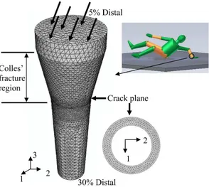
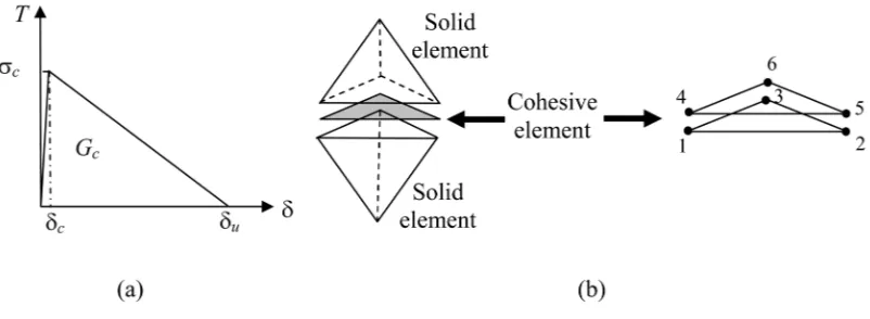
In this study, two idealized human radius bone geometries (Table 1) developed in previous studies (Ural, 2009) based on measurements in the literature (Hsu et al., 1993) were used to assess the effect of strain rate on Colles’ fracture load (Figure 1). The load was applied on the top surface of the model (75° dorsiflexion of the wrist and 10° internal rotation) following the experimental Colles’ fracture studies (Myers et al., 1991; Augat et al., 1998). The proximal ends of the models were fixed in all directions. The crack plane was tiled with cohesive elements (Figure 1) following a traction-displacement relationship (Figure 2). The models were meshed with tetrahedral elements. A mesh sensitivity study was performed (Ural, 2009) that confirmed that the results are independent of mesh size. Implicit nonlinear analyses were performed using ABAQUS (version 6.8, 2008, Simulia, Providence, RI) (For additional information see Supplementary Material).
The strain rate effects on Colles’ fracture load were captured through the variation of the material properties as a function of strain rate. This approach was chosen instead of applying the load at different rates with constant material properties as the cohesive model used in the current study is not rate dependent. Incorporating the variation of the fracture toughness and strength of bone with increasing strain rate in the models effectively simulates the change in the fracture load since these parameters are the underlying material properties that determine the fracture behavior of bone.
NIH-PA Author Manuscript
NIH-PA Author Manuscript
The experimental data on elastic modulus, tensile strength, and energy per unit volume were obtained under 1–200 mm/s loading rate from 25 test specimens (four to six specimens for each loading rate) creating strain rates between 0.08–18 s−1 (Hansen et al., 2008; Zioupos et al., 2008). The data collected from each individual test was incorporated in the finite element models resulting in 25 different sets of material parameters. This allowed capturing the variability of the parameters at each strain rate. The elastic modulus measured in the experiments was assigned to the bulk material, whereas ultimate tensile strength and the energy to fracture were used to define the critical strength (σc) and critical energy release
rate (Gc) of the cohesive model.
The fracture load was identified at the point where the first cohesive element broke. This fracture criterion was chosen based on our previous study that showed only 1–4% increase in fracture load if failure of all cohesive elements were considered (Ural, 2009). The fracture loads were compared at strain rate values corresponding to daily activities (≤0.01 s−1), controlled falling from a standing height (≤0.29 s−1) (Foldhazy et al., 2005) and impact cadaver experiments (range: 2.05–6.78 s−1) (Bass et al., 1997; Duma et al., 1999). Our present approach allows the analysis of fracture scenarios which show both size and strain rate variability.
RESULTS
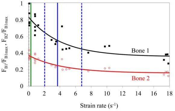
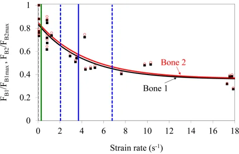
The simulation results showed that the Colles’ fracture load decreased with increasing strain rate and demonstrated a steeper change in lower strain rates (Figure 3 and 4). The fracture loads obtained from the simulations varied between 2288–8364 N for the larger bone (Bone 1) and 1033–3650 N for the smaller bone (Bone 2). The variation in the fracture loads in Figure 3 and 4 corresponding to a single strain rate value is due to the use of individual test results rather than an average fit as outlined in the Methods section. In order to demonstrate the relative change, the fracture loads were first normalized with respect to the overall largest fracture load from all simulations (Figure 3) and then normalized with respect to the individual largest fracture load for each bone (Figure 4) to eliminate the geometric effects. The variation of fracture load with strain rate is captured by an exponential equation (Figures 3 and 4, Table 2).
The fracture load decreased by 3.7% at ̇ ~0.29 s−1 (maximum strain rate measured at controlled falling) compared to the strain rate value that corresponds to daily activities ( ̇ ~0.01 s−1). It reduced further in a drastic way by 34% at ̇~3.7 s−1 (corresponding to mean impact loading). Meanwhile, even higher strain rates ( ̇ ~ up to 18 s−1) only lead to an additional 22% reduction in the fracture load (Table 3).
The results also showed, as it would be expected, that the fracture load at all strain rates was higher for the bone geometry that had larger cortical area and polar moment of inertia (Figure 3). However, the fracture loads for both geometries were reduced at the same rate with increasing strain rate irrespective of their geometrical properties (Figure 4).
DISCUSSION
The current study investigated the effect of strain rate on Colles’ fracture load under varying strain rates. The study is based on recent work from our labs which produced a validated model combining cohesive FEM with experimental measurements (Ural 2009; Ural et al., 2011). The results provide new information on the change in Colles’ fracture load with strain rate which has not been reported in previous studies. Strain rate and size are two confounding factors which can be easily analyzed by our simulation method and provide valuable insight into the fracture scenarios. The results appeared to identify a critical range
NIH-PA Author Manuscript
NIH-PA Author Manuscript
of loading rates (10–50 mm/s) above daily activities that can dramatically increase the risk of Colles’ fracture and this may be particularly important for the improvement of fracture risk assessment and low energy falls in the elderly.
The fracture load predictions found in this study lie within the range of Colles’ fracture loads reported in the literature obtained at varying loading rates. (Augat et al., 1996; Augat et al., 1998; Muller et al., 2003; Ashe et al., 2006; Gdela et al., 2008; Varga et al., 2009) The results showed that Colles’ fracture load decreased substantially more at low strain rate range (low to intermediate rates) compared to higher values (intermediate to high rates). The transition from controlled to impact falling corresponds to the strain rate range where the most drastic reduction in fracture load occurs.
In the current literature, noninvasive fracture risk is evaluated by comparing the fall load calculated based on impact forces to static FEM fracture load predictions (Muller et al., 2003; Melton et al., 2007). The current results indicate that inclusion of the strain rate effects can help prevent the underestimation of fracture risk (by overestimating the actual fracture load). Given that the calculated fracture risk is usually close to 1 (Muller et al., 2003; Melton et al., 2007), potential reduction in the predicted fracture load due to increasing strain rate may have a significant effect on the fracture risk predictions.
The results also highlight the importance of landing during a fall. Events such as grabbing or hitting an object before landing may reduce the fracture risk (Keegan et al., 2004) by not only decreasing the fall load but also by reducing the strain rate induced in the bone which affect the intrinsic fracture resistance of the bone.
Supplementary Material
Refer to Web version on PubMed Central for supplementary material.
Acknowledgments
DV acknowledges NIH Grant AG20618. PZ acknowledges support provided by DACMT (Defence Academy -College of Management and Technology) to study ‘stress fractures in young army recruits’.
References
Adharapurapu RR, Jiang F, Vecchio KS. Dynamic fracture of bovine bone. Materials Science and Engineering: C. 2006; 26:1325–1332.
Ashe MC, Khan KM, Kontulainen SA, Guy P, Liu D, Beck TJ, McKay HA. Accuracy of pQCT for evaluating the aged human radius: An ashing, histomorphometry and failure load investigation. Osteoporosis International. 2006; 17:1241–1251. [PubMed: 16683179]
Augat P, Reeb H, Claes LE. Prediction of fracture load at different skeletal sites by geometric properties of the cortical shell. Journal of Bone and Mineral Research. 1996; 11:1356–1363. [PubMed: 8864911]
Augat P, Iida H, Jiang Y, Diao E, Genant HK. Distal radius fractures: Mechanisms of injury and strength prediction by bone mineral assessment. Journal of Orthopaedic Research. 1998; 16:629– 635. [PubMed: 9820289]
Bass, CR.; Duma, SM.; Crandall, JR.; Morris, R.; Martin, P.; Pilkey, W.; Hurwitz, S.; Khaewpong, N.; Eppinger, R.; Sun, E. The interaction of air bags with upper extremities. Paper presented at: 41st Stapp Car Crash Conference; Warrendale, Pennsylvania. 1997.
Behiri JC, Bonfield W. Crack velocity dependence of longitudinal fracture in bone. Journal of Materials Science. 1980; 15:1841–1849.
NIH-PA Author Manuscript
NIH-PA Author Manuscript
Behiri JC, Bonfield W. Fracture mechanics of bone--the effects of density, specimen thickness and crack velocity on longitudinal fracture. Journal of Biomechanics. 1984; 17:25–34. [PubMed: 6715385]
Boutroy S, Van Rietbergen B, Sornay-Rendu E, Munoz F, Bouxsein ML, Delmas PD. Finite element analysis based on in vivo HR-pQCT images of the distal4 radius is associated with wrist fracture in postmenopausal women. Journal of Bone and Mineral Research. 2008; 23:392–399. [PubMed: 17997712]
Buchanan D, Ural A. Finite element modeling of the influence of hand position and bone properties on the Colles’ fracture load during a fall. Journal of Biomechanical Engineering. 2010; 132:081007. [PubMed: 20670056]
Burghardt AJ, Kazakia GJ, Ramachandran S, Link TM, Majumdar S. Age- and gender-related differences in the geometric properties and biomechanical significance of intracortical porosity in the distal radius and tibia. Journal of Bone and Mineral Research. 2010; 25:983–993. [PubMed: 19888900]
Duma SM, Schreiber PH, McMaster J, Crandall JR, Bass CR, Pilkey WD. Dynamic injury tolerances for long bones of the female upper extremity. Journal of Anatomy. 1999; 194:463–471. [PubMed: 10386782]
Evans GP, Behiri JC, Vaughan LC, Bonfield W. The response of equine cortical bone to loading at strain rates experienced in vivo by the galloping horse. Equine Veterinary Journal. 1992; 24:125– 128. [PubMed: 1582390]
Foldhazy Z, Arndt A, Milgrom C, Finestone A, Ekenman I. Exercise-induced strain and strain rate in the distal radius. Journal of Bone and Joint Surgery-British Volume. 2005; 87:261.
Gdela K, Pietruszczak S, Lade PV, Tsopelas P. On Colles' fracture: An experimental study involving structural and material testing. Journal of Applied Mechanics. 2008; 75:031002–031010. Hansen U, Zioupos P, Simpson R, Currey JD, Hynd D. The effect of strain rate on the mechanical
properties of human cortical bone. Journal of Biomechanical Engineering. 2008; 130:011011. [PubMed: 18298187]
Holmberg AH, Johnell O, Nilsson PM, Nilsson J, Berglund G, Akesson K. Risk factors for fragility fracture in middle age. A prospective population-based study of 33,000 men and women. Osteoporosis International. 2006; 17:1065–1077. [PubMed: 16758143]
Hsu ES, Patwardhan AG, Meade KP, Light TR, Martin WR. Cross-sectional geometrical properties and bone mineral contents of the human radius and ulna. Journal of Biomechanics. 1993; 26:1307– 1318. [PubMed: 8262992]
Keegan THM, Kelsey JL, King AC, Quesenberry CP, Sidney S. Characteristics of fallers who fracture at the foot, distal forearm, proximal humerus, pelvis, and shaft of the tibia/fibula compared with fallers who do not fracture. American Journal of Epidemiology. 2004; 159:192. [PubMed: 14718222]
Kulin R, Jiang F, Vecchio K. Aging and loading rate effects on the mechanical behavior of equine bone. JOM Journal of the Minerals, Metals and Materials Society. 2008; 60:39–44.
Kulin R, Jiang F, Vecchio KS. Effects of age and loading rate on equine cortical bone failure. Journal of the Mechanical Behavior of Biomedical Materials. 2011a; 4:57–75. [PubMed: 21094480] Kulin RM, Jiang F, Vecchio KS. Loading rate effects on the R-curve behavior of cortical bone. Acta
Biomaterialia. 2011b; 7:724–732. [PubMed: 20883834]
MacNeil JA, Boyd SK. Bone strength at the distal radius can be estimated from high-resolution peripheral quantitative computed tomography and the finite element method. Bone. 2008; 42:1203–1213. [PubMed: 18358799]
Melton LJ 3rd, Amadio PC, Crowson CS, O'Fallon WM. Long-term trends in the incidence of distal forearm fractures. Osteoporosis International. 1998; 8:341–348. [PubMed: 10024904]
Melton LJ 3rd, Riggs BL, van Lenthe GH, Achenbach SJ, Muller R, Bouxsein ML, Amin S, Atkinson EJ, Khosla S. Contribution of in vivo structural measurements and load/strength ratios to the determination of forearm fracture risk in postmenopausal women. Journal of Bone and Mineral Research. 2007; 22:1442–1448. [PubMed: 17539738]
Muller ME, Webber CE, Bouxsein ML. Predicting the failure load of the distal radius. Osteoporosis International. 2003; 14:345–352. [PubMed: 12730737]
NIH-PA Author Manuscript
NIH-PA Author Manuscript
Myers ER, Sebeny EA, Hecker AT, Corcoran TA, Hipp JA, Greenspan SL, Hayes WC. Correlations between photon absorption properties and failure load of the distal radius in vitro. Calcified Tissue International. 1991; 49:292–297. [PubMed: 1760774]
O'Neill TW, Cooper C, Finn JD, Lunt M, Purdie D, Reid DM, Rowe R, Woolf AD, Wallace WA. Incidence of distal forearm fracture in british men and women. Osteoporosis International. 2001; 12:555–558. [PubMed: 11527052]
Owen RA, Melton LJ 3rd, Johnson KA, Ilstrup DM, Riggs BL. Incidence of Colles' fracture in a North American community. American Journal of Public Health. 1982; 72:605–607. [PubMed: 7072880] Pietruszczak S, Gdela K, Webber CE, Inglis D. On the assessment of brittle-elastic cortical bone
fracture in the distal radius. Engineering Fracture Mechanics. 2007; 74:1917–1927.
Pietruszczak S, Gdela K. Inelastic analysis of fracture propagation in distal radius. Journal of Applied Mechanics. 2010; 77:011009.
Pistoia W, van Rietbergen B, Lochmuller EM, Lill CA, Eckstein F, Ruegsegger P. Estimation of distal radius failure load with micro-finite element analysis models based on three-dimensional peripheral quantitative computed tomography images. Bone. 2002; 30:842–848. [PubMed: 12052451]
Stein EM, Liu XS, Nickolas TL, Cohen A, Thomas V, McMahon DJ, Zhang C, Yin PT, Cosman F, Nieves J, Guo XE, Shane E. Abnormal microarchitecture and reduced stiffness at the radius and tibia in postmenopausal women with fractures. Journal of Bone and Mineral Research. 2010; 25:2572–2581. [PubMed: 20564238]
Ural A. Prediction of Colles' fracture load in human radius using cohesive finite element modeling. Journal of Biomechanics. 2009; 42:22–28. [PubMed: 19056085]
Ural A, Zioupos P, Buchanan D, Vashishth D. The effect of strain rate on fracture toughness of human cortical bone: A finite element study. Journal of the Mechanical Behavior of Biomedical
Materials. 2011; 4:1021–1032. [PubMed: 21783112]
Varga P, Baumbach S, Pahr D, Zysset PK. Validation of an anatomy specific finite element model of Colles' fracture. Journal of Biomechanics. 2009; 42:1726–1731. [PubMed: 19467661]
Varga P, Pahr DH, Baumbach S, Zysset PK. HR-pQCT based FE analysis of the most distal radius section provides an improved prediction of colles' fracture load in vitro. Bone. 2010; 47:982–988. [PubMed: 20692389]
Zioupos P, Hansen U, Currey JD. Microcracking damage and the fracture process in relation to strain rate in human cortical bone tensile failure. Journal of Biomechanics. 2008; 41:2932–2939. [PubMed: 18786670]
NIH-PA Author Manuscript
NIH-PA Author Manuscript
Figure 1.
Schematics of a falling configuration and a sample 3D finite element mesh of radius bone with the crack plane tiled with cohesive elements. The distal articular surface of the radius was not modeled explicitly. The applied load is transformed to the local coordinate system of the radius assuming uniform distribution. The proximal ends of the models were fixed in all directions. The models were meshed with tetrahedral elements.
NIH-PA Author Manuscript
NIH-PA Author Manuscript
Figure 2.
(a) Traction-displacement relationship defining the cohesive zone model. The parameters that define the cohesive model are the critical strength of the material (σc), the opening
displacement at fracture ( u), and the energy needed for opening the crack (Gc). In the
current simulations, σc, and Gc were used to define the cohesive model. The values of these
parameters were based on experimental measurements reported in the literature (Hansen et al., 2008; Zioupos et al., 2008). (b) Tetrahedral solid elements and the compatible wedge shaped cohesive element with six nodes.
NIH-PA Author Manuscript
NIH-PA Author Manuscript
Figure 3.
Normalized fracture load vs. strain rate for both bone geometries. Note that fracture loads for both geometries are normalized with respect to the overall largest fracture load. The exponential curve fits of the form F = a + be−c ̇ are summarized in Table 2. FB1 and FB2
denote the fracture load for Bone 1 and 2, respectively. The gray dotted line corresponds to strain rates measured during daily activities (0.01 s−1). The green solid line is the maximum strain rate (0.29 s−1) measured in vivo during controlled falling to the ground (Foldhazy et al., 2005). The solid blue line is the average impact strain rate (3.7 s−1) whereas the dotted blue lines define the minimum and maximum impact strain rates measured (2.05–6.78 s−1) in experiments (Bass et al., 1997; Duma et al., 1999).
NIH-PA Author Manuscript
NIH-PA Author Manuscript
Figure 4.
Normalized fracture load vs. strain rate for both bone geometries. Note that fracture loads for both bones are normalized with respect to their individual largest fracture load. The exponential curve fits of the form F = a + be−c ̇ are summarized in Table 2. F
B1 and FB2
denote the fracture load for Bone 1 and 2, respectively. The gray dotted line corresponds to strain rates measured during daily activities (0.01 s−1). The green solid line is the maximum strain rate (0.29 s−1) measured in vivo during controlled falling to the ground (Foldhazy et al., 2005). The solid blue line is the average impact strain rate (3.7 s−1) whereas the dotted blue lines define the minimum and maximum impact strain rates measured (2.05–6.78 s−1) in experiments (Bass et al., 1997; Duma et al., 1999).
NIH-PA Author Manuscript
NIH-PA Author Manuscript
NIH-PA Author Manuscript
NIH-PA Author Manuscript
NIH-PA Author Manuscript
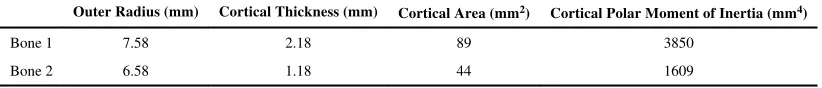
Table 1
Geometric properties at the crack plane for both radius bone models.
Outer Radius (mm) Cortical Thickness (mm) Cortical Area (mm2) Cortical Polar Moment of Inertia (mm4)
Bone 1 7.58 2.18 89 3850
NIH-PA Author Manuscript
NIH-PA Author Manuscript
NIH-PA Author Manuscript
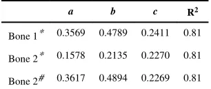
Table 2
Coefficients of the exponential fit parameters F = a + be−c ̇ for 3D human radius simulations where F represent the normalized fracture load and ̇ represents the strain rate.
a b c R2
Bone 1* 0.3569 0.4789 0.2411 0.81
Bone 2* 0.1578 0.2135 0.2270 0.81
Bone 2# 0.3617 0.4894 0.2269 0.81
*
Normalized with respect to maximum fracture load for Bone 1
#
NIH-PA Author Manuscript
NIH-PA Author Manuscript
NIH-PA Author Manuscript
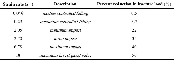
Table 3
Percent change in Colles’ fracture load with increasing strain rate compared to the maximum strain rate (0.01 s−1) measured for daily activities. The reported values correspond to experimentally measured strain rates for controlled falling (Foldhazy et al., 2005) and impact loading experiments (Bass et al., 1997; Duma et al., 1999). The results are based on Bone 1, however, the percent changes were almost the same for Bone 2 with less than 1% difference.
Strain rate (s−1) Description Percent reduction in fracture load (%)
0.046 median controlled falling 0.5
0.29 maximum controlled falling 3.7
2.05 minimum impact 22
3.70 mean impact 34
6.78 maximum impact 46