Possible involvement of a 72-kDa polypeptide in nucleotide
excision repair of
Chlorella pyrenoidosa
identified by affinity
adsorption and repair synthesis assay
Todd Hsu *, Reou-Ching Sheu, Yi-Show Lai
Institute of Marine Biotechnology,National Taiwan Ocean Uni6ersity,Keelung 20224, Taiwan,ROC
Received 9 November 1999; received in revised form 31 January 2000; accepted 23 February 2000
Abstract
A DNA repair synthesis assay monitoring nucleotide excision repair (NER) was established in cell-free extracts of unicellular alga Chlorella pyrenoidosausing cisplatin- or mitomycin C-damaged plasmid DNA as the repair substrate. The algal extracts promoted a damage-dependent increase in32P-dATP incorporation after normalization against an internal control. To identify the proteins responsible for NER, a biotin-labeled duplex 27 mer (2mg) irradiated with or without UV (27 kJ m−2) was immobilized on streptavidin-conjugated agarose beads and incubated with C.pyrenoidosaextracts (50mg) to pull down repair proteins. The extracts post incubation with beads carrying unirradiated 27 mer promoted an eightfold increase in repair synthesis in plasmid DNA (1mg) damaged by 16.8 pmol of cisplatin. The extracts obtained after affinity adsorption with UV-damaged DNA ligand, however, failed to repair plasmid DNA treated with cisplatin, reflecting that some proteins crucial to NER had been sequestered by the damaged 27 mer. A polypeptide 70 – 72 kDa in molecular mass was found to bind much more strongly to the damaged
DNA than to the control DNA after analyzing the proteins bound to the beads by SDS-PAGE, and this polypeptide is believed to play a role in NER inC.pyrenoidosa. © 2000 Elsevier Science Ireland Ltd. All rights reserved.
Keywords:Affinity; Algae; Cisplatin;Chlorella pyrenoidosa; Nucleotide excision repair; Ultraviolet light
www.elsevier.com/locate/plantsci
1. Introduction
Nucleotide excision repair (NER) is an ATP-de-pendent and a multistep DNA repair pathway that plays an important role in removing DNA damage produced by UV irradiation, alkylating agents like cisplatin or reactive oxygen species [1,2]. In NER, DNA lesions are first recognized by damage-recognition proteins, and an endonucleolytic inci-sion is introduced at both sides of the damaged region. The incised DNA fragment is then excised
and the gap generated after incision and excision is filled by synthesis of new DNA that is finally joined to preexisting DNA [1,3,4]. The proteins responsible for NER inEscherichia coli, yeast and human cells have been well characterized [1,3].
Based on the excision of UV-induced pyrimidine dimers from plant DNA, NER has been found to operate in Arabidopsis thaliana [5], unicellular green alga Chlamydomonas reinhardtii [6], carrot protoplasts [7] and some water plants [8]. UV-spe-cific endonucleases acting on pyrimidine dimer-containing DNA have been partially purified from spinach and suspension cultures of the carrotDau
-cus carota [9,10]. Recently, a cDNA homologous
to the human Xeroderma pigmentosum group B (XPB) gene was cloned fromA. thaliana[11]. XPB gene encodes a DNA-dependent ATPase whose DNA helicase motifs are known to participate in Abbre6iations: 6-4PPs, (6-4)photoproducts; CPDs, cyclobutane
pyrimidine dimers; ds DNA, double-stranded DNA; DTT, dithiothre-itol; MMC, mitomycin C; NER, nucleotide excision repair; PMSF, phenylmethylsulfonyl fluoride; SDS-PAGE, sodium dodecyl sulfate polyacrylamide gel electrophoresis; UV, ultraviolet light.
* Corresponding author. Tel.: +886-2-24622192, ext. 5503; fax:
+886-2-24622320.
E-mail address:[email protected] (T. Hsu)
unwinding the damaged DNA region before the dual incision step. Moreover, two isoforms of yeast rad23 gene were identified in a carrot cDNA library when this library was used to complement a UV-sensitive yeast mutant that is deficient in a single-stranded DNA binding protein associated with DNA unwinding [12]. Although some infor-mation regarding NER proteins in higher plants has been obtained, little is known about the nature of NER proteins in lower plants and the way they interact with each other.
In vitro assays detecting damage-specific inci-sion of DNA (the DNA inciinci-sion assay) and dam-age-dependent DNA repair synthesis (the repair synthesis assay) developed in cell-free extracts have been recognized as valuable tools for study-ing the biochemical mechanisms, includstudy-ing the cutting site introduced by the 5% or 3% incision endonuclease, the size of the excised fragment, the cofactors required for repair and the substrate specificity, of NER in E. coli and human cells [13 – 17]. Unicellular algae are ideal model systems for exploring the biochemical mechanisms of NER in lower plants, because they can be cultured and handled easily in the laboratory. We have devel-oped an in vitro DNA incision assay in cell-free extracts of unicelluar alga C. pyrenoidosa, which clearly revealed the cutting position introduced at the 3% side of a pyrimidine dimer by the extracts [18]. Following the development of incision assay, a UV-damaged-DNA binding activity composed of three polypeptides, p72, p80 and p90, was purified from C. pyrenoidosa extracts, and these three polypeptides were found to bind directly to UV-damaged DNA as they could be extracted from gel shift bands produced by the crude ex-tracts or more purified protein fractions [19]. This binding activity may participate in the damage-recognition step of NER, since it recognized both UV and cisplatin-damaged DNA in the absence of ATP. The goal of this research was to identify the proteins involved in NER in C. pyrenoidosa by functional analysis. An in vitro DNA repair syn-thesis assay monitoring NER was established and an affinity adsorption of binding proteins from the algal extracts was performed with the same UV-damaged DNA probe [19] immobilized on a solid phase. The effects of affinity adsorption on the capacity of NER were determined by the repair synthesis assay and a polypeptide 72 kDa in molecular mass that preferentially binds to
dam-aged DNA is believed to play a crucial role in NER.
2. Materials and methods
2.1. Plant materials and cell-free extracts
C. pyrenoidosa kindly provided by Dr
Jiunn-Tzong Wu (Institute of Botany, Academia Sinica, Taipei, Taiwan, ROC) was grown in a synthetic salt medium [20] at 25°C under illumination of a fluorescent light and gentle shaking. Algal growth was monitored by absorbance at 685 nm. Algal cells (1000 ml) at mid-log growth (5.5 – 7.0×10⁷
cells/ml) were collected by centrifugation at 5000×g and the pellet was washed twice with ice-cold distilled water and once with extraction buffer (20 mM Tris – HCl (pH 8.0), 1 mM EDTA, 1 mM DTT, 10% (v/v) glycerol). Algal cells were then suspended in 4 ml hypotonic buffer (40 mM Tris – HCl (pH 8.0), 1 mM EDTA, 1 mM DTT) containing protease inhibitors (2 mM PMSF, 4 mg/ml leupeptin, 1 mg/ml pepstatin), and the swol-len cells were broken on ice by pulsed sonication. After centrifugation at 14 000×g for 10 min at 4°C, the supernatant was transferred to a beaker on ice and nucleic acids were removed by stirring in the presence of 1.5% (w/v) streptomycin sulfate for 15 min. After centrifugation at 16 000×g for 90 min, the supernatant was withdrawn and am-monium sulfate was added to 55% saturation to fractionate repair proteins. The protein precipitate was dissolved in 1 ml dialysis buffer (20 mM Tris – HCl (pH 8.0), 1 mM EDTA, 2 mM DTT, 0.1 M KCl, 12 mM MgCl2, 17% (v/v) glycerol),
and dialyzed against the same buffer for 12 h at 4°C. The dialyzed protein solution was concen-trated with Centricon-10 (Amicon, USA) and used as the cell-free extract for the repair synthesis assay. The protein concentration in the extract was measured by a protein assay kit (Bio-Rad, USA) based on the method developed by Brad-ford [21].
2.2. Preparation of immobilized DNA ligands for
affinity adsorption
A 27-mer oligonucleotide, 5%-GAC CGA GCT
GGG TTA CGA CGC GAC GCC-3% and its
la-beled with biotin at the 5% end by a commercial source. Both strands at equal concentrations (1
mg/ml) were mixed and the mixture was placed in a
PCR thermocycler (Perkin Elmer, Norwalk, CT, USA). The temperature was raised to 95°C and decreased to 25°C at a rate of 1°C/min. After filtering the mixture on a microcon-10 concentra-tor (Amicon, Beverly, MA, USA), the annealed 27 mer retained on the membrane was collected for preparing UV-damaged DNA. The duplex 27 mer was pipetted onto a piece of parafilm, and UV (254 nm) irradiation was performed in an XL-1500 UV crosslinker (Spectronics, Westbury, NY, USA). UV irradiation was expected to induce the formation of CPDs and 6-4PPs between adjacent TT or CT on the 27 mer [22]. UV-irradiated or unirradiated 27 mer (2 mg) was then incubated with a suspension (20 ml) of streptavidin-conju-gated agarose beads (1 – 3 mg streptavidin/ml, Merck, Germany), and the oligonucleotide was immobilized on the beads through the strong affinity between biotin and streptavidin.
2.3. Affinity adsorption of repair proteins
To pull down repair proteins from cell-free ex-tracts of C. pyrenoidosa, the algal extract contain-ing 50 mg proteins in 20 ml dialysis buffer was incubated at 30°C for 20 min with 20 ml agarose beads carrying 2mg UV (27 kJ m−2)-irradiated 27
mer. The suspension was centrifuged at 5000×g
at 4°C, and a small fraction of the extract superna-tant was taken for the determination of protein concentration. An aliquot of the extract contain-ing 30-mg proteins was used for the repair synthe-sis assay. The repair capacity in the extract incubated with beads carrying unirradiated 27 mer or no 27 mer was also tested as a comparison.
2.4. DNA substrates for the repair synthesis assay
Plasmid DNA damaged by the alkylating agent cisplatin or MMC was used as the substrate for the repair synthesis assay. pBR 322 (4.3 kb) and pGEM IV (2.8 kb) plasmid DNA were isolated from E. coli JM 109 by a plasmid DNA prepara-tion kit (Qiagen, Valencia, CA, USA), and the closed circular form DNA was purified by CsCl gradient centrifugation. The concentration of DNA in TE buffer was determined by the ab-sorbance at 260 nm. Alkylating agent-damaged
DNA was prepared by incubating pGEM IV plas-mid (0.1mg/ml in TE buffer) with the same volume of aqueous solution containing different amount of cisplatin or mitomycin C at 37°C overnight in the dark. The damaged DNA was precipitated with ethanol and solubilized in distilled water. Untreated pBR 322 was used as an internal con-trol in each repair reaction mixture.
2.5. In 6itro DNA repair synthesis assay
The standard repair reaction mixture (50 ml) contained 45 mM Tris – HCl (pH 7.8), 4 mM EDTA, 70 mM KCl, 4 mM MgCl2, 1 mM DTT,
40 mM creatine phosphate-Tris (pH 7.7), 2.5 mg phosphocreatine kinase, 2 mM ATP, 18mg bovine serum albumin, 50 mM each of dGTP, dCTP, dTTP, dATP, 2 mCi [a-32P]dATP (3000 Ci/mmol),
1 mg pBR 322 plasmid DNA, 1 mg control or
damaged pGEM IV plasmid DNA, and 30 mg
algal extract proteins. After incubating the reac-tion mixture at 30°C for 2 h, the repair reacreac-tion was terminated by the addition of EDTA to a final concentration of 25 mM. The extract proteins were removed by proteinase K (200 mg/ml) diges-tion in the presence of 0.5% SDS. The mixture was then extracted with phenol/chloroform/isoamyl al-cohol (25:24:1), and DNA products were precipi-tated with ethanol. Purified DNA products were linearized with Pst I and electrophoresed on a 1.2% agarose gel. After ethidium bromide staining, the gel was vacuum dried and exposed to a Kodak XAR-5 X-ray film with an intensifying screen. Band intensities on the autoradiograph were quan-titated using a Amersham/Pharmacia Imagemaster documentation system. Damage-stimulated repair synthesis was expressed as repair factor deter-mined by dividing the band intensity of the dam-aged DNA by that of the internal control.
2.6. SDS-PAGE analysis of proteins captured by
affinity adsorption
To analyze the proteins that were captured on the beads, agarose beads were washed thoroughly with 20 mM KH2PO4(pH 7.5) containing 0.15 M
silver staining. Non-specific binding proteins washed from the beads were also analyzed on the same gel for comparison.
2.7. Chemicals
PMSF, leupeptin, pepstatin, streptomycin sul-fate, cisplatin and mitomycin C were purchased from Sigma (St Louis, MO, USA).Other reagent grade chemicals were obtained from J.T. Baker (Phillipsburg, NJ, USA). DTT and streptavidin-conjugated agarose beads were obtained from Boehringer Mannheim (Mannheim, Germany).
[a-32P]dATP (3000 Ci mmol−1) was a product of
Amersham (Little Chalfont, Amersham, Bucking-hamshire, UK). Biotin-labeled oligonucleotides were synthesized by Perkin Elmer (Norwalk, CT, USA).
3. Results
3.1. Damage-stimulated DNA repair synthesis in
cell-free extracts of C. pyrenoidosa
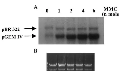
When cisplatin-damaged DNA was incubated with algal protein extracts, a clear dose-dependent increase in dAMP incorporation into damaged DNA was detected after normalization against the internal control, indicating the presence of a major body of NER proteins in the extracts (Fig. 1). Quantitative analysis of DNA band intensities showed that the repair factors for plasmid DNA damaged by cisplatin at 0, 2.8, 5.6, 8.4, 11.2 and 16.8 pmol were 1.0, 2.1, 4.4, 6.2, 6.8 and 7.1, respectively. A linear increase in repair factor was found for pGEM IV DNA treated with 0 – 8.4 pmol cisplatin and NER capacity in the extracts seemed to be saturated by DNA lesions induced by higher concentrations of cisplatin. The excision repair of cisplatin-damaged DNA was absolutely protein-dependent as no repair synthesis could be detected in the absence of extract proteins. Dam-age-specific dAMP incorporation was increased with increasing amount of extract proteins and all our repair assays were performed in the presence of 30 mg extract proteins because proteins at this level already gave a significant degree of damage-dependent repair synthesis. The optimal level of repair synthesis was found in the reaction mixture containing 7 – 10 mM Mg2+ (data not shown). An MMC dose-dependent increase in repair synthesis was also promoted by C. pyrenoidosa protein ex-tracts (Fig. 2). The ratios of damage-stimulated to control dAMP incorporation were 1.1, 3.4, 7.3, 7.7
Fig. 1. Cisplatin dose-dependent DNA repair synthesis in cell-free extracts of C. pyrenoidosa. Cisplatin-damaged or non-damaged pGEM IV plasmid DNA (1mg) was incubated at 30°C for 2 h with algal extract proteins (30mg) in a 50-ml reaction mixture containing dNTPs, [a-32P]dATP, ATP, an ATP-regenerating system and all other required cofactors. pBR 322 DNA (1 mg) was included in each assay as an internal control. The repair reaction was terminated by the addition of EDTA and proteinase K. The DNA products were purified by phenol/chloroform/isoamyl alcohol extrac-tion and ethanol precipitaextrac-tion. The purified DNA products were linearized with Pst I, and electrophoresed on a 1.2% agarose gel. (A) Autoradiogram of the dried gel and (B) ethidium bromide staining of the gel are shown.
Fig. 3. In vitro excision repair of cisplatin (16.8 pmol)-dam-aged DNA in the algal extracts collected after affinity adsorp-tion. The extract (50mg) was incubated at 30°C for 20 min with UV (27 kJ m−2)-irradiated or unirradiated 27 mer dsDNA immobilized on the beads, and the beads were spun down at 5000×g at 4°C for 10 min. An aliquot of the supernatant containing 30 mg proteins was tested for its ability to repair cisplatin-treated DNA. The extract (30 mg) incubated with only streptavidin-conjugated beads was also subjected to the repair synthesis assay as a comparison. (A) Autoradiogram of the dried gel and (B) ethidium bromide staining of the gel are shown.
3.2. Affinity adsorption of repair proteins
To test if the repair proteins in cell-free extracts
ofC.pyrenoidosacould be pulled down by affinity
adsorption, excision repair of cisplatin-damaged DNA was monitored in the extracts post incuba-tion at 30°C for 20 min with UV (27 kJ m−2
)-irra-diated or unirradiated 27 mer ds DNA
insolubilized on the beads. This incubation condi-tion had been proved to be appropriate for specific recognition proteins in C. pyrenoidosa extracts to interact with UV-damaged DNA [19]. Extracts incubated with only streptavidin-conjugated agarose beads and extracts collected after incuba-tion with beads carrying unirradiated 27 mer
ds-DNA displayed a significant level of
damage-dependent repair synthesis. Both extracts showed about an eight to ninefold increase in repair factor after incubation with DNA damaged by cisplatin at 16.8 pmol. In contrast, cisplatin-stimulated repair was almost undetectable in the extracts post incubation with beads carrying UV-damaged 27 mer ds DNA as the repair factor was only 1.2, which signified that very little or no damage-stimulated incorporation of radioactive nucleotides could be detected (Fig. 3, Table 1). When untreated pGEM IV plasmid DNA was incubated with the extracts post incubation with beads carrying UV-irradiated DNA, unirradiated DNA or no ligand as the control for cisplatin-in-dependent semiconservative DNA synthesis, the level of radioactive dAMP incorporated into this plasmid was very close to that incorporated into pBR322 DNA (Fig. 3) with ratios ranging from 0.9 to 1.2 in several experiments. We occasionally found that the extracts preincubated with beads carrying UV-damaged 27 mer gave a compara-and 8.3 for plasmid DNA damaged by MMC at 0,
1, 2, 4 and 6 nmol, respectively. MMC has been shown to react almost exclusively with the N-2 of guanines to form monoadducts and crosslinks. Unlike the DNA crosslinks formed by cisplatin, the crosslinks induced by MMC do not signifi-cantly disturb the DNA helical structure [23]. Therefore, plasmid DNA had to be treated with MMC in the nanomole range in order to generate a detectable repair signal for in vitro DNA repair synthesis assay.
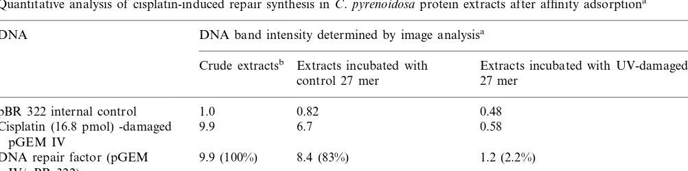
Table 1
Quantitative analysis of cisplatin-induced repair synthesis in C.pyrenoidosaprotein extracts after affinity adsorptiona
DNA DNA band intensity determined by image analysisa
Crude extractsb Extracts incubated with Extracts incubated with UV-damaged control 27 mer 27 mer
1.0 0.48
pBR 322 internal control 0.82
0.58
9.9 6.7
Cisplatin (16.8 pmol) -damaged pGEM IV
DNA repair factor (pGEM 9.9 (100%) 8.4 (83%) 1.2 (2.2%) IV/pBR 322)
aEach data point represents the average of three separate determinations.
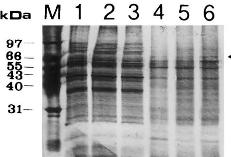
Fig. 4. SDS-PAGE analysis of the proteins captured by affinity adsorption. Biotin-labeled 27 mer irradiated with 0 or 27 kJ m−2 of UV were linked to streptavidin-conjugated agarose beads, and the beads were incubated at 30°C for 20 min with algal extracts containing 50 mg protein. After cen-trifugation at 5000×gat 4°C for 10 min, the supernatant was taken for the repair synthesis assay. The beads were first washed with 20 mM KH2PO4 (pH 7.5) containing 0.15 M NaCl at room temperature, and then boiled in 2× SDS gel loading buffer for 10 min. The proteins in the boiled mixtures were electrophoresed on a 12.5% SDS-polyacrylamide gel and the gel was silver stained. The proteins washed from ligand-free beads and from beads carrying unirradiated or UV-irra-diated 27 mer dsDNA are shown in lanes 1, 2 and 3, respectively. M indicates a protein marker composed of proteins with known molecular weights. The proteins bound to ligand-free beads and to beads carrying control or dam-aged DNA ligand are shown in lanes 4, 5 and 6, respectively.
aged DNA (Fig. 4, lane 6, indicated by an arrow) as this polypeptide p72 was captured only on the beads carrying UV-damaged DNA (Fig. 4, lanes 4 – 6). Since the extracts collected after affinity adsorption with immobilized damaged DNA showed a dramatic decrease in NER capacity, the polypeptide p72 was highly speculated to play a role in NER. Although a polypeptide of 65 kDa was also found to bind preferentially to the dam-aged DNA ligand after affinity adsorption, this preferential binding was not stably detected.
4. Discussion
NER is a wide-spectrum DNA repair pathway found in almost all living organisms. Studies of the mechanisms of NER have focused largely on bac-teria, yeast and humans, with very minor work on plants. Efficient DNA repair systems, however, must have evolved in plants due to the inability of most plants to minimize environmental exposure to noxious agents like UV [24]. Thus, we are interested in revealing the mechanism of NER in plant cells using unicellular alga C. pyrenoidosaas the model system. Since NER requires a coordi-nated action of several proteins, the repair process would be interfered with if any component of the repair system were lacking. In this study UV-dam-aged duplex DNA immobilized on agarose beads was incubated with cell-free extracts of C.
pyrenoidosato pull down damage-dependent
bind-ing proteins and the importance of these proteins in NER was determined by monitoring the differ-ence in NER capacity in the extracts post incuba-tion with insolubilized control and damaged DNA.
Proteins other than those involved in NER, like photolyases responsible for the photoreactivation repair and high mobility group (HMG) proteins, have been shown to bind preferentially to UV-irra-diated DNA containing CPDs and 6-4PPs [25,26]. Photoreactivation repair functions in many prokaryotic and eukaryotic cell types, specifically eliminating CPDs or 6-4PPs present in UV-dam-aged DNA [26]. In this repair pathway, pho-tolyases bind the dipyrimidine photoproducts in the dark, and the covalent bonds connecting two adjacent pyrimidines are opened via the action of photolyases activated by light stimulation. HMG proteins are abundant eukaryotic chromosomal tively lower level of background incorporation
into pBR 322 DNA (Fig. 3), implying that a certain fraction of DNA polymerases might have been adsorbed onto the damaged DNA.
3.3. SDS-PAGE analysis of proteins captured by
affinity adsorption
UV-dam-proteins, which bind more tightly to cisplatin-damaged DNA than to unmodified DNA [27] except the preferential binding of some HMG proteins to UV-damaged DNA [25]. Insolubilized UV-irradiated DNA was selected as the ligand to pull down NER proteins from the algal ex-tracts because cisplatin-treated DNA was not an-ticipated to give a better specificity in capturing NER proteins. Cisplatin-damaged DNA, how-ever, was considered as a more specific substrate than UV-damaged DNA for monitoring NER by the repair synthesis assay, since radiolabel incor-poration detected in UV-damaged DNA may re-sult from repair synthesis linked to UV excision repair and base excision repair. UV excision re-pair (UVER) is a DNA rere-pair system recently discovered in fungi and bacteria that cleaves only UV-induced CPDs and 6-4PPs [28]. Like NER, the gap produced after incision and exci-sion in UVER is filled by DNA polymerization. DNA polymerization also functions in base exci-sion repair to fill the abasic sites generated after excising minor DNA lesions such as hydrated and ring-fragmented pyrimidines from UV-irradi-ated DNA by DNA glycosylases [29,30]. In con-trast, cisplatin-induced DNA damage was found to be removed primarily by NER in E. coli and human cell extracts [31,32], and no repair en-zymes specifically cutting cisplatin-damaged DNA have been reported at present. Cisplatin-damaged plasmid DNA was therefore employed as the major repair substrate in this study to avoid detecting repair activity other than NER. The algal extracts post incubation with beads carrying UV-irradiated ds DNA showed no re-pair capacity for cisplatin-damaged plasmid DNA, indicating that some NER factors had been depleted by UV-damaged DNA fixed on the beads. Therefore, affinity adsorption with in-solubilized UV-damaged DNA should be a sim-ple and an efficient way to capture NER proteins from a cell-free system. When the proteins bound to the insolubilized DNA were analyzed by SDS-PAGE, a polypeptide 72 kDa in molecular mass displayed an apparently higher affinity for damaged DNA than for non-damaged DNA. We previously purified a UV-damaged-DNA binding activity composed of three polypeptides, p72, p80 and p90 from cell-free extracts of C. pyrenoidosa [19]. Because the
irradiated DNA used for affinity adsorption was the same one used as the probe for mobility shift assay detecting the binding activity except the biotin labeled at the 5% end, the polypeptide p72 captured by the immobilized DNA should be no different from the p72 contained in the UV-damaged-DNA binding activity. The extracts post incubation with UV-damaged DNA tended to promote a lower absolute radiolabel incorpo-ration into the control pBR 322 DNA than those incubated with unirradiated DNA, suggest-ing that a fraction of DNA polymerases were pulled down along with the polypeptide p72.
Previous studies revealed that NER in an eu-karyotic system like human cells is initiated by the binding of damage-recognition proteins XPA and RPA to the damaged region without the presence of ATP, and the localization of these proteins recruits the binding of a DNA helicase that utilizes the energy released from ATP to unwind the damaged region and the binding of incision proteins to introduce a dual incision at both sides of a lesion [1,3]. The exact function of the polypeptide p72 in NER remains to be deter-mined, yet the inverse correlation between the increase in the amount of p72 captured by the damaged ligand and the drastic reduction in cell-free NER capacity after affinity adsorption strongly suggests its participation in NER. The preferential binding of this polypeptide to the damaged DNA within a short incubation period in the absence of ATP implies that it may play a role in the stage of damage recognition. The UV-damaged DNA ligand used for affinity ad-sorption has also been shown to be a suitable substrate for C. pyrenoidosa extract proteins to introduce a NER-like nucleotide incision in the presence of ATP. The combination of affinity adsorption with in vitro assays detecting DNA incision should be very helpful in identifying the nucleotide incision complex composed of dam-age-recognition proteins and incision proteins in
C. pyrenoidosa extracts.
Acknowledgements
References
[1] R.D. Wood, DNA repair in eukaryotes, Annu. Rev. Biochem. 65 (1996) 135 – 167.
[2] P. Møller, H. Wallin, Adduct formation, mutagenesis and nucleotide excision repair of DNA damage pro-duced by reactive oxygen species and lipid peroxidation product, Mutat. Res. 410 (1998) 271 – 290.
[3] A. Sancar, Excision repair in mammalian cells, J. Biol. Chem. 270 (1995) 15915 – 15918.
[4] A.S. Balajee, A. May, V.A. Bohr, Fine structural analy-sis of DNA repair in mammalian cells, Mutat. Res. 404 (1998) 3 – 11.
[5] A.B. Britt, J.-J. Chen, D. Wykoff, D. Mitchell, A UV-sensitive mutant ofArabidopsisdefective in the repair of pyrimidine-pyrimidinone (6-4) dimers, Science 261 (1993) 1571 – 1574.
[6] G.D. Small, Repair systems for nuclear and chloroplast DNA Chlamydomonas reinhardtii, Mutat. Res. 181 (1987) 31 – 35.
[7] G.P. Howland, Dark-repair of ultraviolet-induced pyrim-idine dimers in the DNA of wild carrot protoplasts, Nature 254 (1975) 160 – 161.
[8] N. Degani, E. Ben-Hur, E. Riklis, DNA damage and repair: induction and removal of thymine dimers in ultraviolet light irradiated intact water plants, Pho-tochem. Photobiol. 31 (1980) 31 – 36.
[9] A.G. McLennan, A.C. Eastwood, An endonuclease ac-tivity from suspension cultures of Daucus carotawhich acts upon pyrimidine dimers, Plant Sci. 46 (1986) 151 – 157.
[10] P.W. Doetsch, W.H. McCray Jr, M.R. Valenzuela, Par-tial purification and characterization of an endonuclease from spinach that cleaves ultraviolet light-damaged du-plex DNA, Biochim. Biophys. Acta 1007 (1989) 309 – 317.
[11] D.T. Ribeiro, C.R. Machado, R.M.A. Costa, U.M. Praekelt, M.A. Van Sluys, C.F.M. Menck, Cloning of a cDNA from Arabidopsis thaliana homologous to the human XPB gene, Gene 208 (1998) 207 – 213.
[12] A. Strum, S. Lienhard, Two isoforms of plant RAD23 complement a UV-sensitive rad23 mutant in yeast, Plant J. 13 (1998) 815 – 821.
[13] K. Sugasawa, C. Masutani, F. Hanaoka, Cell-free repair of UV-damaged simian virus 40 chromosomes in human cell extracts I. Development of a cell-free system detect-ing excision repair of UV-irradiated SV 40 chromo-somes, J. Biol. Chem. 268 (1993) 9098 – 9104.
[14] S. Tateishi, N. Hori, E. Ohtsuka, M. Yamaizumi, Hu-man nucleotide excision nuclease incises synthetic dou-ble-stranded DNA containing a pyrimidine dimer at the phosphodiester linkage 3%to the pyrimidine dimer, Bio-chemistry 32 (1993) 1541 – 1547.
[15] E. Braithwaite, X. Wu, Z. Wang, Repair of DNA lesions induced by polycyclic aromatic hydrocarbons in human cell-free extracts: involvement of two excision repair mechanisms in vitro, Carcinogenesis 19 (1998) 1239 – 1246.
[16] J.-C. Huang, D.L. Svoboda, J.T. Reardon, A. Sancar, Human nucleotide excision nuclease removes thymine
dimers from DNA by incising the 22nd phosphodiester bond 5% and the 6th phosphodiester bond 3% to the photodimer, Proc. Natl. Acad. Sci. USA 89 (1992) 3664 – 3668.
[17] E. Seeberg, J. Nissen-Meyer, P. Strike, Incision of ultra-violet-irradiated DNA by extracts of E. coli requires three different gene products, Nature 263 (1976) 524 – 526.
[18] C.-W. Chang, J.-C. Ho, T. Hsu, Thymine-dimer depen-dent incision on ultraviolet light damaged-DNA in cell-free extracts of Chlorella pyrenoidosa, Biosci. Biotech. Biochem. 60 (1996) 490 – 492.
[19] T. Hsu, J.-C. Ho, C.-C. Chao, Purification of a UV-dam-aged-DNA binding activity from cell-free extracts of unicellular alga Chlorella pyrenoidosa, Plant Sci. 138 (1998) 137 – 147.
[20] J.D. Weinstein, R.W. Howell, R.D. Leverette, S.Y. Grooms, P.S. Brignola, S.M. Mayer, S.I. Beale, Heme inhibition ofd-aminolevulinic acid synthesis is enhanced by glutathione in cell-free extracts of Chlorella, Plant Physiol. 101 (1993) 657 – 665.
[21] M.M. Bradford, A rapid and sensitive method for quan-titation of microgram quantities of protein utilizing the principle of protein-dye binding, Anal. Biochem. 72 (1976) 248 – 254.
[22] D.E. Brash, UV mutagenic photoproducts inEscherichia coliand human cells: a molecular genetics perspective on human skin cancer, Photochem. Photobiol. 48 (1988) 59 – 66.
[23] A.J. Warren, M.A. Ihnat, S.E. Ogdon, E.E. Rowell, J.W. Hamilton, Binding of nuclear proteins associated with mammalian DNA repair to the mitomycin C-DNA in-terstrand crosslink, Environ. Mol. Mutagen. 31 (1998) 70 – 81.
[24] E.J. Vonrax, H.L. Mitchell, R. Karthikeyan, I. Chatter-jee, B.A. Kunz, DNA repair in higher plants, Mutat. Res. 400 (1998) 187 – 200.
[25] G.B. Sancar, DNA photolyases: physical properties, ac-tion mechanism and roles in dark repair, Mutat. Res. 236 (1990) 147 – 160.
[26] E.A. Pasheva, I.G. Pashev, A. Favre, Preferential bind-ing of high mobility group 1 protein to UV-damaged DNA, J. Biol. Chem. 273 (1998) 24730 – 24736.
[27] P.M. Pil, S.J. Lippard, Specific binding of chromosomal protein HMG 1 to DNA damaged by the anticancer drug cisplatin, Science 256 (1992) 234 – 237.
[28] A. Yasui, S.J. McCready, Alternative repair pathways for UV-induced DNA damage, Bioessays 20 (1998) 291 – 297.
[29] R.P. Cunningham, DNA glycosylases, Mutat. Res. 383 (1997) 189 – 196.
[30] R. Stephen Lloyd, Base excision repair of cyclobutane pyrimidine dimers, Mutat. Res. 408 (1998) 159 – 170. [31] I. Husain, S.G. Chaney, A. Sancar, Repair of
cis-plat-inum-DNA adducts by ABC excinuclease in vivo and in vitro, J. Bacteriol. 163 (1985) 817 – 823.
[32] S. Ullah, I. Husain, W. Carlton, A. Sancar, Human nucleotide excision repair in vitro: repair of pyrimidine dimers, psoralen and cisplatin adducts by HeLa cell-free extract, Nucleic Acids Res. 17 (1989) 4471 – 4484.