Low Levels of Vitamin D correlate with Hemoglobin A1c and Interleukin-10 Levels
in Pediatric Type 1 Diabetes Mellitus Patients
Desy Wulandari1*
, Haryudi A. Cahyono2
, Edi Widjajanto3
, Anik Puryatni4
1
Department of Child Health Faculty of Medicine Brawijaya University/Saiful Anwar Hospital, Malang, 65145, Indonesia 2Endocrinology Division, Department of Child Health Faculty of Medicine Brawijaya University/Saiful Anwar Hospital,
Malang, 65145, Indonesia
3Department of Clinical Pathology Faculty of Medicine, Brawijaya University, Malang, 65145, Indonesia
4Nutrition and Metabolic Diseases Division, Department of Child Health Faculty of Medicine Brawijaya University/Saiful Anwar
Hospital, Malang, 65145, Indonesia
ABSTRACT
Vitamin D is reported to affect immune system and prevent autoimmunity. Some studies show that low vita-min D levels in patients with type 1 diabetes are associated with glycemic control and inflammatory status. The study was cross sectional design with subjects T1DM patients aged 1-18 years, and healthy subjects with similar age. Plasma levels of vitamin D (25(OH)D3) was measured using ELISA. Glycemic control measured by hemoglo -bin A1c (HbA1c). Inflammatory status measured by examination of IL-10 using ELISA method. Comparison of vitamin D levels, HbA1c levels and IL10 levels were analyzed by independent samples ttest. The relationship be -tween vitamin D levels, HbA1c, and IL-10 were analyzed by Pearson's correlation. Our subjects were 20 T1DM patients and 20 healthy controls. Our study showed that vitamin D (25(OH)D3) levels was significantly lower than in healthy controls (20.82±5.53 ng/ml vs 33.14±2.17 ng/ml; p=0.000), HbA1c levels was significantly higher than in healthy controls (10.08±5.02% vs 5.02±0.18%; p=0.000), IL-10 levels was significantly lower than in healthy controls (16.50±4.57 ng/ml vs 73.52±7.11 ng/ml; p=0.000). Vitamin D levels were correlated with HbA1c (p= 0.000; r= -0.871), correlated with IL-10 (p= 0.000; r= 0.853). HbA1c levels were correlated with IL-10 levels (p= 0.000; r= -0.878). Low vitamin D levels are common in T1DM patients. There were significant differences in vitamin D levels, IL-10, and HbA1c among T1DM patients compared to normal subjects. Vitamin D levels were associated with HbA1c levels and IL-10 levels in T1DM.
Keywords: HbA1c, interleukin 10, vitamin D, type 1 diabetes mellitus
Type 1 diabetes results from autoimmune destruc-tion of insulin-producing β cells in the islets of the pancreas [1]. Failure mechanisms of immunoregulator cause autoreactive T cells, inflammatory process in the islets and damage the pancreatic β cell [2,3]. Vitamin D deficiency may increase the risk of developing au-toimmune diseases including type 1 diabetes mellitus. This occurs due to loss of modulation of vitamin D on the immune system and inflammatory reactions in dia-betes [4]. The role of vitamin D as an immunomodula-tor can be explained by several mechanisms that
vitamin D plays a role in dendritic cells that play a cen-tral role in the immune response mediated T cells acti-vation, modifying T cell differentiation, induce the se-cretion of cytokines, which decrease the sese-cretion of pro-inflammatory cytokines such as interleukin-12 (IL-12) and increased anti-inflammatory cytokines such as interleukin-10 (IL-10). Vitamin D also acts directly on T cells to promote IL-10 secreting Tregs. IL-10 is an anti inflammatory cytokine that has the potential downregulation activity of the inflammatory process [5,6].
Indonesia is a tropical climate country where In-donesian people will not come through vitamin D defi-ciency [7]. When there is a relationship between vita-min D deficiency and glycemic control of type 1 dia-betes mellitus, Indonesian children should have good glycemic control, whereas today they found poor
INTRODUCTION
*Corresponding author: Desy Wulandari
glycemic control [8]. Therefore the aim of this study was to determine the relationship between vitamin D levels with HbA1c level and IL-10 level in T1DM.
This cross sectional study was conducted in T1DM children attending the Pediatric Endocrinology Clinic, Department of Pediatrics, Saiful Anwar Hospital Malang from January to March 2014. Twenty children with established T1DM and 20 healthy subject matched age were recruited as controls. The study was approved by the ethics committee of Brawijaya Univer-sity School of Medicine, Saiful Anwar Hospital Malang, Indonesia and informed consent was obtained from all participants. Diabetic children with impair-ment of liver function, renal function, anemia, infec-tion, other autoimmune disease or those on vitamin D supplementation were excluded. Likewise, healthy chil-dren who were on vitamin D were excluded. All pa-tients underwent baseline investigation for hematologic and biochemical laboratory parameters.
Vitamin D measurement
Vitamin D (25(OH)D3) level was assesed using En-zyme Linked Immunosorbent Assay in accordance with the manufacturer's instruction (Orgentec, Alegria, Ger-many). The Alegria® assay features barcoded 8-well-microstrips, called Alegria® Test Strips. Each strip is designed for a single determination of one patient sam-ple. The Alegria® Test Strip holds a complete set of reagents. Included are enzyme conjugate, enzyme sub-strate, sample buffer and a test specific control. Two wells of the Alegria® Test Strip are coated with a 25-OH vitamin D3/D2 antibody and serve as reaction
wells for one control and one patient sample. Two more wells of the Alegria® Test Strip are coated with a 25-OH vitamin D tracer or a 25-OH vitamin D control respectively. The determination is based on a competi-tive enzyme linked immunosorbent assay (ELISA) with the following steps: The sample is pipetted into well No 1. Inside the Alegria® Random Access Analyser the sample is mixed with tracer reagent and the 25-OH vitamin D3/D2 is delivered from vitamin D binding protein. 25-OH vitamin D and tracer reagent coated in well No 2 are suspended with buffer. Sample and con-trol are then transferred to the reaction wells No 3 and No 4 where 25-OH vitamin D3/D2 and 25-OH vita-min D tracer reagent compete for binding to the coated 25-OH vitamin D3/D2 antibody. Complexes are formed between 25-OH vitamin D3/D2 and antibody or 25-OH vitamin D tracer reagent and antibody. After incubation, a first washing step removes unbound and unspecifically bound molecules. Subsequently added enzyme conjugate binds to the immobilized tracer-anti-body complexes. After incubation, a second washing step removes unbound enzyme conjugate. Addition of enzyme substrate solution results in hydrolisation and color development during incubation. The intensity of the blue color can be measured photometrically at 650 nm. Vitamin D insufficiency defined as a level 20-30 ng/ml, vitamin D deficiency as level < 20 ng/ml, whereas normo vitamin D as level > 30 ng/ml [9].
HBA1c measurement
HbA1c level was assesed by BioRad D-10. Pro-gram utilizes principles of ion exchange high perfor-mance liquid chromatography (HPLC). The samples are automatically diluted on the D-10 and injected into
MATERIALS AND METHODS
the analytical cartridge. The D-10 delivers a pro-grammed buffer gradient of increasing ionic strength to the cartridge, where the hemoglobins are separated based on their ionic interactions with cartridge mate-rial. If there is less than 2 ml of sample in the tube, then the sample must be prediluted. Before pipetting, thoroughly mix the sample by gently inverting the tube. To predilute, pipet 1.5 ml of wash/ diluent solu-tion into a labeled 1.5 ml vial, followed by 5 µL of the whole blood sample. Cap the sample vial and mix thor-oughly. The separated hemoglobins then pass through the flow cell of the filter photometer, where changes in the absorbance at 415 nm are measured.
IL-10 measurement
Levels of 10 were measured using Human IL-10 Quantikine ELISA kits according to the R&D Sys-tems (Minneapolis, MN, USA) manufacturer’s instruc-tions.This assay employs the quantitative sandwich en-zyme immunoassay technique. A monoclonal antibody specific for IL-10 has been pre-coated onto a mi-croplate. Standards and samples are pipetted into the wells and any IL-10 present is bound by the immobi-lized antibody. After washing away any unbound sub-stances, an enzyme-linked monoclonal antibody
spe-cific for IL-10 is added to the wells. Following a wash to remove any unbound antibody-enzyme reagent, a substrate solution is added to the wells and color devel-ops in proportion to the amount of IL-10 bound in the initial step. The color development is stopped and the intensity of the color is measured. Samples were ana-lyzed and compared with control samples.
Statistical analysis
The results were analyzed using appropriate statis-tical tests on Statisstatis-tical Package for Social Sciences (SPSS) software. Quantitative data was expressed as mean ± 2 standard deviation (SD). Statistical signifi-cance of quantitative variables between different cate-gories was analysed using independent sample t-test. Pearson’s correlation coefficient (r) was used to indi-cate significant linear relationship among quantitative variables. A p value <0.05 was considered as significant.
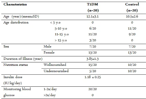
Of the total number of children surveyed, 13 T1DM and 13 of the healthy children were females. The mean age for diabetics versus controls was 12.1±3.1 years versus 10.5±2.6 years (Table 1).
The result of hematological and biochemical
labora-RESULTS AND DISCUSSION
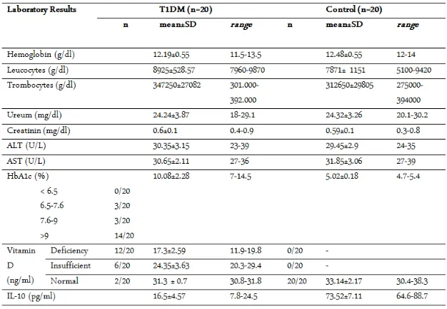
tory measurement are shown in table 2. From the labo-ratory results showed high levels of HbA1c (10.08±2.28), 14 out of 20 had poor metabolic control (HbA1c > 9%), 12 T1DM patients with vitamin D defi-ciency, 6 patients with vitamin D insuffidefi-ciency, and 2 patients with normal level of vitamin D, other result of hematological and biochemical parameters showed as normal level.
Mean of vitamin D (25(OH)D3) level was 33.14±2.17 ng/ml in the normal control, and 20.82±5.53 in T1DM group (p=0.000) (table 3). Mean HbA1c level was 5.02±0.18 % in the normal control, and 10.08±5.02 in T1DM group (p=0.000) (table 3). Mean IL-10 level was 73.52±7.11 pg/ml in the normal control, and 16.50±4.57 in T1DM group (p=0.000) (table 3). There was correlation between 25(OH)D3 level and HbA1c level (p=0.000; r=-0.871), IL-10 level (p=0.000; r= 0.853). HbA1c levels in patients T1DM was correlated with levels of IL-10 (p 0.000; r=-0.878).
This is consistent with previous epidemiological data with peak incidence occurs during puberty [10]. Based on the sex distribution was found that most fe-males (13 out of 20). The incidence of type 1 DM in Indonesia with a sex ratio of female than male 45:55 [11]. The mean insulin dose used in our study was 1.18±0.25. In contrast to the study by Batubara et al11 (1.0±0.3) IU/kg/day, the study group Hvidoere (1.0± 0.3) IU/kg/day [12]. HbA1c levels (HbA1c=10.08± 2.28) was showed poor metabolic control. This is simi-lar to research in Tanzania obtained with a mean HbA1c levels were still high (12.6±3.5 %)13, similarly a study done in Sudan where Elamin et al. found a high incidence of poor glycemic control estimated by HbA1c [14]. Most likely the underlying cause is the as-sociation between limited insulin supply and lack of self-monitoring of blood glucose. Furthermore, in addi-tion to limited insulin supply, patients reduce their in-sulin dose to ensure longer periods of inin-sulin treat-ment. Nevertheless, insulin storage might also influ-ence the effect of the insulin, as many families store the insulin in a pot with cold water, exposing the sulin to high temperatures. Storage of insulin may in-fluence the effects of insulin. Insulin must never be frozen. Direct sunlight or warming (in hot climates) damages insulin. Patients should not use insulins that have changed in appearance (clumping, frosting, pre-cipitation, or discolouration). Unused insulin should be stored in a refrigerator (4-8 0C). After first usage, an insulin vial should be discarded after 3 months if kept at 2-8 0C or 4 weeks if kept at room temperature. However, for some insulin preparations, manufacturers
recommend only 10-14 days of use in room tempera-ture [13-15].
Vitamin D levels of T1DM patients was signifi-cantly lower than healthy controls (p=0.000). Of the 20 subjects of T1DM, 12 out of 20 with a deficiency of vi-tamin D, 6 out of 20 vivi-tamin D insufficiency, and 2 out of 20 with normal levels, whereas all subject in the control group with normal vitamin D levels. Our study similar with study in Switzerland, in a cross-sectional study, 60–84% of T1DM were 25(OH)D3 deficient [16]. In Qatar, in a case control study, 90.6% of T1DM children versus 85.3% of nondiabetic children had vita-min D deficiency [17]. Similarly, in North India in a case-control study, 58% of T1DM and only 32% of controls had 25(OH)D3 deficiency [18]. In Northeast-ern US, in a cross-sectional study, it has been found that 15% of T1DM patients were 25(OH)D3 deficient and 61% were insufficient, findings inversely associated with age Low vitamin D levels in type 1 diabetes is likely to be influenced by immunomodulatory system [19]. Immunomodulatory functions of vitamin D is the ability to inhibit the expression of Th1 cytokines, en-hance the role of Th2 cytokines through directly act on T cells or indirectly through APCs [20]. Vitamin D af-fects Th cell polarization by inhibiting Th1 (IFN-γ pro-duction) and augmenting Th2 cell development (IL-4, IL-5, and IL-10 production). Vitamin D acts directly on Th cells and can, in the absence of APC, enhance the development of a Th2 phenotype and augment the ex-pression of the transcription factors c-maf and GATA-3 [21,22].
There are significant differences in HbA1c levels in T1DM compared to controls and also significant nega-tive correlation between HbA1c levels T1DM and lev-els of vitamin D. High HbA1c levlev-els indicate poor glycemic control. A study in the United States had av-erage high HbA1c levels (9.3±1.9 %) in T1DM with vi-tamin D deficiency. Vivi-tamin D status is one of the fac-tors associated with glycemic control in children and adolescents with T1DM.19 VDR (Vitamin D Receptor) found on pancreatic beta cells, suggesting its role in glycemic control. Vitamin D inhibits inflammatory cy-tokine that is involved in beta cell destruction. Re-search by Magee and colleagues examined the associa-tion between vitamin D and glycemic control, mea-sured by HbA1c in type 1 diabetes in the pediatric population [23].
vitro studies in Turkey whereas levels of IL-10 were lower compared to the control group (p= 0.033) [24]. IL-10 may provide an important part of the anti in-flammatory properties by inhibiting the transcription factor. IL-10 is able to inhibit the nuclear translocation of kB heterodimer. 1,25(OH)2D3 can increase NF-kB inhibitor (INF-kB-α) by increasing mRNA stability and decreasing the phosphorylation of IkB-α. The increase in IkB-α levels reduces nuclear translocation of NF-kB and thereby downgrades its activity [25,26].
There was a significant negative correlation be-tween HbA1c levels and the levels of IL-10. IL-10 was also significantly associated with HbA1c from a study performed by Sawa [27]. IL-10 pathway is reportedly working on phosphatidylinositol 3-kinase (PI3K) which is responsible for insulin action on glucose up-take and suppression of gluconeogenesis [27,28].
In conclusion, the present study revealed that vitamin D deficiency was higher in diabetic children compared to healthy controls. It will be of interest for future studies to investigate whether vitamin D supplementation will improve glycemic control and inflammation status in vitamin D deficient diabetic children.
We would like to thank the Department of Child Health, Faculty of Medicine, University of Brawijaya/ dr.Saiful Anwar General Hospital, Malang, Indonesia for providing the grant to accomplish this research. We also thank to Ajeng S.Si from Pathology Clinics Saiful Anwar General Hospital, and Satuman MSi, from Faal Laboratory of Medical Faculty, Brawijaya University for their good assistance in this research.
1. Achenbach P, Bonifacio E, Koczwara K, Ziegler AG (2005) Natural history of type 1 diabetes. Diabetes. 54(2): 25-31.
2. Rabinovitch A (2004) Roles of Cell-Mediated Immunity and Cytokines in the Pathogenesis of Type 1 Diabetes Mellitus. Diabetes Mellitus: A Fundamental and Clinical Text 3rd Edition. Philadelphia: Lippincott Williams &
4. Mathieu C (2011) Vitamin D and the Immune System:
Getting It Right. International Bone & Mineral Society. 8: 178-186.
5. Mathieu C, Gysemans C, Giulietti A, Bouillon R (2005) Vitamin D and diabetes. Diabetologia. 48(7): 1247-57. 6. Bouillon R, Geert C, Lieve V, Evelyne VE, Annemieke V,
Hilary FL (2008) Vitamin D and Human Health: Lessons from Vitamin D Receptor Null Mice. Endocr Rev. 29(6): 726–776.
7. Handono K, Puspitasari L, Rudijanto A, Wahono S, Kalim H (2013) Vitamin D Serum Level And Disease Ac-tivity In Patients With Systemic Lupus Erythematosus. In-ternational Journal of Pharmaceutical Science Invention. 2(2): 35-40.
8. Cahyono HA (2011) Gambaran Klinis dan Laboratoris Diabetes Melitus Tipe 1 pada Anak. Jurnal Kedokteran Brawijaya. 26(4): 195-198.
9. Holick MF (2007) Vitamin D deficiency. N Engl J Med. 357(3): 266-81.
10. Soltesz G, Patterson CC, Dahlquist G (2007) Worldwide childhood type 1 diabetes incidence–what can we learn from epidemiology?. Pediatric Diabetes. 8(6): 6–14. 11. Batubara JRL (2002) Audit of childhood diabetes control
in Indonesia. Paediatrica Indonesiana. 42(11-12): 280-286. 12. De Beaufort CE, Swift PGF, Skinner CT, Aanstoot HJ,
Aman J, Cameron F (2007) The Hvidoere study group on childhood diabetes. Continuing stability of cener differ-ences in pediatric diabetes care : Do advances in diabetes treatment improve outcome?. Diabetes Care. 9: 2245-50. 13. Majaliwa ES, Munubhi E, Ramaiya K, Mpembeni R,
Sanyiwa A, Mohn A, Chiarelli F (2007) Survey on Acute and Chronic Complications in Children and Adolescents with Type 1 Diabetes at Muhimbili National Hospital in Dar es Salaam, Tanzania. Diabetes Care. 30(9): 2187-92. 14. Elamin A, Hussein O, Tuvemo T (2006) Growth, puberty
and final height in children with Type 1 diabetes. J Dia-betes Complications. 20(4): 252-6.
15. ISPAD (2011) Diabetes in Childhood and adolescence. IS-PAD Clinical Practice Consensus Guidelines. p 61 16. Janner M, Ballinari P, Mullis PE, Flick CE (2010) High
prevalence of vitamin D deficiency in children and adoles-cent with type 1 diabetes. Swiss Med Wkly. 140(7): 1-6. 17. Bener A, Alsaied A, Al-Ali M, Al-Kubaisi A, Basha B,
Abraham A, Guiter G, Mian M (2009) High prevalence of vitamin D deficiency in type 1 diabetes mellitus and healthy children. Acta Diabetologica. 46: 183–9.
18. Borkar VV, Devidayal VS, Bhalla AK (2010) Low levels of vitamin D in North Indian children with newly diagnosed type 1 diabetes. Pediatric Diabetes. 11(5): 345–50. 19. Svoren BM, Volkening LK, Wood JR, Laffel LMB (2009)
Significant vitamin D deficiency in youth with type 1 dia-betes mellitus. J of Peds. 154(1): 132–134.
ACKNOWLEDGMENT