Pigmented Oral Lichen Planus:
A Case Report
Firstine Kelsi Hartanto1, Thomas George Kallarakal2
1Department of Oral Medicine, Faculty of Dentistry, Trisakti University–Indonesia
2Department of Oro-maxillofacial Surgical & Medical Sciences, Faculty of Dentistry, University of Malaya–Malaysia
‘Corresponding Author: Firstine Kelsi Hartanto, Faculty of Dentistry, Trisakti University –Indonesia. Email: [email protected]
Received date:July 05, 2017.Accepted date:September 11, 2017.Published date:September 29, 2017.
Copyright: ©2017 Hartanto FK, Kallarakal TG. This is an open access article distributed under the terms of the Creative Commons Attribution License, which permits unrestricted use, distribution, and reproduction in any medium provided the original author and sources are credited.
Background: Lichen planus is a chronic muccocutaneous inflammatory disorder. Oral lichen planus (OLP) has certain specific characteristics in its clinical presentation, but can also be presented in forms resembling other diseases. This may introduce difficulty in the diagnostic process. It has been reported that OLP affects between 0.1 to 4% individuals, predominantly women and people over 40 years old.Case Report:A 46-year-old woman complained of a non-healing ulcer present for 1.5 months. Intraorally, multiple white striae with prominent central areas of brownish hyperpigmentation were apparent on the right and left buccal mucosa, right and left buccal sulcus, and lateral and dorsum of the tongue. A biopsy was completed and the histopathology features confirmed the diagnosis of OLP. For the initial treatment, a topical corticosteroid was prescribed, followed by steroid mouthwash. Response to this treatment was positive. OLP has been frequently reported to affect women over the age of 40, with psychological stress as a primary predisposing factor. The clinical presentation is characteristic of Wickham’s striae with erosive areas. However, a biopsy and histopathological examination is mandatory to confirm the diagnosis. The clinical feature of pigmented OLP has been reported and confirmed by microscopic finding of band-like lymphocytic appearance which is the pathognomonic features of OLP, along with basal cell liquefaction degeneration, and melanin in continence at the lamina propria.Conclusion:Pigmented OLP is a variant of erosive OLP. Anamnesis, clinical presentation, and histopathological examination confirm diagnosis.
Keywords : hyperpigmentation, oral lichen planus
Lichen planus is a relatively common
inflammatory disorder of the skin and mucous
distribution and distinct clinical presentation, but may also present as a form or pattern is similar to other diseases. This makes diagnosing based exclusively on clinical features is challenging.1
In 1869, Erasmus Wilson first described Lichen planus and reported the oral involvement of the lesion.2
In general, the etiopathogenesis of lichen planus is complex, including interactions between and among, genetic, environmental, and lifestyle factors.1 Systemic
medications, dental materials, chronic liver disease, the hepatitis C virus, stress, tobacco chewing, and the graftand graft-versus host diseases have also been identified as factors contributing to the etiology of OLP.3
Studies have shown that OLP affects between 0.1 to 4% of individuals.1 It occurs more frequently in
women than men, with a ratio of 1.4:1, and in adults over the age of 40, although it can also affect younger adults and children.4 Intraorally, OLP might present
itself as white striae, white papules, white plaques, erythema, erosions, or blisters. The buccal mucosa is the most common site affected by OLP, followed by the tongue, gingiva, and hard palate. The lesions are usually bilateral, and in the majority of cases, two or more sites are involved.5,6,7
Pigmentation associated with OLP is frequently observed. Cawson (2008) reported that the degenerative changes in basal keratinocytes frequently led to pigmentary incontinence. The melanin in pigment is ingested by macrophages in the superficial corium, resulting in a brownish-pigmented area in the mucosa, which can persist long after the OLP has resolved.8
A 46-year-old woman complained about non-healing ulcers on her right and left inner cheeks, persisting for approximately 1.5 months. The ulcers were associated with a burning sensation when subjected to spicy foods and minty toothpaste. Past medical history revealed ongoing follow-up treatments for a nodular goiter. She was not taking any medications. Extaroral examination showed no skin lesions, and the right and left submandibular
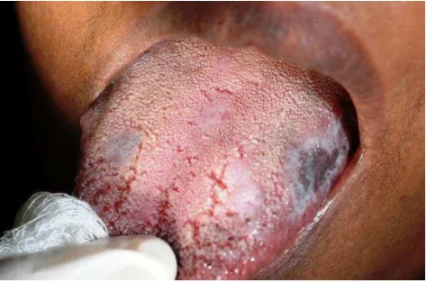
lymph nodes were not palpable. Intraorally, multiple white striae with prominent central areas of brownish hyperpigmentation were apparent on the right and left buccal mucosa (Fig.1). Whitish plaque-like lesions with hyperpigmentation lines radiating from peripheral to central were observed on the dorsum and lateral borders of the tongue (Fig.2). A similar whitish lesion was also detected at the right and left lower posterior buccal sulcus. This evidence led to a clinical diagnosis of OLP; an incisional biopsy was taken from the lesion on the left buccal mucosa to confirm the diagnosis.
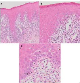
Microscopic findings showed a surface of parakeratinized stratified squamous epithelium exhibiting liquefaction degeneration of basal cells with thickening of the basement membrane zone in focal areas, and numerous civatte bodies within the lower 1/3rdepithelium. The underlying fibrous
connective tissue showed a mildly diffuse, band-like appearance of mixed acute and chronic inflammatory infiltrate consisting of eosinophils, lymphocytes, and plasma cells. Scattered melanin incontinence in the lamina propria was also observed (Fig.3).
Based on the clinical and histopathological
examination, the diagnosis of OLP was
determined and confirmed. For treatment, the patient was given 1 mg Dexamethasone oral paste to apply to the lesions, and was advised to avoid spicy foods. At the one-month follow-up, the lesion had not regressed, thus a use of a steroid mouth rinse (Dexamethasone 0.5 mg to dissolve in 15 ml water) for two weeks was prescribed. Non-sodium lauryl sulphate toothpaste was also suggested.
Figure 1. White anular striae with a central area of brownish hyperpigmentation and erythematous background on the right (A) and left (B) buccal mucosa, which also extended to buccal sulcus.
hyper-Figure 3.(A) Oral epithelium exhibits basal cells liquefaction degeneration with a band-like inflammatory infiltrate in the lamina propria, (B) Scattered melanin in continence observed within the inflammatory cells, (C) Civatte bodies (arrow) (H&E, original magnification Ax10, Bx20, Cx40).
Lichen planus is well known as a chronic
complex mucocutaneous disease of unknown
etiology, uncertain pathogenesis, and varied histopathological features. It can occur across the lifespan, including infants under 6 months through adults above the age of 80 years. Although both men and women are affected2, there are somewhat higher
prevalence rates in women.6 In our case, the patient
was a female in the 4th decade of life. This age
coincides with research by Chitturi et al that OLP occurs mostly between the ages of 41 and 70 years.5
There are six recognized clinical types of OLP including reticular, papular, plaque, atropic, ulcerative, and erosive.9 Among these types, the
reticular form is the most common.5, 10, 11Our patient
also presented with the reticulo-annular type, as well
as the plaque-like type. Buccal mucosa is a common
site affected by this disease for both types of reticular and erosive OLP, while the tongue, gingiva, lip and palate are predominantly affected by erosive type.12
Although the etiopathogenesis is uncertain, physical stress appeared to be an important etiologic factor in 90% of OLPs.8 In addition,
foods, dental procedures, systemic diseases, and poor oral hygiene have also been indicate as precipitating factors of OLP.9,13 In our case,
we observed stress is the etiology and spicy foods as the precipitating factor.
be demonstrated by Periodic Acid Schiff (PAS) stain2, can disrupt the basement membrane. In
our case, these features were observed.
A study by Anjum et al., showed melanin pigmentation in the basal layer with evidence of extension to the upper part of the lamina propria2Our
case also showed melanin pigmentation in the basal layer and upper part of the lamina propria. Another study showed clinical observation of 67.24% hyperpigmentation in the OLP5. There are various
reasons for oral mucosal pigmentation, such as race, smoking, stress, anxiety, Adison’s disease, and post-inflammatory changes.14 A study found that
96.97% patients with history of anxiety disorders showed OLP associated with hyperpigmentation.
The proposed mechanism of hyperpigmentation is related to the GABA imbalance that occurs during stress and anxiety. This imbalance occurs in anxiety disorders and might be passed through the cranial nerve, stimulating production of melanocytes, resulting in excessive deposition in OLP.2Other work
suggested that hyperpigmentation was caused by post-inflammatory changes in recurrence flare-up and healing, which was mostly found in the chronic reticular form of OLP.5 Unfortunately, we could not
exactly determine the mechanism of hyper-pigmentation in our case due to lack of information from the history.
Studies that focused on the skin suggested that the increased stimulation of melanocytes to produce melanin occurred after an inflammatory reaction, and this, along with the presence of melanophages contributed to the clinical presentation of hyper-pigmentation in OLP.15, 16, 17The exact mechanism of
hyperpigmentation in OLP remains unclear, but it has been suggested that the cytokines released by the band of lymphocytes stimulate the melanocytes.18 Further
studies are needed to comprehend the occurrence of hyperpigmentation in OLP through histopathological, immunohistochemistry, and biological aspects . The limitation in our case is we could not provide sufficient information regarding the etiology of hyperpigmentation present in OLP. However, it is important in every case of erosive OLP to confirm the clinical diagnosis with histopathology result.
I express my deep gratitude to Prof. Rosnah Zain, Prof. Siar Chong Huat, and my colleagues at Department of Oro-maxillofacial Surgical and Medical Sciences, University of Malaya for the guidance and encouraging me during my training.
1. Y., Griffiths, M., Holmstrup, P., Mutlu, S., Porter, S. & Wray, D.. Update On Oral Lichen Planus: Etiopathogenesis and Management. Crit Rev Oral Biol Med.1998;9;86-122.
2. Anjum R, Singh J & Kudva S. A
Clinicohistopathologic Study and Probable Mechanism of Pigmentation in Oral Lichen Planus. World J Dent. 2012;3 :330-334.
3. Ismail, S. B., Kumar, S. K. S. & Zain, R. B. Oral lichen planus and lichenoid reactions: etiopathogenesis, diagnosis, management and malignant transformation. J Oral Sci .2007;49:89-106.
4. Sugerman, P. B., Savage, N. W., Walsh, L. J., Zhao, Z. Z., Zhou, X. J., Khan, A., Seymour, G. J. & Bigby, M. The Pathogenesis of Oral Lichen Planus. Crit Rev Oral Biol Med2002;13;350-365. 5. Chitturi RT, Sindhuja P, Parameswar RA, et al. A
clinical study on oral lichen planus with special emphasis on hyperpigmentation. J Pharm Bioallied
Sci.2015;7(Suppl 2):S495-S498.
doi:10.4103/0975-7406.163513.
6. Shen ZY, Liu W, Zhu LK, Feng JQ, Tang GY, Zhou ZT. A retrospective clinicopathological study on oral lichen planus and malignant transformation: Analysis of 518 cases. Med Oral Patol Oral Cir Bucal.2012;17:e943–7
7. Budimir V, Richter I, Andabak-Rogulj A,
Vucicevic-Boras V, Budimir J, Brailo V. Oral lichen planus – Retrospective study of 563 Croatian patients. Med Oral Patol Oral Cir Bucal.2014;19:e255–60.
8. Cawson, R.A & Odel, E.W. Essentials of Oral Pathology and Oral Medicine. 8th ed. Churchill
Livingstone.Edinburgh. 2008
10. Bajaj DR, Khoso NA, Devrajani BR, Matlani BL, Lohana P. Oral lichen planus: A clinical study. J Coll Physicians Surg Pak. 2010;20:154–7.
11. Ingafou M, Leao JC, Porter SR, Scully C. Oral lichen planus: A retrospective study of 690 British patients. Oral Dis. 2006;12:463–8.
12. Bagán-Sebastián, J. V., Milián-Masanet, M. A., Peñarrocha-Diago, M. & Jiménez, Y. 1992. A clinical study of 205 patients with oral lichen planus. J Oral Maxillofac Surg, 50,116-118. 13. Eisen, D. The clinical features, malignant potential,
and systemic associations of oral lichen planus: A study of 723 patients. J Am Acadof Dermatol 2002;46,207-214.
14. Kauzman A, Pavone M, Blanas N, Bradley G. Pigmented lesions of the oral cavity: Review, differential diagnosis, and case presentations. J Can Dent Assoc. 2004;70:682–3
15. Patsakas A, Demetriou N, Angelopoulos A.
Melanin pigmentation and inflammation in
human gingiva J Periodontol. 1981;52:701–4 16. Morelli JG, Norris DA. Influence of inflammatory
mediators and cytokines on human melanocyte function. J Invest Dermatol. 1993;100:191S–5 17. Nordlund JJ. Postinflammatory
hyperpigmen-tation. Dermatol Clin. 1988;6:185–92