Introduction
Creatinine is produced in the muscles by the nonenzymatic changes of creatine and phosphor creatinine. The liver has a momentous role in the assembly of creatinine through methylation of guanidine aminoacetic acid. The normal serum creatinine level is 0.5 to 1.0 mg/dL according to diurnal and menstrual variations, pursuit, and diet. Creatinine is used to assess renal function, however, serum creatinine level will not increase until renal function has decreased by at least 50% (Hamilton et al., 1972). In chronic renal failure there is a steady and continued decrease in renal clearance or glomerular filtration rate (GFR), which leads
to the gathering of urea, creatinine and other chemicals in the blood (Leveyet al., 2005).
Hemodialysis is one of the renal replacement therapy. In this technique, body waste product like urea, creatinine and free water are removed from the blood, when the kidneys are impaired. The principle of hemodialysis is the diffusion of solutes through a semi permeable membrane (Abrahamet al., 2012). Early dialysis membranes were based on cellulose, with cuprophane (a copper substituted cellulose) being one of the most commonly used early membranes. These were cheap to produce and had advantages of being thinwalled. However, they had the disadvantage of being immunoreactive. They incited an inflammatory response in the patients with activation of complement, neutrophil superoxide production and cytokine induction (such as interleukin (IL) 1) and tumor necrosis factor. This contributed to dialysis symptoms and intolerance, and in the long term may have contributed to dialysis
Study of Creatinine Transport through
Chitosan/Pectin/Poly(Vinyl Alcohol) Blend Membranes
Ni Putu Sri Ayuni
1,*, Ni Wayan Yuningrat
11Faculty of Mathematics and Natural Sciences, Universitas Pendidikan Ganesha,
Jalan Udayana No.11 Singaraja, Bali 81116, Indonesia
Abstract
Creatinine was the final product of creatine metabolism inskeletal muscle. Increasing of creatinine showed the decreasing of kidney function. Kidney function decreased could be treated by hemodialysis (HD). One of the natural polymer was cellulose which often used as hemodialysis membrane. Chitosan as natural membrane was used as membrane in this research because the structure was almost similar to cellulose. Chitosan was complexed by pectin and poly(vinil alcohol) (PVA) to fixed mechanical characteristic of a membrane. The objective of this research was to synthesize, characterize, and know efficiency of creatinine transport using chitosan/pectin/PVA blend membranes. The functional groups of synthesized membrane were characterized by FTIR spectrophotometer. The efficiency of optimum creatinine transport was observed by using membrane with chitosan and pectin ratio 70:30 while PVA used 0.5%; 1.0% and 1.5%. The source and acceptor phase resulted were complexed by picric acid and analyzed byUltravioletVisible(UVVis) Spectrophotometer. The result of membrane synthesized which was analyzed by FTIR Spectrophotometer showed that there is a band broadening on wave number 3448.72 cm1. It is indicated that there are an overlapped stretching of hydrogen bond OH on PVA and
NH2 on chitosan. The optimum creatinine 70 mg/L transported using a membrane with 1.5% PVA addition is 59%.
Keywords: Chitosan, creatinine, membrane, pectin, poly(vinyl alcohol)
*Corresponding author: Ni Putu Sri Ayuni
Faculty of Mathematics and Natural Sciences, Universitas Pendidikan Ganesha, Jalan Udayana No.11 Singaraja, Bali 81116, Indonesia
related cachexia. They were also only available in lowflux form. They only allowed the passage of small molecules with very few molecules above a molecularweight (MW) of 5000 gaining passage through the membrane (Kerr and Huang, 2010).
Reported by Ayuni (2013) that the weakness of cellulose membrane characteristic was solved by using chitosanpectin PEC membrane which yielded transportefficiency 25.24%. Chitosan is a second abundant natural polysaccharide after cellulose. Chitosan [poly (b1/4)2amino2deoxyDglucopyranose] is a collective name for a group of partially and fully deacetylated chitin compounds. Due to its biological characteristics unique, including biodegradability, nontoxicity and antimicrobial. Pectin is an edible and water soluble polysaccharide which consists primarily of dgalacturonic acid with a part of the carboxyl groups being methoxylated. It is widely used in food industry, and its potential for biomedical applications has been also investigated. Ionic interactions occur between polyanions and polycations, leading to the formation of a polyelectrolyte complex (PEC). The other types of interactions that can form between amino groups (chitosan) and carboxyl groups (pectin), such as hydrogen bonds and covalent bonds formed using specific conjugating chemicals (Richertet al., 2004). There is only ionic interaction in chitosanpectin PEC membrane so that the membrane is not selective to creatinine.This weakness can be solved by synthetic polymer addition like PVA. PVA is a water soluble synthetic polymer. Due to the characteristics of easy preparation, good biodegradability, excellent chemical resistance, and good mechanical properties, PVA has been used in many biomaterial applications (Park and Ruckenstein, 2001). Chitosan contains hydroxyl and amine groups, which is potentially miscible with PVA due to the formation of hydrogen bonds (Miya et al., 1983). The crosslinking reaction of chitosan with pectin is done in order to incorporate the reactive COOH groups of pectin to the backbone chain of chitosan. The substitution is expected to increase the active sites on the
surface of chitosan membrane, through creatinine can be transported via hydrogen bonds formation. The mechanical properties of the membrane can be improved by decreasing the chitosan cross linked with pectin hydrophilicity. It can be achieved by mixing the composite with the PVA. The appropriate ratio of the hydrophilic groups and the hydrophobic groups is expected to produce a membrane with high mechanical strength and flexibility (Lusianaet al., 2016). The objective of this research was to synthesize, characterize, and measure the efficiency of creatinine transport using chitosan/pectin/PVA membrane. The functional groups of synthesized a membrane were characterized by FTIR Spectrophotometer. The efficiency of optimum creatinine transport was observed by using membrane with chitosan and pectin ratio70:30 while PVA addition 0.5%; 1.0% and 1.5%. The source and acceptor phase resulted were complexed with picric acid and analyzed by UVVisible Spectrophotometer.
Materials and Methods
Materials
Chitosan, poly (vinyl alcohol), pectin, aquadest, 0.4 M acetic acid solution (CH3COOH), 0.4 M sodium hydroxyde (NaOH) solution, 8.8 mM picric acid, 0.1 M hydrochloric acid, phosphate buffer solution pH 7.4. All chemicals were analytical grade and purchased from P.T. Merck.
Methods
Synthesis of chitosan/pectin/PVA blend membranes
dissolved in 4 mL aquadest, and then pectin solution was added to 0.07 g chitosan and PVA solution (Table 1). This mixture was stirred for 24 h at room temperature. About 10 mL of the solution was put into petri dish and dried in the oven at temperature 70°C. Afterwards, 0.4 M NaOH was added to remove the membrane from petri dish. The membrane was washed by distilled water until neutral and then dried in open air.
Characterizations of chitosan/pectin/PVA blend membranes
The properties of the membrane were determined by analyzing the material (chitosan, pectin, PVA) before and after blending. Functional groups were obtained by using FTIR Spectrophotometer (Shimadzu).
Water uptake test
To measure the water uptake capability of the porous membranes, the wet weight (Wwet) of a membrane was measured, and then the wet membrane was dried in an oven at 60ºC for 24 h, followed by vacuum drying for 24 h. The dry weight (Wdry) of the membrane was immediately measured. The water uptake was calculated using Eq. 1.
Study of creatinine transport with
chitosan/pectin/PVA blend membranes
Transport test was performed using a transport device with the membrane sited in the middle of device. Transport process experiment was done by using the following creatinine concentration 70, 100, and 130 mg/L. The source phase contained 50 mL creatinine standard solution with phosphate buffer addition, while the acceptor phase only contained 50 mL aquadest and phosphate
buffer. The transport tests were carried out for 6 h with samples from source and acceptor phase taken out every 2 h for analysis using UVVis spectrophotometer. For the purpose of analysis, creatinine was initially complexed with picric acid. This complex solution was measured at 516 nm. Each membrane and concentration variation was performed twice repetition. The transport efficiency creatinine (E) was calculated using Eq. 2.
Results and Discussion
Synthesis and characterization of chitosan/ pectin/PVA blend membranes
The membranes were synthesized using phase inversion method, in which the liquid phase is converted to a solid phase by evaporating the solvent by gradual heating at a temperature range of 40–70°C for 24–48 h (Lusiana et al., 2016). Membranes was characterized of its functional groups by FTIR spectrophotometer and water uptake.
FTIR spectra analysis
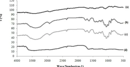
The membrane functional groups were characterized by using FTIR spectrophotometer (Figure 1). In the IR spectra
Table 1. Composition of chitosan, pectin, and PVA.
chitosan, stretching vibration of the amide group of chitosan appears at 1604.77 cm1. The change in the characteristic shape of the chitosan spectrum as well as shifting of peak to a lower frequency range due to hydrogen bonding between –OH of PVA and –OH or –NH2of chitosan is evidentin membrane. In the spectrum of membrane two relatively broadbands appear in the region 2000–1600 cm1 which can be ascribed to the overlapping of bands due to amino and carboxylic groups of the interacting molecules. A strong peak at 1635.64 cm1 (asymmetric stretching vibration of carboxylate) also indicates the interchain or intermolecular ionic salt bonds, i.e. polyelectrolyte complex (PEC) between amino groups of chitosan and carboxyl groups of pectin (Rashidovaet al., 2004). In the other words, it has been assumed that PEC formation proceeded at the expense of electrostatic interaction between the positively charged amino groups on C2 of the chitosan pyranose ring and the negatively charged carboxyl groupson the C5 of the pectin pyranose ring. Absorption at 1273.30 and 1327.07 cm1wave numbers only appear on the membrane spectrum. It is thought to be derived from an ester group showing alginate and PVA crosslinked (Lusianaet al., 2013). From the FTIR results, the hypothetical structure of the membrane is presented in Figure 2.
Water uptake
For materials contacting human blood, the balance between hydrophilic and hydrophobic was important (Lusianaet al., 2016). Water absorption of the membrane depends on the structure and composition of the polymer membrane (Huangfu et al., 2009). In this study, membranes were made
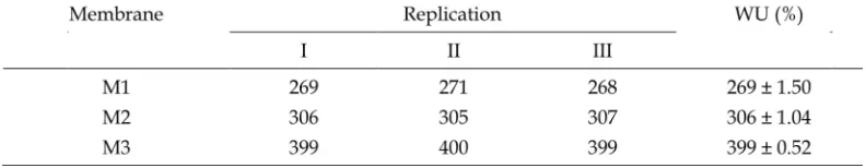
of three different PVA addition 0.5% (M1), 1% (M2), and 1.5% (M3). The water uptake (Table 2 and Figure 3) indicated that membranes with 1.5% PVA have the highest water absorption capacity, which amount to 399%.
This value relates to the ability of water diffusion to all parts of the membrane.The appropriate ratio of the hydrophilic groups of chitosan and the hydrophobic groups of PVA is expected to produce a membrane with high mechanical strength and flexibility(Lusianaet al., 2016). This is due to the fact that when a biopolymer contacts with the liquid, there is
Figure 2. Hypothetical structure of the membrane.
Table 2. Water uptake percentage with different composition membrane.
a development due to the corresponding thermodynamics and the presence of tensile forces caused by the cross effect that occurs in the polymer chain. As the membrane expands, the mobility of the polymer chain increases to facilitate the penetration of the solvent, in addition to the small ions trapped within the diffused membrane leaving the membrane thus providing a greater opportunity for the solvent to fill in the abandoned empty spaces. The development of the chitosanalginate membrane is probably due to the ionic interaction between the NH3+group of chitosan and the COOions of the membrane alginate (Dash et al., 2011; Sagnella and MaiNgam, 2005). The blend membranes usually exhibited higher wáter absorption degree then the
chitosanpectin membrane (255%) (Ayuni, 2013), indicating a more flexible membrane structure (Huangfuet al., 2009; Gaoet al., 2014).
Study of creatinine transport with chitosan/ pectin/PVA blend membranes
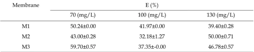
From Figure 46 and Table 3, it can be seen that the longer the transport time, the higher the transport percentage. Furthermore, the transport efficiency percentage tends to increase with increasing amount of PVA in the membrane. It can be seen from Figure 3, that the greater content PVA in blended membranes will increase the ability water uptake of membrane.
In other words, increasing PVA content in the membrane so that increasing
Figure 4. Transport efficiency creatinine 130 mg/L. Figure 5. Transport efficiency creatinine 100 mg/L.
Figure 6. Transport efficiency creatinine 70 mg/L.
Table 3. Creatinine efficiency transport for membranes at transport time 6 h.
the hydrophilicity of the membrane. It is also correlated with an increase in creatinine transport capability. This might be due to the fact that the addition of PVA into chitosanpectin membranes is able to improve membrane hydrophilicity. It can also be seen from Figure 7 that membrane composition with 1.5% PVA addition (M3) is the highest percentage transport efficiency of creatinine.
The right blend of hydrophilic groups and hydrophobic membrane will result in a certain hydrophilicity values with optimum performance. Appropriate range values of hydrophilicity can make the membrane work in the transport process optimally. Hydrophilicity of the membrane plays a role in determining transport efficiency of a membrane. If membrane hydrophilicity achieves right composition so that the membrane will expand according to optimal water content capacity. Water contained in the membrane transport process is a means to permeate through the membrane. The optimum equivalent ratio of chitosanpectin with PVA addition were determined by using the membranes for creatinine transport and water uptake percentage.
Conclusions
Chitosan/pectin/poly(vinyl alcohol) blend membranes were prepared successfully by inverse phase method. FTIR spectra at 1635.64 cm1of the membrane have indicated the interchain or intermolecular ionic salt bonds, i.e. polyelectrolyte complex (PEC) between amino groups of chitosan and carboxyl groups of pectin. Absorption at 1273.30 and 1327.07 cm1wave numbers only appears on the membrane spectrum, this is thought to be derived from an ester group showing crosslinking between alginate and PVA. Creatinin 70 mg/L which is transported by a membrane with 1.5% PVA addition yields optimum transport efficiency 59%.
Acknowledgements
This research was financially supported by the Technology Research and Higher Education through the Beginner Lecturer
Research scheme 2016.
References
Abraham, S., Venu, A., Ramachandran, A., Chandran, P.M., and Raman, S. 2012. Assessment of quality of life in patients on hemodialysis and the impact of counseling. Saudi J. Dis. Transpl., 23, 953–957.
Ayuni, N.P.S, and Siswanta, D.A.S. 2013. Kajian transpor kreatinin menggunakan membran kompleks polielektrolit (PEC) kitosanpektin. Wahana Matematika dan Sains., 8(1), 88–96.
Dash, M., Chiellini, F., Ottenbrite, R.M., and Chiellini, E. 2011. Chitosan A versatile semisynthetic polymer in biomedical applications. Prog. Polym. Sci., 36, 981–1014.
Gao, A., Liu, F., and Xue, L. 2014. Preparation and evaluation of heparinimmobilized poly (lactic acid) (PLA) membrane for hemodialysis. J. Membr. Sci., 452, 390–399.
Hamilton, R.W., Gardner, A., Penn, A.S., and Goldberg, M. 1972. Acute tubular necrosis caused by exerciseinduced myoglobinuria. Ann. Intern. Med., 77, 77–82.
Huangfu, P., Gong, M., Zhang, C., Yang, S., Zhao, J., and Gong, Y. 2009. Cell outer membrane mimetic modification of a crosslinked chitosan surface to improve its hemocompatibility. Colloids Surf., B., 71, 268–274.
Kerr, P.G., and Huang, L. 2010. Review: membranes for haemodialysis. Nephrology., 15, 381–385.
Levey, A.S., Eckardt, K.U., Tsukamoto, Y., Levin, A., and Coresh, J. 2005. Definition and classification of chronic kidney disease: a position statement from kidney disease: improving global outcomes (KDIGO)z’. Kidney Int., 67, 2089–2100.
Appl., 7, 186–189.
Lusiana, R.A., Siswanta, D., and Hayashita, T. 2013. The influence of PVA. cl. citric acid/chitosan membrane hydrophicility. Indo. J. Chem., 13, 262–270.
Lusiana, R. A., Siswanta, D., and Mudasir. 2016. Preparation of citric acid crosslinked chitosan/poly(vinyl alcohol) blend membranes for creatinine transport. Indo. J. Chem., 16, 144–150. Miya, M., Yoshikawa, S., Iwamoto, R., and
Mima, S. 1983. Mechanical properties of poly(vinyl alcohol)chitosan blend films. Kobunshi Ronbunshu. 645–651. Park, J.S., Park, J.W., and Ruckenstein, E. 2001. Thermal and dynamic mechanical analysis of PVA/MC blend hydrogels. Polymer., 42, 4271–4280.
Rashidova, S.S., Milusheva, R.Y., Semenova, L.N., Mukhamedjanova, M.Y., and Voropaeva, N.L. 2004. Characteristics of interactions in the pectinchitosan system. Chromatographia., 59, 779–782. Richert, L., Boulmedais, F., Lavalle, P., Mutterer, J., and Ferreux, E. 2004. Improvement of stability and cell adhesion properties of polyelectrolyte multilayer films by chemical cross linking. Biomacromolecules., 5, 284–294.