www.elsevier.com / locate / bres
Research report
Vasopressin impairs K
ATPand K
cachannel function after brain injury
*
Alana Salvucci, William M. Armstead
Departments of Anesthesia and Pharmacology, University of Pennsylvania, Philadelphia, PA 19104, USA
Accepted 3 October 2000
Abstract
This study was designed to characterize the role of vasopressin in impaired pial artery dilation to activators of the ATP sensitive K (KATP) and calcium sensitive K (K ) channel following fluid percussion brain injury (FPI) in newborn pigs equipped with a closedca cranial window. Topical vasopressin was coadministered with the KATP and Kca channel agonists cromakalim and NS1619 in a
1
concentration approximating that observed in CSF following FPI. Vasopressin so administered attenuated pial artery dilation to these K channel activators under conditions of equivalent baseline diameter during non injury conditions (1361 and 2361 vs. 461 and 1062%
28 26
for cromakalim 10 , 10 M before and after vasopressin, respectively). Attenuated responses were fully restored when these agonists were coadministered with vasopressin and the vasopressin antagonist [l-(b-mercapto-b,b-cyclopentamethylene propionic acid) 2-(o-methyl)-Tyr-AVP] (MEAVP). Cromakalim and NS1619 induced pial artery dilation was attenuated following FPI and MEAVP preadministration partially prevented such impairment (1361 and 2361, sham control; 261 and 561, FPI; and 961 and 1562%,
28 26
FPI-MEAVP pretreated for responses to cromakalim 10 , 10 M, respectively). These data show that vasopressin blunts KATPand Kca channel mediated cerebrovasodilation. These data suggest that vasopressin contributes to impaired KATP and Kca channel function after brain injury. 2000 Elsevier Science B.V. All rights reserved.
Theme: Disorders of the nervous system
Topic: Trauma
1
Keywords: Newborn; Cerebral circulation; K Channel; Vasopressin; Brain injury
1. Introduction activators and inhibitors have provided functional evidence
1 1
that K channels, especially ATP sensitive K (KATP) and
1 12
Relaxation of blood vessels can be mediated by several calcium sensitive K (Kca ) channels, regulate tone of
1
mechanisms, including cGMP, cAMP, and K channels cerebral blood vessels in vitro and in vivo [16]. While
1
[16]. Membrane potential of vascular muscle is a major several recent studies have characterized the role of K
1
determinant of vascular tone, and activity of K channels channels in cerebrovascular control under physiological is a major regulator of membrane potential [20]. Activation conditions, less is known concerning their contributions
1
or opening of these channels increases K efflux, thereby under pathological conditions.
producing hyperpolarization of vascular muscle. Mem- Traumatic brain injury is one of the major causes of brane hyperpolarization closes voltage dependent calcium morbidity, mortality, and pediatric intensive care unit channels and thereby causes relaxation of vascular muscle admissions of children today [22,25]. Although the effects [19,20]. Direct measurements of membrane potential and of traumatic brain injury have been well described for
1
K current in vitro indicate that several different types of adult animal models [12,17,18,27], few have investigated
1
K channels are present in cerebral blood vessels. In these effects in the newborn. To reproduce some of the addition, a number of pharmacological studies using biomechanical aspects of closed head injury, fluid percus-sion brain injury (FPI) has been used in the adults of several species [17,18]. More recently, Prins et al. [21] *Corresponding author. Tel.: 11-215-573-3674; fax: 1
1-215-349-characterized the effects of FPI on several parameters 5078.
E-mail address: [email protected] (W.M. Armstead). including mortality, intracranial pressure, and mean arterial
blood pressure in the developing and adult rat. Other CSF. Pial arterial vessels were observed with a dissecting earlier studies had compared the cerebral hemodynamic microscope, a television camera mounted on the micro-effects of FPI in newborn (1–5 days old) and juvenile scope, and a video output screen. Vascular diameter was (3–4 weeks old) pigs. For example, it was observed that measured with a video microscaler.
pial vessels constricted more, and that regional cerebral Methods for brain FPI have been described previously blood flow decreased and remained depressed longer, in [27]. A device designed by the Medical College of newborns than in juveniles [8]. Virginia was used. A small opening was made in the Vasopressin contributes to the regulation of cerebral parietal skull contralateral to the cranial window, also 0.5 hemodynamics in the piglet [9–11]. Vasopressin is released cm from begma. A metal shaft was sealed into the opening into CSF by FPI and also contributes to impaired pial on top of intact dura. This shaft was connected to the artery dilation to opioids such as dynorphin following such transducer housing, which was in turn connected to the an insult in the piglet [5]. Dynorphin elicits pial vasodilat- fluid percussion device. The device itself consisted of an ion via activation of KATP and Kca channels [3,24]. Since acrylic plastic cylindrical reservoir 60 cm long, 4.5 cm in KATPand Kcachannel function is impaired after FPI [2,4], diameter, and 0.5 cm thick. One end of the device was altered dilation to this opioid could relate to such impaired connected to the transducer housing, whereas the other end
1
K channel function. Interestingly, vasopressin has been had an acrylic plastic piston mounted on O-rings. The observed to block the KATP channel in porcine coronary exposed end of the piston was covered with a rubber pad. artery smooth muscle cells [26]. However, vasopressin did The entire system was filled with 0.9% saline. The not appear to have any direct effect on the Kca channel in percussion device was supported by two brackets mounted the above studies [26]. on a platform. FPI was induced by striking the piston with Therefore, the present studies were designed to char- a 4.8-kg pendulum. The intensity of the blow (usually acterize the role of vasopressin in impaired pial artery 1.9–2.3 atm with a constant duration of 19–23 ms) was dilation to activators of KATP and Kca channels following controlled by varying the height from which the pendulum FPI in the newborn pig. was allowed to fall. The pressure pulse of the blow was recorded on a storage oscilloscope triggered photoelectri-cally by the fall of the pendulum. The amplitude of the
2. Methods pressure pulse was used to determine the intensity of the
injury. Thirty-five newborn (1–5 days old, 1.3–2.1 kg) pigs of
either sex (19 male, 16 female) were used in these 2.1. Protocol experiments. All protocols were approved by the
Institu-tional Animal Care and Use Committee. Animals were Two types of pial arterial vessels, small arteries (resting sedated with isoflurane (1–2 MAC). Anesthesia was diameter 120–160 mm) and arterioles (resting diameter maintained with a-chloralose (30–50 mg / kg, sup- 50–70mm), were examined to determine whether segmen-plemented with 5 mg / kg per h iv). A catheter was inserted tal differences in the effects of FPI could be identified. into a femoral artery to monitor blood pressure and to Typically, 2–3 ml of CSF were flushed through the sample for blood gas tensions and pH. Drugs to maintain window over a 30-s period, and excess CSF was allowed anesthesia were administered through a second catheter to run off through one of the needle ports.
placed in a femoral vein. The trachea was cannulated, and Five types of experiments were performed: (1) sham the animals were mechanically ventilated with room air. A control (n56), (2) FPI (n56), (3) FPI pretreated with heating pad was used to maintain the animals at 37–398C. MEAVP (n56), (4) vasopressin with and without MEAVP
A cranial window was placed 0.5 cm from the bregma pretreatment (n512), and (5) time control (n55). and the mid sagittal line in the parietal skull of these In the FPI experiments, responses of arterial vessels to
28
anesthetized animals. This window consisted of three the synthetic KATP channel agonist (2) cromakalim (10 ,
26
parts: a stainless steel ring, a circular glass coverslip, and 10 M, Smith Kline Beechman), the endogenous KATP
three ports consisting of 17-gauge hypodermic needles channel activator calcitonin gene related peptide (CGRP
28 26
attached to three precut holes in the stainless steel ring. For 10 , 10 M, Sigma Chemical) and the synthetic Kca
28 26
20 min after the highest concentration of one drug was diameter and systemic arterial pressure values were ana-washed off before a different drug was infused. The lyzed using ANOVA for repeated measures. If the value percent change in artery diameter values were calculated was significant, the data were then analyzed by Fisher’s on the basis of the diameter measured in the control period protected least significant difference test. An alpha level of for each drug before injury for preinjury (control) values, P,0.05 was considered significant in all statistical tests. whereas the diameter present in the control period before Values are represented as means6S.E. of the absolute the drug administration after injury was used for brain values or percent changes from control values.
injury values.
In the vasopressin experiments, the responses of arterial
vessels to cromakalim, CGRP and NS1619 were obtained 3. Results
in the absence of vasopressin, in the presence of
vas-opressin (40 and 400 pg / ml), concentrations observed in 3.1. Effect of vasopressin on KATP and Kca channel CSF after FPI, and in the presence of vasopressin and agonist induced pial artery dilation under non injury MEAVP (5mg / kg i.v.). Because vasopressin is a vasodilat- conditions
or, U46619 (0.3 ng / ml), a vasoconstrictor, was
coadminis-28 26
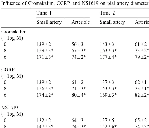
tered with vasopressin to make certain that pial artery Cromakalim, CGRP, and NS1619 (10 , 10 M) diameter was equivalent in the absence and presence of elicited reproducible pial small artery 120–160 mm) and vasopressin. The vehicle for all agents was 0.9% saline, arteriole (50–70 mm) vasodilation (Table 1). In these which had no effect on pial artery diameter. Confirmation statistical comparisons, there were 5 degrees of freedom of vasopressin receptor blockade was determined by for each agonist and the level of significance was P,
comparing responses to this agonist before and after 0.001. Vasopressin (40 pg / ml) increased pial small artery antagonist administration. Time control experiments were diameter from 14765 to 16067mm, n56. During coad-conducted in a separate series of animals and were ministration of U46619 (0.3 ng / ml) with vasopressin (40 designed to obtain responses to drugs initially and then 1 h pg / ml), there was no net change in pial artery diameter later (designated as time 1 and time 2 in Table 1). (150614 vs. 155614 mm). Under such conditions of equivalent baseline diameter, vasopressin / U46619 coad-2.2. Statistical analysis ministered with cromakalim, CGRP or NS 1619 attenuated
1
pial small artery dilation to these K channel agonists As described above, two different size pial arteries were (Fig. 1). Attenuated responses were fully restored when measured for each intervention. In time control experi- these agonists were coadministered with vasopressin and ments, the reproducibility of such measurements was 63 the vasopressin antagonist MEAVP (5mg / kg, i.v.) (Fig. 1).
mm for vessels between 120 and 160mm and 61mm for In these statistical comparisons, there were 4 degrees of vessels between 50 and 70 mm in diameter. Pial arteriolar freedom for each agonist and the level of significance was P,0.001. Similar effects were observed in pial arterioles. Since the concentration of substances detected in CSF reflect but may not be equivalent numerically to such
Table 1 changes within cells, the influence of a higher
concen-a
Influence of Cromakalim, CGRP, and NS1619 on pial artery diameter tration of vasopressin (400 pg / ml) on K and K
ATP ca
channel agonist induced vasodilation was also investigated.
Time 1 Time 2
These experiments were also conducted under conditions Small artery Arteriole Small artery Arteriole
of equivalent baseline diameter. Therefore, during coad-Cromakalim
ministration of U46619 (0.3 ng / ml) with vasopressin (400 (21og M)
pg / ml), there was no net change in pial artery diameter
0 13962 5663 14363 6162
(143618 vs. 148618 mm). Under such conditions of 8 15963* 6763* 16363* 7362*
6 17163* 7462* 17764* 7962* equivalent baseline diameter, vasopressin / U46619 coad-ministered with cromakalim, CGRP, and NS1619 further
CGRP 1
attenuated pial small artery dilation to these K channel (21og M)
agonists (1161 and 2261 vs. 161 and 362% for
0 13962 6162 13763 6261
28 26
cromakalim 10 , 10 M in the absence and presence of 8 15663* 7163* 15363* 7361*
6 17462* 8064* 16963* 8262* vasopressin, respectively n56). Similar effects were ob-served in pial arterioles.
NS1619 (21og M)
3.2. Role of vasopressin in impaired KATP and Kca
0 13262 6463 13765 6562
channel agonist induced pial artery dilation following 8 14763* 7463* 15266* 7463*
6 15664* 8063* 16267* 8063* FPI
a
Values aremm6S.E.M., n55. *
Fig. 1. Influence of coadministered vasopressin (40 pg / ml) or vasopressin and the vasopressin antagonist MEAVP (5mg / kg, i.v.) on pial small artery
28 26
dilation to cromakalim, CGRP, and NS1619 (10 , 10 M) under conditions of equivalent baseline diameter, n56. *P,0.05 compared to control.
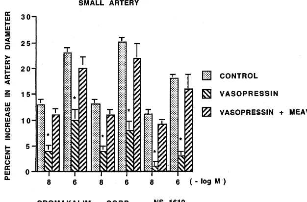
artery dilation was attenuated within 1 h post FPI (Figs. 261% for vasopressin 40 and 400 pg / ml before and after 2–4). In animals pretreated with the vasopressin antagonist MEAVP, respectively, n56). MEAVP did not have any MEAVP, such impaired vasodilation was partially pre- significant effect on pial artery diameter.
vented, though responses were still attenuated compared to control (Figs. 2–4). Similar effects were observed in pial
3.3.1. Blood chemistry and intensity of injury arterioles.
Blood chemistry values were obtained at the beginning and the end of all experiments. These values were: 3.3. Confirmation of effective receptor blockade
7.4660.02, 3465, and 9569 mmHg vs. 7.4560.02, 3566, and 9468 mmHg for pH, P , and P respectively
Vascular responses to vasopressin were blocked by CO2 O2
MEAVP (5 mg / kg i.v.) (961 and 1561 vs. 161 and before and after injury. Administration of MEAVP did not
28 26 28 26
Fig. 2. Influence of cromakalim (10 , 10 M) on pial small artery Fig. 3. Influence of CGRP (10 , 10 M) on pial small artery diameter diameter before (control), after fluid percussion brain injury (FPI), and before (control), after fluid percussion brain injury (FPI), and after FPI in after FPI in animals pretreated with the vasopressin antagonist MEAVP (5 animals pretreated with the vasopressin antagonist MEAVP (5mg / kg, mg / kg, i.v.), n56. *P,0.05 compared to control. 1P,0.05 compared i.v.), n56. *P,0.05 compared to control. 1P,0.05 compared to
NS1619 induced pial artery dilation to a greater extent than that observed with coadministration of vasopressin (40 pg / ml). Although the precise concentration at the receptor level is uncertain, the lower concentration (e.g., 40 pg / ml) is approximately the one observed for vasopressin in cortical periarachnoid CSF 1 h after FPI [5]. Since CSF concentrations reflect but are not equivalent to changes in substance concentration at the receptor level (where the effective concentration presumably is higher), these data support the functional significance of the interaction
1
between vasopressin and K channel activators.
Another series of experiments were designed to further investigate the functional significance of the above
de-1
scribed modulatory role of vasopressin in K channel mediated vasodilation. In particular, cromakalim, CGRP, and NS1619 induced pial artery dilation was attenuated within 1 h of FPI, consistent with previous studies [2,4]. New data from the present study show that MEAVP
1
partially prevented such diminished K channel agonist vasodilation post insult. These data suggest that vasopres-sin contributes to KATP and Kca channel function impair-ment observed following FPI. Previous studies showing
28 26 that MEAVP attenuated pial artery vasoconstriction in-Fig. 4. Influence of NS1619 (10 , 10 M) on pial small artery diameter
before (control), after fluid percussion brain injury (FPI), and after FPI in duced by FPI (5) indicate that vasopressin contributes to animals pretreated with the vasopressin antagonist MEAVP (5mg / kg, impaired cerebral hemodynamics following brain injury. In i.v.), n56. *P,0.05 compared to control. 1P,0.05 compared to
that systemic MEAVP blocked the vascular action of absence of MEAVP.
topical vasopressin without affecting the response to other substances (10), these data indicate that this antagonist was selective for vasopressin and that it crosses the blood brain significantly affect blood chemistry values. The amplitude barrier in sufficient quantity.
of the pressure pulse, used as an index of injury intensity, Cellular sites of origin for vasopressin detected in CSF
was 1.960.1 atm. in previous studies [5] include neurons, glia, vascular
smooth muscle, and endothelial cells. The present ex-perimental design, however, does not allow any conclusion
4. Discussion to be drawn with respect to cellular sites of origin for
vasopressin. Because MEAVP had no significant effect on Results of the present study show that coadministration baseline pial artery diameter, vasopressin probably has of vasopressin with cromakalim, CGRP, or NS1619 at- minimal contribution to cerebrovascular tone during
phy-1
tenuated vasodilation to these K channel agonists in a siologic conditions. In that pial small arteries exhibited non injury state. Because vasopressin had a vascular effect about the same percentage decrease in responsiveness to
1
of its own, U46619 was coadministered with vasopressin K channel activators following FPI as that observed with in a concentration that resulted in a no net change in pial pial arterioles, these data also suggest that there are artery diameter. Therefore, vasopressin, in effect, was probably minimal regional segmental vascular differences
1 1
coadministered with K channel activators under con- in altered K channel agonist activity following FPI. ditions of equivalent baseline diameter. Because previous Previous studies have investigated the selectivity of the studies have shown that U46619, in concentrations higher agents used as probes for KATP and Kca channel activation than that used in the present study (1 vs. 0.3 ng / ml), did induced pial artery dilation. Cromakalim induced pial not have any effect on pial dilation to cromakalim and artery dilation has been observed to be blocked by CGRP (2), vasopressin alone appears to exert these glibenclamide and unchanged by iberiotoxin, KATPand Kca
inhibitory effects. Similar unpublished studies have ob- channel antagonists respectively [7]. Conversely, NS1619 served that U46619 (1 ng / ml) did not affect NS1619 induced pial artery dilation was blocked by iberiotoxin and induced pial artery dilation. Additional experiments were unchanged by glibenclamide [3,4,7]. These data suggest performed to determine if there was a dose response that cromakalim and NS1619 are selective KATP and Kca
1
hyperpolari-zation of cerebral vascular muscle in vitro [23], and cross tributes to impaired KATP and Kca channel function after selectivity experiments have similarly been performed brain injury.
supportive of its selectivity for the KATP channel in the piglet [7]. Inclusion of data for CGRP in the present study,
therefore, lends physiologic functional perspective to re- Acknowledgements
sults indicative of vasopressin’s modulatory role in KATP
channel vascular function. However, it has also been The authors thank Miriam Kulkarni and John Ross for observed that NS1619 may additionally possess calcium technical assistance with performance of the experiments. channel antagonistic activity and, therefore, may not be This research was supported by grants from the National useful as a probe for Kca channel activation [14]. In Institutes of Health.
contrast, recent observations in the piglet show that vasoconstrictor responses to the calcium channel agonist
Bay K8644 were unchanged in the presence of NS1619 References [3]. These results suggest that NS1619 has no calcium
channel blocking activity and, therefore, may be consid- [1] W.M. Armstead, Superoxide generation links protein kinase C
1
ered to be selective for activation of Kca channels in the activation to impaired ATP-sensitive K channel function after brain injury, Stroke 30 (1999) 153–159.
newborn pig. 1
[2] W.M. Armstead, Brain injury impairs ATP-sensitive K channel Previous studies have observed that vasopressin is
function in piglet cerebral arteries, Stroke 28 (1997) 2273–2280.
released into CSF and contributes to altered dilation to the 1
[3] W.M. Armstead, Role of activation of calcium sensitive K channels opioid dynorphin following FPI in the newborn pig [5]. and cAMP in opioid-induced pial artery dilation, Brain Res. 747 Results of the present study extend the latter observations (1997) 252–258.
1
[4] W.M. Armstead, Role of impaired cAMP and calcium sensitive K to indicate that vasopressin also modulates KATP and Kca
channel function in altered cerebral hemodynamics following brain channel agonist mediated vascular activity following FPI,
injury, Brain Res. 768 (1997) 177–184.
suggestive of more distal signal transduction impairment [5] W.M. Armstead, Role of vasopressin in altered pial artery responses post insult. Although the mechanism for such vasopres- to dynorphin and b endorphin following brain injury, J.
Neuro-1
sinergic modulation of K channel function following FPI trauma 13 (1996) 115–123.
[6] W.M. Armstead, Influence of brain injury on vasopressin-induced is uncertain, one possibility could relate to a change in the
pial artery vasodilation: role of superoxide anion, Am. J. Physiol. vascular response to vasopressin following FPI, resulting
270 (1996) H1272–H1278.
1
in physiologic antagonism of K channel induced vasodi- 1
[7] W.M. Armstead, Role of ATP-sensitive K channels in cGMP-lation. Specifically, vasopressin reverses from a dilator to a mediate pial artery vasodilation, Am. J. Physiol. 270 (1996) H423– vasoconstrictor following FPI [6]. Such vasoconstriction H426.
[8] W.M. Armstead, C.D. Kurth, Different cerebral hemodynamic could, therefore, oppose the ability of KATP and Kca
responses following fluid percussion brain injury in the newborn and channel agonists to vasodilate.
juvenile pig, J. Neurotrauma 11 (1994) 487–497.
1
Alternatively, vasopressin could also indirectly alter K [9] W.M. Armstead, C.W. Leffler, D.W. Busija, R. Mirro, Vasopressin channel agonist vasodilation. Support for this position and prostanoid mechanisms in control of cerebral blood flow in comes from the observation that vasopressin impairs KATP hypotensive newborn pigs, Am. J. Physiol. 258 (1990) H408–H413. [10] W.M. Armstead, R. Mirro, D.W. Busija, C.W. Leffler, Vascular and Kca channel mediated vasodilation even in the absence
responses to vasopressin are tone dependent in the cerebral circula-of traumatic brain injury (and under conditions circula-of
equiva-tion of the newborn pig, Circ. Res. 64 (1989) 136–144.
lent baseline diameter). While cellular studies have shown [11] W.M. Armstead, R. Mirro, S.L. Zuckerman, C.W. Leffler, Vas-a similVas-ar modulVas-atory role of vVas-asopressin for the KATP opressin modulates cerebrovascular responses to opioids in newborn channel in isolated pig coronary artery smooth muscle cells pigs, J. Pharmacol. Exp. Ther 260 (1992) 1107–1112.
[12] D.S. DeWitt, D.C. Prough, C.L. Taylor, M. Whitley, Reduced [26], results of the present study are the first to
demon-cerebral blood flow, oxygen delivery, and electroencephalographic strate such a relationship between vasopressin and the Kca activity after traumatic brain injury and mild hemorrhage in cats, J.
channel. In unrelated studies, endothelin-1 has been ob- Neurosurg. 76 (1992) 812–821.
served to contribute to KATPchannel mediated vasodilator [13] L. Edvinsson, R. Ekman, I. Jansen, J. McCulloch, R. Uddman, impairment following FPI via a protein kinase C (PKC) Calcitonin gene related peptide and cerebral blood vessels: dis-tribution and vasomotor effects, J. Cereb. Blood Flow Metab. 7 dependent generation of superoxide anion [1,15]. The role
(1987) 720–728. of PKC activation in vasopressin induced impairment of
[14] M. Holland, P.D. Langton, N.B. Standen, J.P. Boyle, Effects of the KATPand Kcachannel mediated vasodilation following FPI BK channel activator N1619, on rat cerebral artery smooth muscle,
ca
is uncertain. Additionally, brain injury could alter the Br. J. Pharmacol. 117 (1996) 119–129.
1 1
number or binding of K channels available for activation, [15] T. Kasemsri, W.M. Armstead, Endothelin impairs ATP-sensitive K channel function after brain injury, Am. J. Physiol. 273 (1997) the degree of hyperpolarization that subsequently occurs or
H2639–H2647.
the ultimate response to hyperpolarization itself. 1
[16] T. Kitazono, D.D. Heistad, F.M. Faraci, Role of ATP-sensitive K In conclusion, results of the present study show that channels in CGRP-induced dilation of basilar artery in vivo, Am. J. vasopressin blunts KATP and Kca channel mediated cere- Physiol. 265 (1993) H581–H585.
Endogenous opioids may mediate secondary damage after ex- [23] A. Saito, T. Makaki, Y. Uchiyama, T.J.F. Lee, K. Goto, Calcitonin perimental brain injury, Am. J. Physiol. 253 (1987) E565–E574. gene related peptide and vasodilator nerves in large cerebral arteries [18] T.K. McIntosh, R. Vink, L. Noble, I. Yamakami, S. Fernyak, H. of cats, J. Pharmacol. Exp. Ther. 248 (1989) 455–462.
Soares, A.I. Faden, Traumatic brain injury in the rat: Characteriza- [24] V. Shankar, W.M. Armstead, Opioids contribute to hypoxia-induced
1
tion of a lateral fluid percussion model, Neuroscience 28 (1989) pial artery dilation through activation of ATP-sensitive K channels,
233–244. Am. J. Physiol. 269 (1995) H997–H1002.
[19] M.T. Nelson, J.B. Potlak, J.F. Worley, N.B. Standen, Calcium [25] J.J. Tepas, M.L. Dokler, Critical care of the injured child, Semin. channels, potassium channels and voltage dependence of arterial Pediatr. Surg. 4 (1995) 120–127.
1
smooth muscle tone, Am. J. Physiol. 259 (1990) C3–C18. [26] T. Wakatsuki, Y. Nakaya, I. Inoue, Vasopressin modulates K [20] M.T. Nelson, J.M. Quayle, Physiological roles and properties of channel activities of cultured smooth muscle cells from porcine
potassium channels in arterial smooth muscle, Am. J. Physiol. 268 coronary artery, Am. J. Physiol. 263 (1992) H491–H496. (1995) C799–C822. [27] E.P. Wei, W.D. Dietrich, J.T. Povlishock, R.M. Navari, H.A. Kontos, [21] M.L. Prins, S.M. Lee, C.L.Y. Cheng, D.P. Becker, D.A. Hovda, Fluid Functional, morphological, and metabolic abnormalities of the percussion brain injury in the developing and adult rat: a compara- cerebral microcirculation after concussive brain injury in cats, Circ. tive study of mortality, morphology intracranial pressure and mean Res. 46 (1980) 37–47.
arterial blood pressure, Dev. Brain. Res. 95 (1996) 272–282. [22] J.G. Rodriguez, S.T. Brown, Childhood injuries in the United States,